
Acute coronary syndrome for Emd emergency medicine exam should cover everything
Reading File
Now let me search the library in parallel for all ACS-related sections:
Finding Sources
Finding Sources
Excellent! I have the key sources. Let me now read the main ACS sections from Rosen's Emergency Medicine and Tintinalli's (the two primary EM textbooks) plus Frameworks for Internal Medicine (which has a comprehensive overview), all in parallel:
Finding Sources
Reading File
Reading File
Good. Now let me get the main ACS content from the Frameworks for Internal Medicine book (which has the most comprehensive overview) and search for specific topics in Tintinalli's and Rosen's:
Reading File
Finding Sources
Finding Sources
Reading File
Finding Sources
Reading File
Finding Sources
Reading File
Finding Sources
Excellent - I have rich content from Rosen's block 11. Let me now get the management/treatment sections and Tintinalli complications in parallel:
Reading File
Reading File
I now have rich content from multiple emergency medicine textbooks. Let me get the final key section - the management/treatment details from Rosen's:
Reading File
Finding Sources
I now have comprehensive content. Let me get the final ECG images and fetch one key image while also getting the Tintinalli Tintinalli ACS treatment section:
Loading Image
Loading Image
I now have all the content I need. Let me compile the comprehensive ACS notes:Here is the comprehensive ACS guide for your EMD Emergency Medicine exam:
ACUTE CORONARY SYNDROME (ACS) - Complete EMD Exam Guide
Sources: Rosen's Emergency Medicine, Tintinalli's Emergency Medicine, Frameworks for Internal Medicine
1. DEFINITION & CLASSIFICATION
ACS describes a spectrum of clinical syndromes resulting in myocardial ischemia with or without infarction. Diagnosis relies on three pillars: history + ECG + biomarkers.
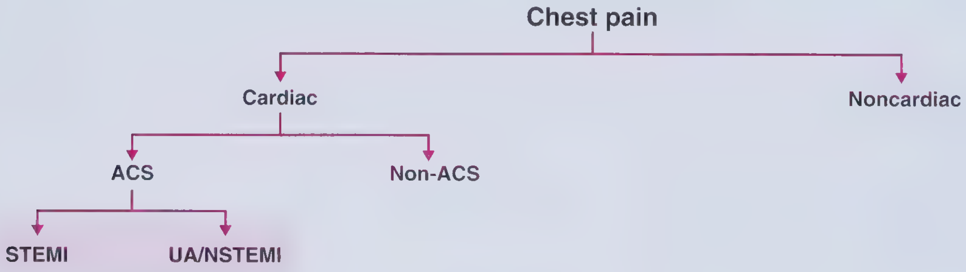
Classification of Chest Pain:
| Subtype | ECG | Troponin | Mechanism |
|---|---|---|---|
| STEMI | ST elevation | Elevated | Complete occlusion |
| NSTEMI | ST depression / T-wave inversion / normal | Elevated | Partial occlusion |
| Unstable Angina (UA) | ST depression / T-wave inversion / normal | Normal | Partial occlusion/spasm |
Key distinction: STEMI vs UA/NSTEMI matters because only STEMI benefits from immediate reperfusion (PCI or fibrinolytics). UA vs NSTEMI are distinguished solely by troponin - UA has no myocardial necrosis.
- Frameworks for Internal Medicine, p. 33-34
2. PATHOPHYSIOLOGY
Most common mechanism: Rupture of an unstable/vulnerable atherosclerotic plaque - the plaque cap ruptures, exposing the lipid core, triggering platelet aggregation and thrombus formation.
Other causes:
- Stent thrombosis - in patients who recently underwent PCI
- Coronary artery embolism - traveling clot
- Coronary artery vasospasm - including Prinzmetal (variant) angina
- Provoked by: cocaine, amphetamines, marijuana, 5-fluorouracil, sumatriptan
- Active smokers constitute the majority of spontaneous vasospasm cases
- Type 2 MI - supply/demand mismatch (anemia, tachycardia, hypotension)
Plaque features that predispose to rupture:
-
Large lipid core
-
Thin fibrous cap
-
High macrophage/foam cell content
-
Low smooth muscle content
-
Frameworks for Internal Medicine, p. 34
3. CLINICAL PRESENTATION
Typical Symptoms
- Substernal chest pain/pressure/heaviness
- Radiation to upper extremity, jaw, or epigastrium
- Duration typically >20 minutes
- Associated: diaphoresis, nausea, syncope
Atypical Presentations (MEMORIZE - exam favorite)
"Angina equivalents" include:
- Dyspnea (especially in women and elderly)
- Fatigue, weakness
- Abdominal pain (especially in women >65)
- Delirium/altered mental status (elderly)
- Back or shoulder pain
High-risk groups for atypical presentation:
- Women - most likely to present with atypical symptoms
- Diabetics - autonomic neuropathy blunts pain
- Elderly - classic chest pain occurs in only ~50% of patients ≥85 years
- Post-operative patients
Pearl: One-third of women >65 with acute MI present with abdominal pain alone. Do NOT miss this.
- Rosen's Emergency Medicine, p. 4015-4017
4. ECG INTERPRETATION
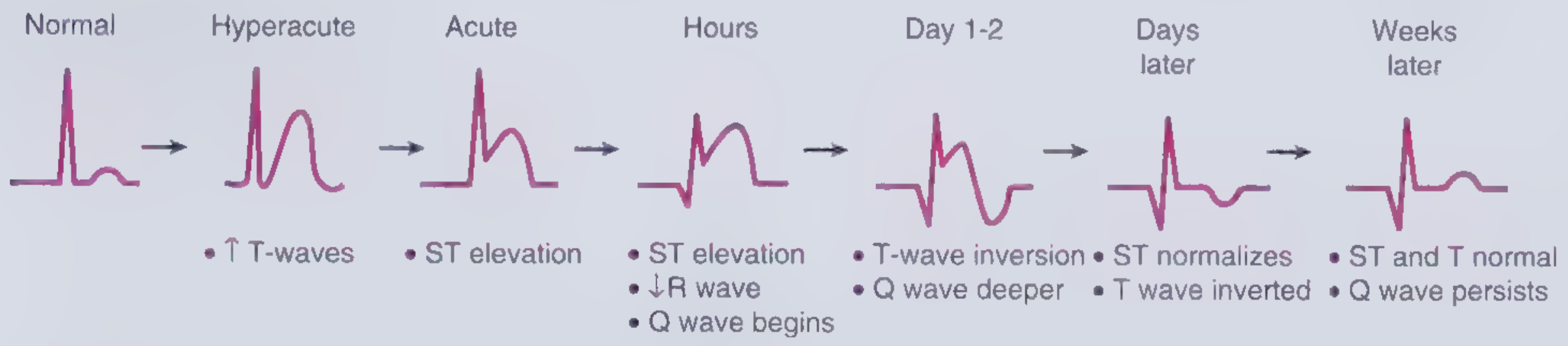
ECG Evolution of STEMI
| Stage | ECG Finding |
|---|---|
| Hyperacute (minutes) | Tall, broad-based, symmetrical T-waves |
| Acute (hours) | ST elevation + Q wave begins + R wave decreases |
| Hours later | Persistent ST elevation, Q waves |
| Day 1-2 | T-wave inversion, Q waves deeper |
| Days later | ST normalizes, T still inverted |
| Weeks later | ST and T normal, Q waves persist |
STEMI Diagnostic Criteria (J-point elevation in ≥2 contiguous leads)
| Lead group | Required elevation |
|---|---|
| V2-V3 in men ≥40 yrs | ≥0.2 mV |
| V2-V3 in men <40 yrs | ≥0.25 mV |
| V2-V3 in women | ≥0.15 mV |
| All other leads | ≥0.1 mV |
UA/NSTEMI ECG Findings
- New horizontal or downsloping ST depression ≥0.05 mV in ≥2 contiguous leads
- T-wave inversion ≥0.1 mV in ≥2 contiguous leads with prominent R wave or R/S >1
- ECG may also be entirely normal (does NOT exclude ACS)
Contiguous Lead Groups
- Anterior: V1-V6
- Inferior: II, III, aVF
- Lateral/Apical: I, aVL
- Right ventricular: V3R, V4R
- Posterior (inferolateral): V7-V9
Territory - Artery Correlations
| Territory | Leads | Artery |
|---|---|---|
| Anterior | V1-V4 | LAD |
| Inferior | II, III, aVF | RCA (most), LCx |
| Lateral | I, aVL, V5-V6 | LCx, Diagonal |
| Right Ventricle | V3R-V4R | RCA (proximal) |
| Posterior | V7-V9 (ST depression V1-V3) | RCA/LCx |
False-Positive ST Elevation (STEMI Mimics - EXAM CRITICAL)
- Early repolarization (benign)
- Left ventricular hypertrophy (LVH)
- Left bundle branch block (LBBB) - use Sgarbossa criteria
- Ventricular paced rhythm
- Brugada syndrome
- Acute pericarditis
- Myocarditis
- Subarachnoid hemorrhage
- Hyperkalemia
- Stress cardiomyopathy (Takotsubo)
- Cholecystitis (RCA territory)
False-Negative (Can Miss ACS)
- Prior MI with Q waves + persistent ST elevation
- LBBB
- Ventricular paced rhythm
- The initial ECG is nondiagnostic in ~50% of ED patients ultimately diagnosed with STEMI
- ~20% of patients ultimately diagnosed with AMI have nondiagnostic initial ECGs
KEY RULE: A single normal ECG does NOT exclude ACS. Serial ECGs + serial troponins are required.
- Rosen's Emergency Medicine, p. 411-413; Frameworks for Internal Medicine, p. 33
5. BIOMARKERS
Troponin (I or T) - Gold Standard
- Cardiac-specific (cTnI, cTnT)
- Rise begins: 3-6 hours after onset
- Peak: ~24 hours
- Duration of elevation: 7-10 days (TnI), up to 14 days (TnT)
- High-sensitivity troponin (hsTn): Can detect elevation within 1-3 hours; enables 0h/1h or 0h/2h protocols
- UA = no rise in troponin; NSTEMI = troponin rises
Other Biomarkers
| Marker | Rise | Peak | Return to Normal |
|---|---|---|---|
| Myoglobin | 1-2 hrs | 4-6 hrs | 12-24 hrs |
| CK-MB | 3-6 hrs | 12-24 hrs | 48-72 hrs |
| Troponin I | 3-6 hrs | 12-24 hrs | 7-10 days |
| Troponin T | 3-6 hrs | 12-24 hrs | 10-14 days |
CK-MB usefulness: Can detect reinfarction (re-elevation after initial fall) because it returns to normal faster than troponin.
6. RISK STRATIFICATION SCORES
HEART Score (Designed specifically for ED use)
(Most important for EM exam)
| Variable | 0 | 1 | 2 |
|---|---|---|---|
| History | Non-specific | Mixed elements | Highly suspicious |
| ECG | Normal | Non-specific repolarization changes | Significant ST deviation |
| Age | <45 | 45-65 | >65 |
| Risk factors | No risk factors | 1-2 risk factors | ≥3 or hx of atherosclerosis |
| Troponin | ≤normal limit | 1-3x normal | >3x normal |
HEART Score interpretation:
- 0-3: Low risk - safe for discharge + outpatient workup
- 4-6: Moderate risk - observation + additional testing
- 7-10: High risk - early invasive strategy
TIMI Risk Score (for UA/NSTEMI - 7 variables, 1 point each)
- Age ≥65
- ≥3 CAD risk factors
- Prior coronary stenosis ≥50%
- ST deviation on presenting ECG
- ≥2 anginal events in prior 24h
- Prior aspirin use in past 7 days
- Elevated serum cardiac markers
Score 0-2 = low; 3-4 = intermediate; 5-7 = high
GRACE Score
- Based on: age, HR, SBP, creatinine, Killip class, cardiac arrest at admission, elevated markers, ST deviation
- Predicts in-hospital and 6-month mortality
- Originally derived for inpatients; the HEART score is preferred in the ED
EDACS (Emergency Department Assessment of Chest Pain Score)
-
Another validated ED-specific tool
-
Incorporates: age, sex, known CAD, diaphoresis, radiation, exertional pain
-
Rosen's Emergency Medicine, p. 570-577
7. WORKUP IN THE ED
Step 1 - 12-lead ECG within 10 minutes of arrival (do this first, always)
Step 2 - Serial ECGs: Repeat at 15-30 minute intervals if initial nondiagnostic
Step 3 - Biomarkers: Troponin at 0h and 3h (or 0h/1h for high-sensitivity)
Step 4 - Chest X-ray: Not diagnostic for ACS, but rules out other causes and assesses for pulmonary edema/mediastinal widening (aortic dissection)
Step 5 - Additional imaging if needed:
- Echocardiography - wall motion abnormalities (regional hypokinesis/akinesis) in ischemia
- Coronary CT Angiography (CCTA) - useful in low-to-intermediate risk patients; radiation 2-5 mSv; requires heart rate control; high NPV for CAD
Step 6 - Additional leads: Right-sided leads (V3R, V4R) for inferior STEMI → rule out RV infarction; posterior leads (V7-V9) for posterior MI
8. MANAGEMENT
Initial Universal Management (ALL ACS)
Mnemonic: MONA-B (with caveats)
| Drug | Details | Notes |
|---|---|---|
| Morphine | 2-4 mg IV (up to 0.1 mg/kg); repeat q5-30 min | Use if unresponsive to NTG; caution with hypotension |
| Oxygen | Target SpO2 ≥94% | Do NOT give routinely if SpO2 normal - may increase mortality |
| Nitrates (NTG) | 0.4 mg SL; can repeat x3; then IV 10 mcg/min titrated | CONTRAINDICATED with hypotension, inferior STEMI + RV involvement, recent PDE-5 inhibitor use |
| Aspirin | 162-325 mg chewed immediately (non-enteric coated) | Give to all unless true allergy |
| Beta-blockers | Oral metoprolol preferred; avoid IV in acute phase | Contraindicated: acute decompensated HF, cardiogenic shock, bradycardia, severe bronchospasm |
Antiplatelet Therapy (Dual Antiplatelet - DAPT)
P2Y12 inhibitors (add to aspirin):
| Drug | Loading Dose | Notes |
|---|---|---|
| Clopidogrel | 300-600 mg PO | Prodrug; delayed onset; lower potency |
| Ticagrelor | 180 mg PO | Preferred in ACS (PLATO trial); reversible; faster onset |
| Prasugrel | 60 mg PO | Only with PCI-confirmed anatomy; avoid if >75 yrs, <60 kg, prior stroke/TIA |
Note: For STEMI with planned PCI, give P2Y12 inhibitor at time of PCI or as soon as possible. For fibrinolysis, use clopidogrel (NOT ticagrelor/prasugrel).
Anticoagulation
| Agent | Use | Notes |
|---|---|---|
| UFH (Unfractionated Heparin) | STEMI (PCI), NSTEMI | 60 U/kg bolus (max 4000 U) + 12 U/kg/hr infusion; preferred for renal failure |
| LMWH (Enoxaparin) | NSTEMI preferred | 1 mg/kg SC q12h; do NOT use if CrCl <30 mL/min |
| Fondaparinux | NSTEMI/UA | 2.5 mg SC daily; avoid if PCI (risk of catheter thrombosis) |
| Bivalirudin | STEMI during PCI | Direct thrombin inhibitor; reduces bleeding risk |
9. STEMI-SPECIFIC MANAGEMENT
Time Targets (Exam Critical)
- Door-to-ECG: ≤10 minutes
- Door-to-balloon (primary PCI): ≤90 minutes
- Door-to-needle (fibrinolysis): ≤30 minutes
- If PCI-capable center, primary PCI is preferred over fibrinolysis
Reperfusion Strategy Decision
STEMI confirmed
    ↓
PCI-capable center available within 120 min of first medical contact?
    YES → Primary PCI
    NO  → Fibrinolysis (if no contraindications) within 30 min
Fibrinolytic Therapy
Indications: STEMI with symptom onset <12 hours, no PCI available within 120 min
Agents: tPA (alteplase), reteplase, tenecteplase (TNK-tPA - weight-based single bolus, most commonly used)
Absolute Contraindications:
- Any prior intracranial hemorrhage (ever)
- Ischemic stroke within 3 months
- Known structural cerebrovascular lesion (AVM)
- Known intracranial malignancy
- Active bleeding (not menses)
- Significant closed-head trauma within 3 months
- Suspected aortic dissection
Relative Contraindications:
- SBP >180 or DBP >110 at presentation
- History of chronic severe hypertension
- Ischemic stroke >3 months ago
- Current anticoagulation use
- Traumatic/prolonged (>10 min) CPR
- Major surgery within 3 weeks
- Recent internal bleeding (2-4 weeks)
- Pregnancy
- Active peptic ulcer disease
- Non-compressible vascular punctures
RV Infarction (inferior STEMI with RV involvement)
Triad: Hypotension + Elevated JVP + Clear lung fields
- ECG: ST elevation in V3R-V4R
- Management:
- IV fluids (preload-dependent RV)
- AVOID nitrates (will drop preload and cause profound hypotension)
- AVOID diuretics
- AVOID morphine (preload reduction)
- Consider atropine or pacing for bradycardia
- Early reperfusion is critical
Killip Classification (LV failure severity post-MI)
| Class | Finding | Mortality |
|---|---|---|
| I | No signs of HF | ~5% |
| II | Mild HF, rales <50% lung fields, S3 | ~10% |
| III | Pulmonary edema, rales >50% lung fields | ~40% |
| IV | Cardiogenic shock | ~80-90% |
10. NSTEMI/UA MANAGEMENT
Goal: Stabilize, risk-stratify, decide on invasive vs. conservative strategy.
Initial Management
- DAPT (aspirin + P2Y12 inhibitor)
- Anticoagulation (enoxaparin or UFH preferred; fondaparinux if high bleeding risk)
- Beta-blocker (oral, unless contraindicated)
- Statin (high-intensity: atorvastatin 80 mg or rosuvastatin 40 mg)
- Consider IV nitroglycerin for ongoing ischemia
Invasive vs. Conservative Strategy
Early invasive strategy (cardiac cath within 24-72h) indicated if:
- TIMI ≥3 or GRACE >140
- Refractory angina despite medical therapy
- Hemodynamic instability
- Elevated troponin
- New ST changes
- Signs of heart failure
- High HEART score (7-10)
Conservative (ischemia-guided) approach acceptable if:
- Low TIMI score, low HEART score
- No high-risk features
- Patient preference
11. COMPLICATIONS OF ACS
Dysrhythmias (most common complication, 72-100% of AMI)
Bradydysrhythmias:
| Dysrhythmia | Frequency |
|---|---|
| Sinus bradycardia | 35-40% |
| 1st degree AV block | 4-15% |
| 2nd degree AV block type I (Wenckebach) | 4-10% |
| 2nd degree AV block type II | 0.5-1% |
| 3rd degree (complete) AV block | 5-8% |
| Asystole | 1-5% |
Tachydysrhythmias:
| Dysrhythmia | Frequency |
|---|---|
| VPBs (ventricular premature beats) | 99% |
| Accelerated idioventricular rhythm (AIVR) | 50-70% |
| Non-sustained VT | 60-69% |
| Sinus tachycardia | 30-35% |
| AFib | 4-10% |
| Sustained VT | 2-6% |
| VFib | 4-7% |
AIVR (rate 60-100 bpm) is a reperfusion arrhythmia - often benign, no specific treatment needed.
VFib - most common cause of sudden cardiac death in the early MI period. Defibrillate immediately (200J biphasic).
Mechanical Complications (usually Day 3-7)
| Complication | Features | Treatment |
|---|---|---|
| Free wall rupture | Sudden death or cardiac tamponade; EMD/PEA | Emergency surgery; pericardiocentesis as bridge |
| Ventricular septal defect (VSD) | New harsh holosystolic murmur; step-up in O2 at RV | Emergency surgery; IABP bridge |
| Papillary muscle rupture | Acute severe MR, pulmonary edema; soft/absent murmur | Emergency surgery; IABP, vasodilators |
Cardiogenic Shock
- Most serious complication; mortality ~50-80%
- Defined: SBP <90 mmHg + signs of hypoperfusion
- Management: early PCI/revascularization, intra-aortic balloon pump (IABP), vasopressors (norepinephrine preferred), consider LVAD/Impella
Pericarditis (Dressler Syndrome)
- Post-MI pericarditis (2-10 weeks post-MI)
- Symptoms: fever, pleuritic chest pain, pericardial rub
- Treatment: ASA + colchicine; avoid NSAIDs/steroids in early post-MI period
Left Ventricular Aneurysm
- Persistent ST elevation weeks after STEMI
- Associated with VT, mural thrombus (anticoagulate), HF
12. SPECIAL POPULATIONS & SCENARIOS
Elderly Patients
- Higher morbidity/mortality; independent risk factor for death
- Higher rate of atypical presentation
- ~50% of STEMI patients ≥85 years present with heart failure on admission vs 1.7% in those <65
- No absolute age cutoff for revascularization per AHA guidelines
- Early invasive strategy still preferred, but consider quality of life and goals of care
Cocaine/Amphetamine-Induced ACS
- Mechanism: vasospasm + accelerated atherosclerosis + in-situ thrombosis
- Cocaine inhibits reuptake of catecholamines → vasoconstriction
- Management:
- Benzodiazepines (first-line for cocaine chest pain)
- AVOID beta-blockers (unopposed alpha stimulation → paradoxical vasoconstriction)
- NTG and calcium channel blockers (verapamil, diltiazem)
- Aspirin
- PCI if STEMI criteria met
LBBB and ACS (Sgarbossa Criteria)
Original criteria (treat as STEMI if score ≥3):
- Concordant ST elevation ≥1 mm in a lead with a positive QRS (+5 points)
- Concordant ST depression ≥1 mm in V1-V3 (+3 points)
- Discordant ST elevation ≥5 mm (+2 points)
Modified Sgarbossa (Smith Criteria): Discordant ST/S ratio >0.25 is more sensitive.
Type 2 MI
- Not from plaque rupture; from supply/demand mismatch
- Causes: tachyarrhythmias, anemia, hypotension, sepsis, respiratory failure
- Treat underlying cause; anticoagulation/PCI may not be indicated
13. NITRATE CONTRAINDICATIONS (Exam Must-Know)
Absolute contraindications to nitrates in ACS:
- SBP <90 mmHg (hypotension)
- Inferior STEMI with suspected/confirmed right ventricular infarction
- Use of PDE-5 inhibitors within:
- 24 hours (sildenafil, vardenafil)
- 48 hours (tadalafil)
- Results in severe refractory hypotension
14. BETA-BLOCKER USE IN ACS
Benefits: Reduce HR, BP, myocardial O2 demand; prevent arrhythmias; reduce infarct size
When to use: Oral beta-blockers within 24h in all ACS patients without contraindications
IV beta-blocker: Reserved for tachyarrhythmias or hypertension not controlled by oral agents
Contraindications:
- Signs of acute decompensated heart failure or cardiogenic shock
- High degree AV block (without pacemaker)
- Active bronchospasm/severe COPD
- Bradycardia (HR <60)
- SBP <100 mmHg
15. QUICK SUMMARY TABLE
| Feature | STEMI | NSTEMI | UA |
|---|---|---|---|
| ECG | ST elevation or new LBBB | ST depression, T-wave changes, or normal | ST depression, T-wave changes, or normal |
| Troponin | Elevated | Elevated | Normal |
| Mechanism | Complete occlusion | Partial occlusion | Partial occlusion/vasospasm |
| Reperfusion | Immediate (PCI/lytics) | Within 24-72h (invasive strategy) | Risk-stratify |
| P2Y12 choice | Ticagrelor or prasugrel (PCI) / clopidogrel (lytics) | Ticagrelor or clopidogrel | Same |
16. HIGH-YIELD PEARLS FOR EMD EXAM
- ECG within 10 min - every suspected ACS patient, no exceptions
- Single normal ECG does NOT exclude ACS - repeat serially
- Aspirin 162-325 mg chewed - non-enteric coated, give ASAP
- NTG is contraindicated in RV infarction - this is a classic exam vignette
- Avoid beta-blockers in cocaine-induced chest pain
- HEART score is the validated ED-specific risk tool (not TIMI/GRACE)
- Morphine caution - may increase adverse events in some ACS patients; not withheld universally but use judiciously
- Fibrinolysis absolute contraindication: any prior intracranial hemorrhage - no exceptions
- AIVR post-STEMI = reperfusion arrhythmia - do NOT treat
- Mechanical complications (VSD, papillary rupture, free wall rupture) peak at Day 3-7
- Door-to-balloon ≤90 min / Door-to-needle ≤30 min
- RV infarction triad: hypotension + elevated JVP + clear lungs → give fluids, not diuretics
- Rosen's Emergency Medicine (9780323757898) | Tintinalli's Emergency Medicine (9781260019933) | Frameworks for Internal Medicine (9781496359308)
This is a shared conversation. Sign in to Orris to start your own chat.