Can ultrasound detect appendicitis

Reading File
Searching Images

ultrasound appendicitis appendix sonography
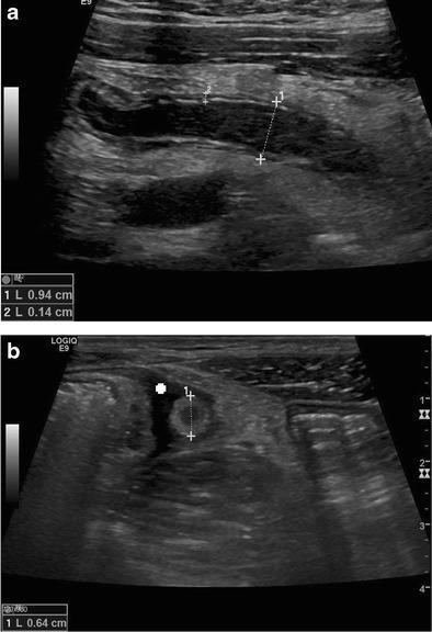
Educational diagnostic ultrasound images demonstrating acute appendicitis in longitudinal (a) and transverse (b) planes. Image (a) shows a longitudinal view of a distended, non-compressible appendix with an anteroposterior diameter of 0.94 cm and mural thickening of 0.14 cm. Hypoechoic free fluid is visible surrounding the structure. Image (b) shows the appendix in a transverse cross-section, displaying the classic 'target sign' (bull’s eye appearance), characterized by alternating hyperechoic and hypoechoic concentric rings representing the fluid-filled lumen, mucosa/submucosa, and muscularis layers. The transverse diameter is measured at 0.64 cm. These findings—specifically a total diameter greater than 6 mm, wall thickening, and periappendiceal fluid—are key diagnostic ultrasound criteria for acute appendicitis. The images serve as an educational example of graded-compression sonography for gastrointestinal pathology.
Educational diagnostic ultrasound images demonstrating acute appendicitis in longitudinal (a) and transverse (b) planes. Image (a) shows a longitudinal view of a distended, non-compressible appendix with an anteroposterior diameter of 0.94 cm and mural thickening of 0.14 cm. Hypoechoic free fluid is visible surrounding the structure. Image (b) shows the appendix in a transverse cross-section, displaying the classic 'target sign' (bull’s eye appearance), characterized by alternating hyperechoic and hypoechoic concentric rings representing the fluid-filled lumen, mucosa/submucosa, and muscularis layers. The transverse diameter is measured at 0.64 cm. These findings—specifically a total diameter greater than 6 mm, wall thickening, and periappendiceal fluid—are key diagnostic ultrasound criteria for acute appendicitis. The images serve as an educational example of graded-compression sonography for gastrointestinal pathology.

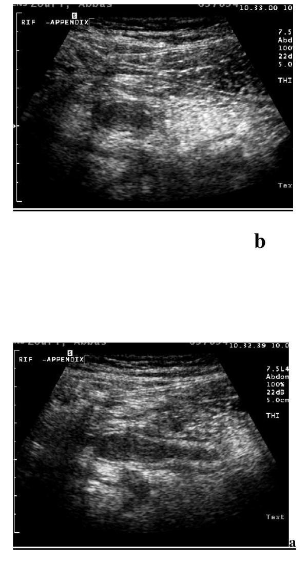
This diagnostic image displays Tissue Harmonic Imaging (THI) sonography of the right iliac fossa (RIF) demonstrating signs of acute appendicitis. The content is divided into two views: (a) a longitudinal scan and (b) a transverse scan, both acquired using a high-frequency (7.5 MHz) linear transducer. In the longitudinal view (a), the appendix appears as a blind-ended, aperistaltic, and non-compressible tubular structure with a significantly thickened wall and a total diameter exceeding 6 mm. In the transverse view (b), the inflamed appendix exhibits the characteristic 'target sign' or 'bull's-eye' appearance, consisting of concentric layers with alternating echogenicity, which corresponds to the histological layers of the appendiceal wall. These findings are hallmarks of appendiceal inflammation. The high-resolution THI technology enhances the visualization of these tissue interfaces compared to conventional ultrasound, aiding in definitive diagnosis for medical students and clinicians.
This diagnostic image displays Tissue Harmonic Imaging (THI) sonography of the right iliac fossa (RIF) demonstrating signs of acute appendicitis. The content is divided into two views: (a) a longitudinal scan and (b) a transverse scan, both acquired using a high-frequency (7.5 MHz) linear transducer. In the longitudinal view (a), the appendix appears as a blind-ended, aperistaltic, and non-compressible tubular structure with a significantly thickened wall and a total diameter exceeding 6 mm. In the transverse view (b), the inflamed appendix exhibits the characteristic 'target sign' or 'bull's-eye' appearance, consisting of concentric layers with alternating echogenicity, which corresponds to the histological layers of the appendiceal wall. These findings are hallmarks of appendiceal inflammation. The high-resolution THI technology enhances the visualization of these tissue interfaces compared to conventional ultrasound, aiding in definitive diagnosis for medical students and clinicians.

**Imaging Modality:** Abdominal Ultrasound (Greyscale sonography).

**Anatomical Region:** Right lower quadrant of the abdomen.

**Observed Pathology:** Acute appendicitis.

**Characteristic Visual Features:**
- **Transverse View:** The image displays a cross-sectional ("target" or "bullseye") appearance of the appendix.
- **Morphology:** There is a non-compressible, blind-ending tubular structure with an enlarged outer diameter measuring 11 mm (exceeding the standard diagnostic threshold of 6–7 mm).
- **Wall Features:** Circumferential wall thickening is visible with a central hypoechoic lumen.
- **Extra-appendiceal Findings:** Prominent hyperechoic (echogenic) periappendiceal fat is observed surrounding the structure, indicative of secondary inflammatory changes (fat stranding).
- **Secondary Cues:** On-screen calipers provide measurement verification of the distended structure.

**Key Diagnostic Features:** Dilated appendix (>6 mm) in the transverse plane combined with increased echogenicity of the surrounding mesenteric fat, classic sonographic hallmarks of acute inflammation.

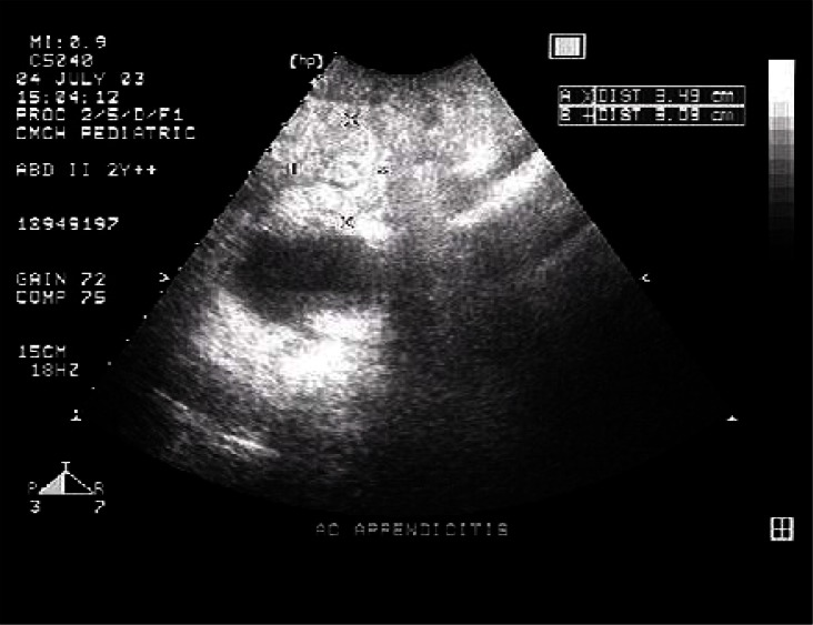
**Imaging Modality:** Abdominal Ultrasound (Greyscale sonography). **Anatomical Region:** Right lower quadrant of the abdomen. **Observed Pathology:** Acute appendicitis. **Characteristic Visual Features:** - **Transverse View:** The image displays a cross-sectional ("target" or "bullseye") appearance of the appendix. - **Morphology:** There is a non-compressible, blind-ending tubular structure with an enlarged outer diameter measuring 11 mm (exceeding the standard diagnostic threshold of 6–7 mm). - **Wall Features:** Circumferential wall thickening is visible with a central hypoechoic lumen. - **Extra-appendiceal Findings:** Prominent hyperechoic (echogenic) periappendiceal fat is observed surrounding the structure, indicative of secondary inflammatory changes (fat stranding). - **Secondary Cues:** On-screen calipers provide measurement verification of the distended structure. **Key Diagnostic Features:** Dilated appendix (>6 mm) in the transverse plane combined with increased echogenicity of the surrounding mesenteric fat, classic sonographic hallmarks of acute inflammation.

**Imaging Modality:** B-mode Ultrasound (Sonography)

**Anatomical Region:** Right lower quadrant of the abdomen

**Observed Pathology:** Acute appendicitis with appendicolith.

**Characteristic Visual Features:**
*   **Transverse View:** The image displays a cross-section of the vermiform appendix appearing as a non-compressible, blind-ending tubular structure.
*   **Target Sign:** A classic "target" or "bull’s-eye" appearance is visible, characterized by concentric layers of varying echogenicity representing the inflamed appendiceal wall layers (mucosa, submucosa, and muscularis propria).
*   **Appendicolith:** A distinct, highly echogenic (hyperechoic) foci is located within the lumen of the appendix, associated with subtle posterior acoustic shadowing, indicative of a fecalith or appendicolith.
*   **Wall Changes:** There is evidence of mural thickening and luminal distension.
*   **Surrounding Tissue:** Increased echogenicity of the periappendiceal fat is noted, suggestive of secondary inflammatory changes (fat stranding).

**Key Diagnostic Features:** The combination of a dilated appendiceal diameter, the presence of an intraluminal obstructing appendicolith, and the distinct target sign in the transverse plane are definitive sonographic markers for acute obstructive appendicitis.
**Imaging Modality:** B-mode Ultrasound (Sonography) **Anatomical Region:** Right lower quadrant of the abdomen **Observed Pathology:** Acute appendicitis with appendicolith. **Characteristic Visual Features:** * **Transverse View:** The image displays a cross-section of the vermiform appendix appearing as a non-compressible, blind-ending tubular structure. * **Target Sign:** A classic "target" or "bull’s-eye" appearance is visible, characterized by concentric layers of varying echogenicity representing the inflamed appendiceal wall layers (mucosa, submucosa, and muscularis propria). * **Appendicolith:** A distinct, highly echogenic (hyperechoic) foci is located within the lumen of the appendix, associated with subtle posterior acoustic shadowing, indicative of a fecalith or appendicolith. * **Wall Changes:** There is evidence of mural thickening and luminal distension. * **Surrounding Tissue:** Increased echogenicity of the periappendiceal fat is noted, suggestive of secondary inflammatory changes (fat stranding). **Key Diagnostic Features:** The combination of a dilated appendiceal diameter, the presence of an intraluminal obstructing appendicolith, and the distinct target sign in the transverse plane are definitive sonographic markers for acute obstructive appendicitis.

**Imaging Modality:** Ultrasound (Grayscale B-mode sonography).

**Anatomical Region:** Right lower quadrant (RLQ) of the abdomen.

**Observed Pathology:** Acute appendicitis.

**Characteristic Visual Features:**
The image displays a longitudinal view of a blind-ending, tubular structure in the right iliac fossa, consistent with the vermiform appendix. Key sonographic findings include:
*   **Increased Caliber:** Significant thickening and distension of the appendiceal diameter.
*   **Wall Thickening:** Distinct mural thickening with preservation of gut signatures (hypoechoic and hyperechoic layers).
*   **Luminal Distension:** The lumen appears fluid-filled and non-compressible.
*   **Periappendiceal Changes:** There is a surrounding zone of increased echogenicity in the adjacent mesenteric fat, indicative of inflammatory fat secondary to the infection.

**Diagnostic Features:** The combination of a non-compressible, blind-ending tubular structure exceeding 6mm in diameter with surrounding hyperechoic inflammatory fat is highly diagnostic of acute appendicitis. The orientation and anatomical marker on the UI confirm the probe placement over the McBurney's point region.
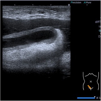
**Imaging Modality:** Ultrasound (Grayscale B-mode sonography). **Anatomical Region:** Right lower quadrant (RLQ) of the abdomen. **Observed Pathology:** Acute appendicitis. **Characteristic Visual Features:** The image displays a longitudinal view of a blind-ending, tubular structure in the right iliac fossa, consistent with the vermiform appendix. Key sonographic findings include: * **Increased Caliber:** Significant thickening and distension of the appendiceal diameter. * **Wall Thickening:** Distinct mural thickening with preservation of gut signatures (hypoechoic and hyperechoic layers). * **Luminal Distension:** The lumen appears fluid-filled and non-compressible. * **Periappendiceal Changes:** There is a surrounding zone of increased echogenicity in the adjacent mesenteric fat, indicative of inflammatory fat secondary to the infection. **Diagnostic Features:** The combination of a non-compressible, blind-ending tubular structure exceeding 6mm in diameter with surrounding hyperechoic inflammatory fat is highly diagnostic of acute appendicitis. The orientation and anatomical marker on the UI confirm the probe placement over the McBurney's point region.

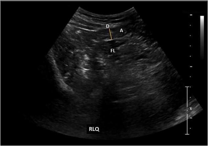
This diagnostic image is a B-mode grayscale abdominal ultrasound focusing on the right lower quadrant (RLQ). The image demonstrates a cross-sectional view of the vermiform appendix (labeled 'A'), appearing as a tubular, hypoechoic structure within the peritoneal cavity. A yellow measurement caliper ('D') indicates an outer diameter of 6 mm. Adjacent to the appendix, a small, localized periappendiceal fluid collection ('FL') is visible as an anechoic to hypoechoic area, suggesting localized inflammatory changes. The image quality allows for the differentiation of the appendix from surrounding soft tissue and bowel loops. This visual is clinically significant for the assessment of acute abdominal pain and the diagnostic evaluation of appendicitis. It serves as an educational example of RLQ sonography, illustrating target signs and fluid presence associated with inflammatory processes in the iliac fossa.

This diagnostic image is a B-mode grayscale abdominal ultrasound focusing on the right lower quadrant (RLQ). The image demonstrates a cross-sectional view of the vermiform appendix (labeled 'A'), appearing as a tubular, hypoechoic structure within the peritoneal cavity. A yellow measurement caliper ('D') indicates an outer diameter of 6 mm. Adjacent to the appendix, a small, localized periappendiceal fluid collection ('FL') is visible as an anechoic to hypoechoic area, suggesting localized inflammatory changes. The image quality allows for the differentiation of the appendix from surrounding soft tissue and bowel loops. This visual is clinically significant for the assessment of acute abdominal pain and the diagnostic evaluation of appendicitis. It serves as an educational example of RLQ sonography, illustrating target signs and fluid presence associated with inflammatory processes in the iliac fossa.

Finding Sources
Searching PubMed

"appendicitis" AND "ultrasound" AND "diagnosis"

Reading File
Reading File
Here is a thorough answer based on the Grainger & Allison's Diagnostic Radiology textbook and recent meta-analyses.

Can Ultrasound Detect Appendicitis?

Yes - ultrasound (US) is a well-established first-line tool for evaluating suspected appendicitis, though it has important limitations compared to CT.

Technique: Graded Compression Sonography

The standard approach is graded compression US: the probe is applied with gradually increasing pressure over the right iliac fossa to displace bowel loops and bring the appendix into view. It is particularly well suited for children and thin patients.

Ultrasound Signs of Acute Appendicitis

These are the key findings to look for (from Grainger & Allison's Diagnostic Radiology, p. 472):
SignDetail
Non-compressible blind-ending tubular structureThe primary finding
Diameter ≥ 7 mmOuter-to-outer wall measurement
No peristalsisAperistaltic on real-time imaging
AppendicolithHyperechoic focus with posterior acoustic shadowing
Hyperechoic surrounding fatReflects periappendiceal inflammation
Peri-appendiceal fluid or abscessIndicates advanced disease
Oedema of the caecal poleSecondary inflammatory change
Maximal tenderness over appendixSonographic McBurney's sign
The classic transverse cross-section appearance is the "target sign" (bull's eye) - concentric rings of alternating echogenicity representing the inflamed appendiceal wall layers.

Diagnostic Performance

  • Sensitivity: 78-98%
  • Specificity: 85-98% (Grainger & Allison's Diagnostic Radiology, p. 472)
A recent 2025 systematic review and meta-analysis (Castro-Luna et al., Pediatr Radiol 2025) confirmed these performance characteristics across pediatric populations comparing US, CT, and MRI. In pregnant women, Wang et al. (J Clin Ultrasound, 2023) found US remains diagnostically useful despite the technical challenge of a gravid uterus.

Pitfalls and Limitations

False negatives can occur with:
  • Focal tip appendicitis (only the tip is inflamed)
  • Retrocaecal appendicitis (obscured by caecum)
  • Gangrenous or perforated appendicitis - the appendix may become compressible after perforation (38% of pediatric perforations, 55% of adult perforations)
  • Gas-filled appendix (obscures the lumen)
  • Obesity and excessive bowel gas (operator-dependent)
False positives can arise from:
  • Dilated fallopian tube
  • Peri-appendicitis / inflammatory bowel disease
  • Inspissated stool mimicking an appendicolith
The biggest practical limitation: in most hands, a normal appendix is not visualised, so a negative US where the appendix is simply not seen is of little diagnostic value. The textbook notes that a multicentre German study of 2,280 patients found US did not result in proven clinical benefit when used routinely.

When US Is Most Useful

US is recommended over CT in these groups (lower radiation concern):
  • Children (when diagnostic doubt exists)
  • Young women (higher incidence of tubal/ovarian pathology mimicking appendicitis)
  • Pregnant patients
  • Clinically indeterminate cases - US adds the most value here, less so when clinical suspicion is already very high or very low
US is also useful for diagnosing conditions that mimic appendicitis: ectopic pregnancy, ovarian cyst/torsion, salpingitis, diverticulitis, mesenteric adenitis, Crohn's disease, and urolithiasis.

US vs. CT

CT remains the gold standard for accuracy (sensitivity/specificity >95%), especially in adults, because it visualises the appendix in virtually all cases and is not operator-dependent. However, it carries radiation exposure. The typical imaging algorithm is: US first (especially in children/young women/pregnancy), then CT if US is non-diagnostic.

Here are real ultrasound images of appendicitis for visual reference:
Appendicitis ultrasound - longitudinal and transverse views with target sign
Longitudinal (a) and transverse (b) views. Note the non-compressible structure measuring 9.4 mm in diameter and the classic "target sign" in the transverse plane.
Appendicitis ultrasound - dilated appendix with periappendiceal fat stranding
Transverse view: dilated appendix measuring 11 mm (>6-7 mm threshold) with surrounding hyperechoic fat stranding - hallmarks of acute appendicitis.
  • Grainger & Allison's Diagnostic Radiology, p. 472-473
This is a shared conversation. Sign in to Orris to start your own chat.