
Explain me ANATOMY of NOSE clearly and in simple words
Reading File
Now let me search the medical library for nose anatomy content in parallel:
Finding Sources
Finding Sources
Searching the Web
anatomy of nose external internal nasal cavity turbinates simple explanation
I have great sources. Let me now read the most relevant anatomy sections in parallel:
Reading File
Reading File
Reading File
Finding Sources
Excellent content found. Let me now read the key anatomy sections and fetch the diagrams in parallel:
Reading File
Reading File
Reading File
Loading Image
Loading Image
Loading Image
Now let me fetch a few more key diagrams and read sections on blood supply, nerves, and Kiesselbach's plexus:
Reading File
Loading Image
Loading Image
I now have rich content and beautiful diagrams from the textbooks. Let me fetch the blood supply diagram and the sinus ostia diagram:
Loading Image
Loading Image
I now have all the content and diagrams needed to write a comprehensive, well-illustrated anatomy of the nose. Here is the full explanation:
Anatomy of the Nose - Simple & Clear
The nose is the entry point of your respiratory system. It does much more than just let air in - it warms, humidifies, and filters every breath you take. Structurally, it has two main parts: the external nose (what you see on your face) and the internal nasal cavity (the space inside).
1. External Nose
The external nose is pyramid-shaped, with its tip (apex) pointing forward. Think of it like a tent held up by a frame of bones and cartilages.
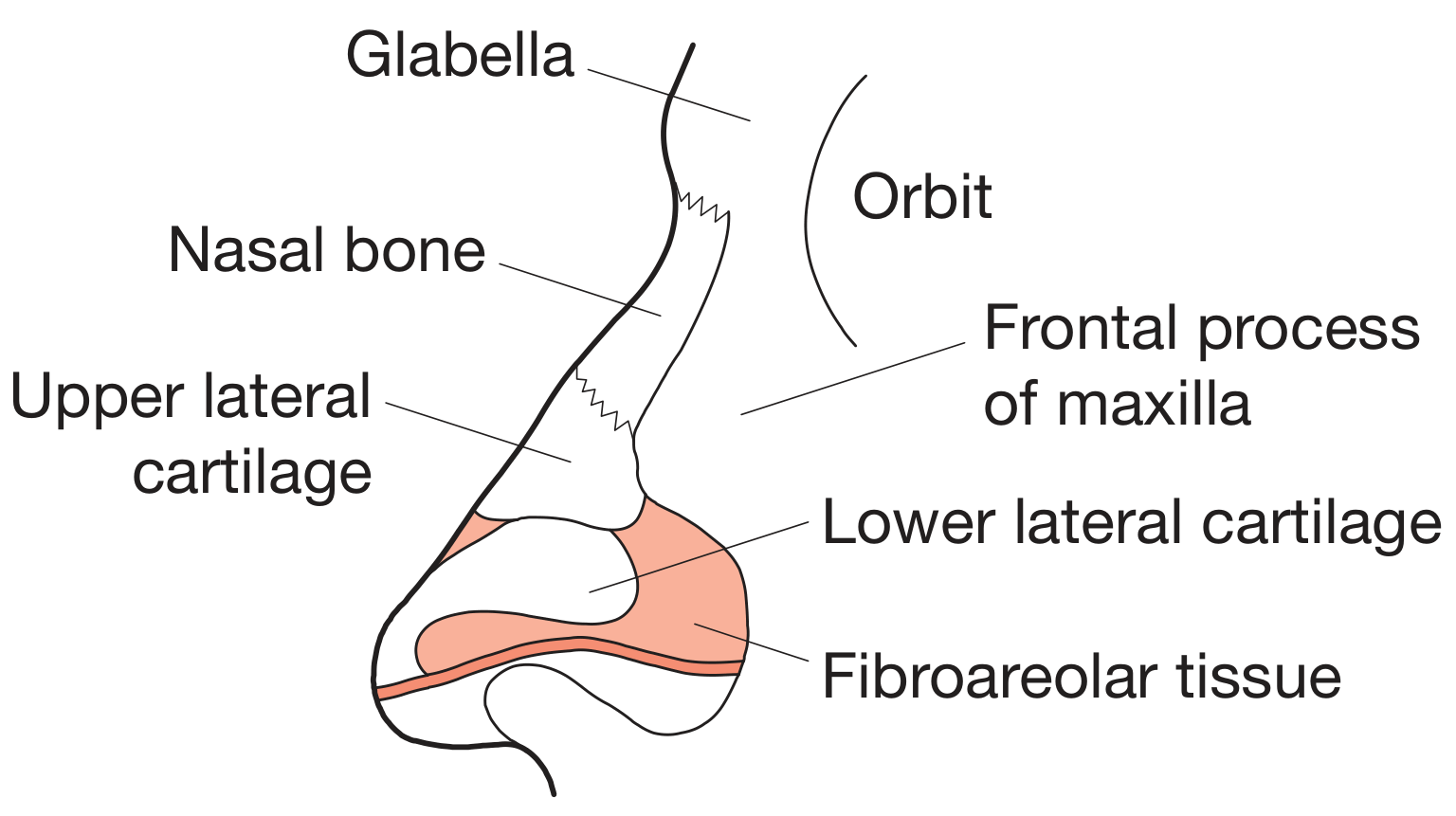
Skeleton of the External Nose:
The upper part is supported by bone, and the lower part is supported by cartilage:
| Part | What Supports It |
|---|---|
| Upper 1/3 (root/bridge) | Nasal bones + frontal bones + maxilla |
| Middle 1/3 | Upper lateral cartilages (lateral process of septal cartilage) |
| Lower 1/3 (tip & nostrils) | Lower lateral cartilages (major alar cartilages), minor alar cartilages, septal cartilage |
The nostrils (nares) are the two openings at the bottom. Just inside each nostril is a small widened space called the nasal vestibule, which is lined with skin and has coarse hairs (vibrissae) that trap large dust particles.
(Source: Gray's Anatomy for Students)
2. Nasal Septum - The Dividing Wall
The nasal septum is a wall in the midline that divides the nose into right and left halves. It has three components:
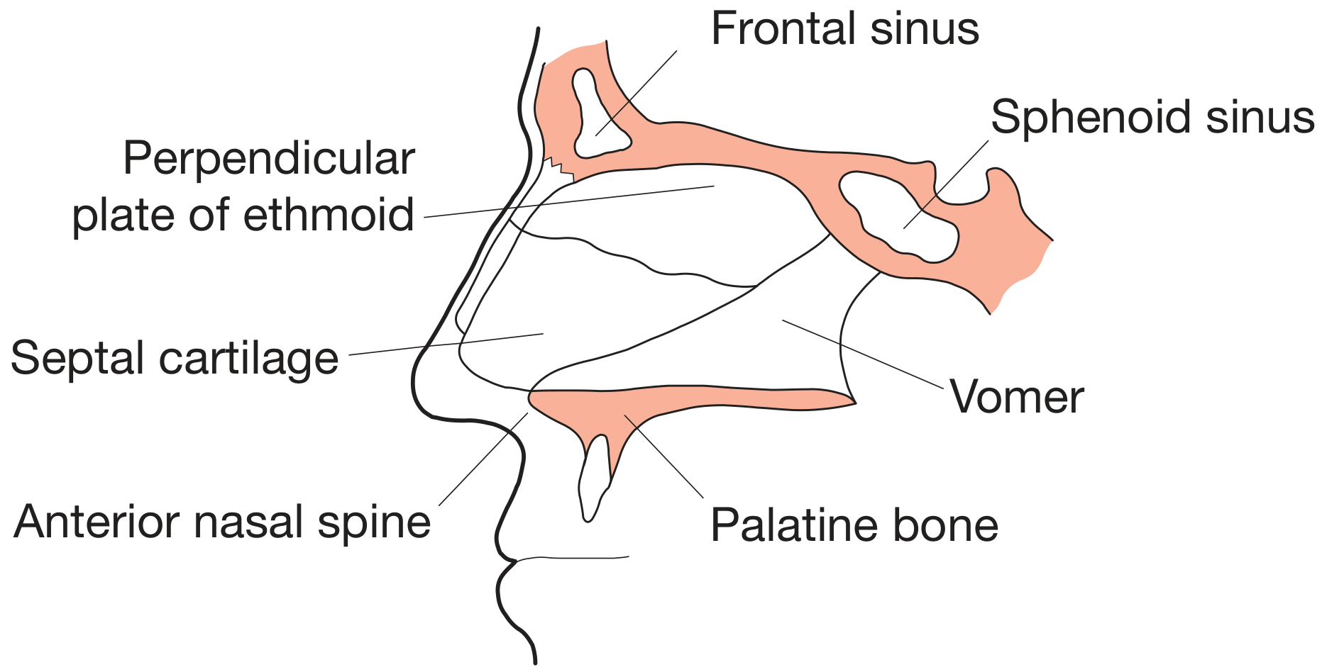
- Septal cartilage - the anterior (front) part - the flexible part you can squeeze
- Perpendicular plate of the ethmoid bone - the upper bony part
- Vomer bone - the lower bony part, sitting on the floor
A deviated septum (when this wall leans to one side) is very common and can cause nasal obstruction.
(Source: Bailey and Love's Short Practice of Surgery)
3. Nasal Cavity - The Inside Space
The nasal cavity runs from the nostrils at the front to the choanae (two openings) at the back, where it connects to the throat (nasopharynx). Each cavity has:
- A roof (ceiling) - formed by the cribriform plate of the ethmoid, which transmits smell nerves
- A floor - the hard palate (the roof of your mouth)
- A medial wall - the nasal septum
- A lateral wall - the most complex side, with the turbinates
4. Turbinates (Conchae) - The Scroll-Like Shelves
The lateral (outer) wall has three curved, shelf-like bony projections called turbinates (or conchae). Think of them like speed bumps that slow down and swirl the air.
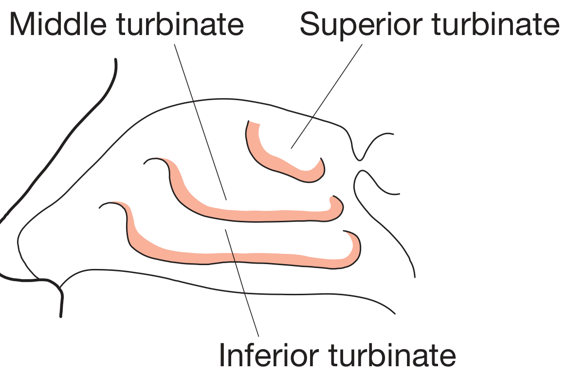
| Turbinate | Location | Main Function |
|---|---|---|
| Inferior turbinate | Largest, bottom shelf | Warms and moistens air; nasolacrimal duct opens below it |
| Middle turbinate | Middle shelf | Key surgical landmark; sinuses drain near here |
| Superior turbinate | Smallest, top shelf | Olfactory region nearby |
Below each turbinate is a groove called a meatus:
- Inferior meatus - nasolacrimal duct (tear duct) opens here (that is why your nose runs when you cry)
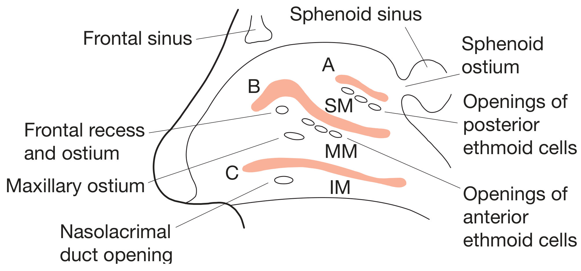
- Middle meatus - receives drainage from frontal, maxillary, and anterior ethmoid sinuses
- Superior meatus - receives drainage from posterior ethmoid sinus
- Spheno-ethmoidal recess (above the superior turbinate) - sphenoid sinus drains here
(Source: Bailey and Love's Short Practice of Surgery)
5. Paranasal Sinuses - The Air Chambers
These are hollow, air-filled spaces inside the skull bones that connect to the nasal cavity. There are four pairs:
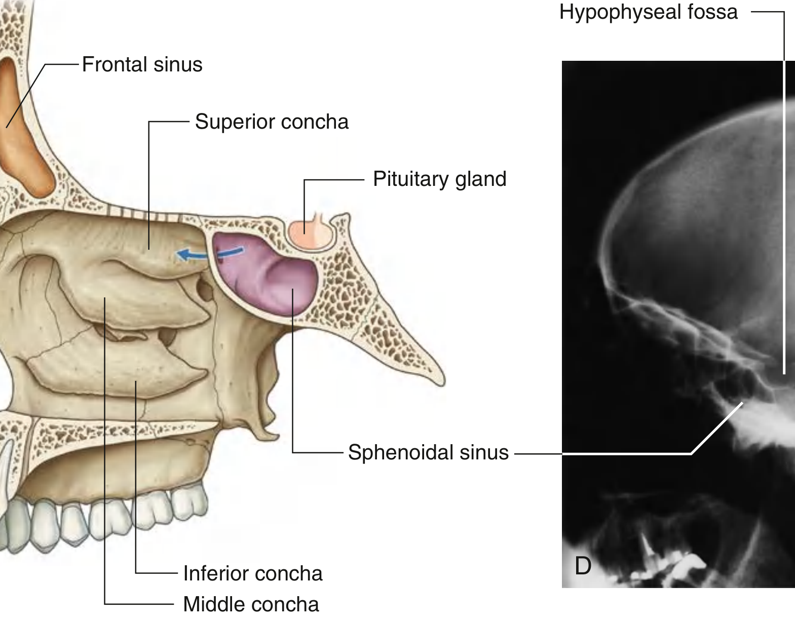
| Sinus | Location | Drains Into |
|---|---|---|
| Maxillary sinus | Inside cheekbones (largest sinus) | Middle meatus |
| Frontal sinus | Inside forehead, above eyebrows | Middle meatus (via frontal recess) |
| Ethmoid sinus | Between eyes, like a honeycomb | Anterior cells → middle meatus; posterior cells → superior meatus |
| Sphenoid sinus | Deep behind the nose, near the pituitary gland | Spheno-ethmoidal recess |
All sinuses are lined with ciliated mucosa that sweeps mucus toward the nasal cavity. When they get blocked (sinusitis), they fill with fluid and cause pressure/pain.
6. Blood Supply - Where Nosebleeds Come From
The nose has a very rich blood supply from two sources:
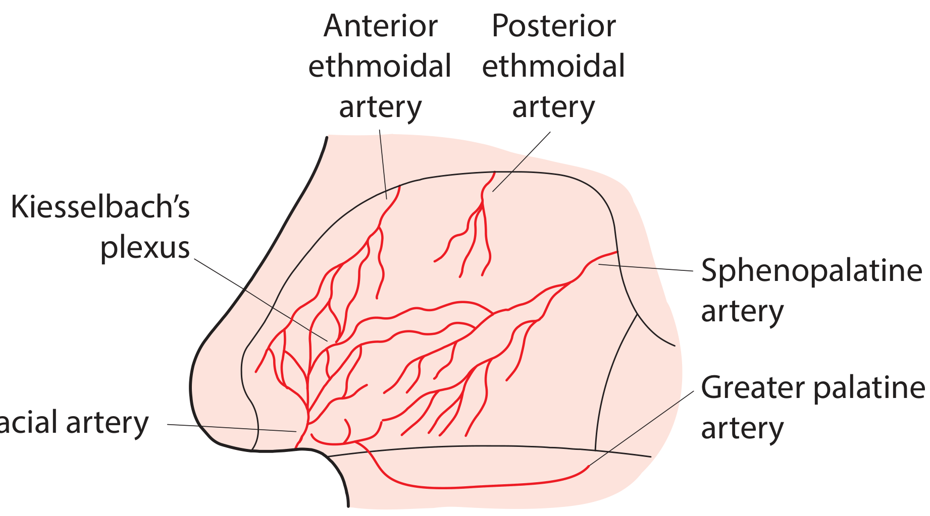
Arterial supply:
- External carotid artery contributes via the sphenopalatine artery (main supply to posterior nose) and the greater palatine artery (reaches the front of the septum via a canal in the floor)
- Internal carotid artery contributes via the anterior and posterior ethmoidal arteries (branches of the ophthalmic artery), which supply the upper and anterior nose
All of these arteries meet and anastomose on the front part of the nasal septum to form Kiesselbach's plexus (also called Little's area). This is the most common site of nosebleeds (epistaxis) because it is superficial, easily traumatized, and has a dense network of vessels.
Venous drainage goes via the ophthalmic and facial veins and the pterygoid plexus. The ophthalmic vein connects to the cavernous sinus, which is why a nasal infection can, rarely, spread to the brain.
(Source: Bailey and Love's Short Practice of Surgery)
7. Nerve Supply - Smell and Sensation
The nose is served by two nerve systems:
Olfactory (smell) - CN I:
- Smell receptors sit in the olfactory epithelium in the roof of the nasal cavity (near the superior concha and cribriform plate)
- Olfactory nerve fibers pass through tiny holes in the cribriform plate to reach the brain
- This is why a head injury that fractures the cribriform plate can destroy your sense of smell
Sensory (touch, pain, temperature) - CN V (trigeminal nerve):
- The anterior ethmoidal nerve (from ophthalmic division, V1) supplies the front of the nasal cavity and the external nose tip
- The sphenopalatine branches (from maxillary division, V2) supply most of the nasal mucosa, septum, and turbinates
- All paranasal sinuses are also innervated by branches of CN V
Autonomic (mucus secretion and vessel tone):
- Parasympathetic fibers (via the greater petrosal nerve and pterygopalatine ganglion) stimulate mucus secretion and cause nasal congestion
- Sympathetic fibers cause vasoconstriction (decongestants mimic this effect)
8. Lining (Mucosa) - The Functional Layer
The nasal cavity is lined by two types of epithelium:
- Olfactory epithelium - in the roof/upper cavity; contains smell receptor cells
- Respiratory epithelium - lining the rest; pseudo-stratified columnar with cilia and goblet cells
The respiratory mucosa does three key jobs:
- Warms inspired air using its rich blood supply
- Moistens air with mucus secretion
- Filters particles - cilia sweep the mucus blanket backward into the throat (mucociliary clearance)
Quick Summary
External Nose
  ├── Bony upper part (nasal bones)
  └── Cartilaginous lower part (alar + septal cartilages)

Nasal Septum (divides left & right)
  ├── Septal cartilage (front)
  ├── Ethmoid (perpendicular plate) (upper back)
  └── Vomer (lower back)

Nasal Cavity (each side)
  ├── Lateral wall → 3 turbinates + 3 meatuses
  ├── Roof → cribriform plate (smell nerves pass here)
  └── Floor → hard palate

Paranasal Sinuses → Maxillary, Frontal, Ethmoid, Sphenoid

Blood Supply → Kiesselbach's plexus (front septum = nosebleed site)

Nerves → CN I (smell) + CN V (sensation) + Autonomic
Sources: Bailey and Love's Short Practice of Surgery (28th Ed.), Gray's Anatomy for Students, Cummings Otolaryngology Head & Neck Surgery, Bailey and Love's Short Practice of Surgery - basic anatomy of the nose and paranasal sinuses, p. 785-786
This is a shared conversation. Sign in to Orris to start your own chat.