
Screening for pda in neonates
Reading File
Now let me search the medical library and PubMed in parallel for PDA screening in neonates.
Finding Sources
Finding Sources
Searching PubMed
"patent ductus arteriosus" AND screening AND neonates
Good results. Now let me read the most relevant textbook sections in parallel - especially neonatology/pediatric sources, and fetch PubMed abstracts for the most relevant recent papers.
Reading File
Reading File
Reading File
Reading Article
PMID: 37899032
Reading Article
PMID: 41749152
Good data. Now let me get the clinical manifestations/screening details from Schwartz's and look for a dedicated neonatology source, plus the echocardiography image and the targeted neonatal echo abstract.
Reading File
Finding Sources
Reading Article
PMID: 40870487
Loading Image
I now have comprehensive information. Let me also do a quick web search for current screening guidelines/pulse oximetry protocols in neonates.
Searching the Web
PDA screening neonates pulse oximetry echocardiography criteria 2024 2025 guidelines
Finding Sources
Excellent - I now have a very strong set of information. Let me read the Harriet Lane Handbook entry on PDA as it specifically mentions VLBW infant incidence and acyanotic CHD screening.
Reading File
Reading Web Page
https://publications.aap.org/pediatrics/article/155/5/e20250…
I now have all the information needed for a comprehensive answer. Let me compile it.
Screening for PDA in Neonates
Background
Patent ductus arteriosus (PDA) is a failure of postnatal closure of the ductus arteriosus - the fetal vessel connecting the pulmonary artery to the descending aorta. At birth, rising oxygen tension triggers smooth muscle contraction and functional closure within 10-15 hours, followed by anatomic fibrosis (ligamentum arteriosum) over days to weeks.
- Incidence: ~1 in 2,000 term births; rises dramatically with prematurity
- 40-60% of VLBW infants have a PDA; reaches 60-75% in neonates <28-30 weeks gestation
- Female predominance (2:1 ratio)
- Untreated isolated PDA carries ~30% mortality, primarily from CHF
- Harriet Lane Handbook, 23rd ed., p. 243; Schwartz's Principles of Surgery, 11th ed., p. 787
Why Screening Matters
A clinically silent or small PDA may still carry risks (infectious endarteritis ~0.45%/year after the second decade). A hemodynamically significant PDA (hs-PDA) in preterm infants causes:
- Left-to-right shunting → left ventricular volume overload
- Pulmonary over-circulation → respiratory distress, BPD risk
- Diastolic "steal" → mesenteric/renal underperfusion, NEC risk
- Intraventricular hemorrhage risk in extreme preterms
Screening Methods
1. Clinical Assessment
Signs suggestive of PDA:
| Feature | Finding |
|---|---|
| Murmur | Continuous "machinery" murmur, grade 1-4/6, loudest at LUSB (left upper sternal border) / left infraclavicular area |
| Pulse | Wide pulse pressure; bounding/waterhammer pulses |
| Precordium | Hyperactive; left ventricular heave |
| Tachycardia/tachypnea | From increased sympathetic discharge |
| Cyanosis | Absent in uncomplicated left-to-right PDA; differential cyanosis (lower limbs more cyanosed than upper) if Eisenmenger reversal occurs |
In neonates, the murmur is often only systolic on the first day of life (audible at upper left sternal border / left infraclavicular area) and becomes continuous as pulmonary vascular resistance falls. In very premature infants, the classic murmur may be absent even with large shunts - clinical signs alone are unreliable.
- Goldman-Cecil Medicine, p. 1064; Schwartz's, p. 788
2. Pulse Oximetry (CCHD Screening)
The AAP 2025 updated algorithm uses pre- and post-ductal SpO₂ to screen for critical congenital heart disease (CCHD):
Timing: At 24-48 hours of age (or just before discharge if <24 hours)
Sites:
- Pre-ductal: Right hand (reflects aortic arch blood, before ductus joins)
- Post-ductal: Either foot
Interpretation:
| Result | Criteria |
|---|---|
| Pass | SpO₂ ≥95% in both sites AND difference ≤3% |
| Rescreen in 1 hr | SpO₂ 90-94% in either site, OR >3% difference |
| Fail (immediate echo) | SpO₂ <90% in either site, OR fails 3 consecutive screens |
A passing pulse oximetry screen does NOT exclude PDA. Isolated left-to-right PDA (without right-to-left shunt) typically passes oximetry. The test is most useful for detecting ductal-dependent cyanotic lesions.
- AAP 2025 CCHD Screening Protocol; CDC CCHD screening page
3. Echocardiography - The Gold Standard
Echocardiography (2D + colour Doppler) is the definitive tool for PDA diagnosis and characterisation.
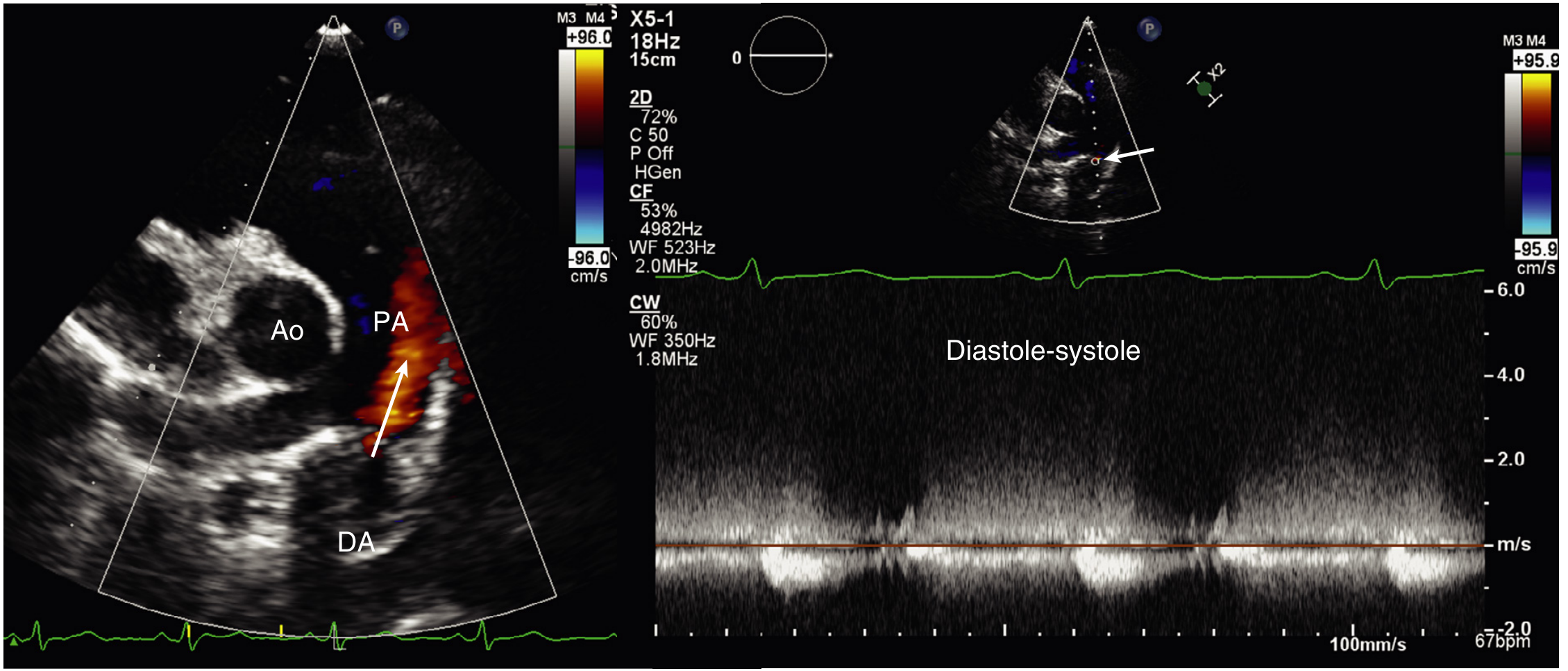
Fig. Parasternal short-axis view (left): colour Doppler jet (arrow) in the PA arising from the descending aorta (DA) in diastole. CW Doppler (right): characteristic continuous high-velocity flow from diastole through systole. (Textbook of Clinical Echocardiography)
Key echocardiographic criteria for hs-PDA:
- Ductal diameter >1.5 mm (or >1.4-1.5 mm/kg in VLBW)
- Left atrium:aortic root (LA:Ao) ratio >1.4-1.5 (indicates volume overload)
- Antegrade diastolic flow reversal in descending aorta
- Left-to-right or bidirectional ductal flow on colour Doppler
- Diastolic ductal flow in the PA has sensitivity 96%, specificity 100% for PDA diagnosis
- LV dilatation / reduced LV function
- Textbook of Clinical Echocardiography, p. 548
Targeted Neonatal Echocardiography (TnECHO)
A 2025 systematic review (PMID 40870487) of 11 studies (PRISMA, Feb 2025) found:
- TnECHO implementation associated with a 49% reduction in PDA ligation rates
- Improved diagnostic precision for shunt significance and myocardial function
- Enables physiology-guided decisions, reducing unnecessary interventions
- The 2024 ASE guidelines from the American Society of Echocardiography updated indications and training requirements for TnECHO/cPOCUS in the NICU
4. Biomarkers
BNP / NT-proBNP (serum or urinary):
- Elevated in hs-PDA due to ventricular volume/pressure overload
- A 2024 systematic review (PMID 37899032) found urinary NT-proBNP has modest sensitivity and specificity for hs-PDA; requires local validation before routine use
- NT-proBNP levels >1800 pg/mL in preterm infants are often used as a threshold, though cutoffs vary by gestational age and postnatal age
5. Near-Infrared Spectroscopy (NIRS) - Emerging
A 2026 meta-analysis (PMID 41749152) of 9 studies found:
- Cerebral rSO₂ (regional oxygen saturation) is significantly lower in PDA infants vs. controls (SMD -0.42, 95% CI -0.74 to -0.10)
- Renal rSO₂ and peripheral SpO₂ did not show significant differences
- Suggests cerebral NIRS may serve as a non-invasive bedside indicator for PDA, but AUC studies are still needed
6. Other Investigations
| Test | Finding in PDA |
|---|---|
| Chest X-ray | Cardiomegaly, increased pulmonary vascular markings, pulmonary oedema in large shunts |
| ECG | Small-moderate PDA: normal or LVH; large PDA: BVH |
| Cardiac catheterization | Reserved only when pulmonary hypertension is suspected |
Screening Strategy by Population
Term Neonates
- Routine newborn examination (auscultation at 6-24 hours and again at discharge)
- Pulse oximetry CCHD screen at 24-48 hours
- Echo only if murmur persists, abnormal oximetry, or clinical concern
Preterm Neonates (<32 weeks or VLBW)
- Routine clinical reassessment daily in NICU (murmur, bounding pulses, hyperdynamic precordium)
- Echocardiography is the standard - many units perform a screening echo at 3-7 days of life in all infants <28 weeks gestation
- Repeat echo if clinical deterioration, increasing FiO₂ requirement, or feeding intolerance
- Consider urinary NT-proBNP or cerebral NIRS as adjuncts where echo is unavailable
Key Points Summary
- Clinical signs are insensitive in very preterm infants - a large PDA can be silent on auscultation.
- Pulse oximetry screens for CCHD broadly, not PDA specifically; a left-to-right PDA will usually pass.
- Echocardiography with colour Doppler is the gold standard; diastolic PA jet has 96% sensitivity, 100% specificity.
- TnECHO by trained neonatologists is now supported by ASE 2024 guidelines and reduces overtreatment.
- Biomarkers (NT-proBNP) and cerebral NIRS are adjuncts, not replacements for echo.
- Management after diagnosis: watchful waiting in asymptomatic, COX inhibitors (indomethacin/ibuprofen/acetaminophen) for hs-PDA in preterms, catheter-based or surgical closure in term infants or those refractory to medical therapy.
This is a shared conversation. Sign in to Orris to start your own chat.