
Tip of penis enlarged diagnosis
glans penis enlargement balanitis penile lesion
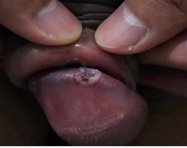
This clinical photograph shows a close-up view of a penile lesion located on the glans penis. The primary finding is a solitary, well-demarcated, circular ulcer approximately 5-10mm in diameter. The lesion exhibits a distinct morphology characterized by a raised, whitish, indurated peripheral border and a central base that is purplish-red or dusky in hue. The surrounding glans tissue appears mildly erythematous and glossy. This visual presentation is characteristic of educational material focused on sexually transmitted infections (STIs), specifically demonstrating an ulcerative condition of the male genitalia. The clinical significance involves the differential diagnosis of genital ulcers, such as primary syphilis (chancre), chancroid, or herpes simplex virus, although the clean-based, sharply defined appearance of the border is particularly illustrative of specific dermatological manifestations of balanitis or infectious ulcers.
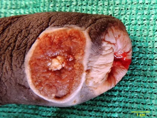
Gross photograph of a total penectomy specimen from a 63-year-old man with a 6-month history of a painless non-healing penile ulcer. The external penis shows chronic balanitis with phimosis. On longitudinal section, a diffusely infiltrative tumor occupies the foreskin and glans penis. The lesion measures approximately 3 cm in maximum diameter, with a variegated tan-brown to gray-white cut surface and focal necrosis. The tumor penetrates the mucocutaneous junction into submucosa; there is limited visible invasion into adjacent penile tissues, though deeper sampling may reveal extension. The specimen was obtained after a clinical diagnosis of penile squamous cell carcinoma, with histopathology confirming invasion beyond the epithelial layer. The case emphasizes the natural history of penile cancer associated with chronic phimosis and uncircumcised status, presenting as a painless ulcer. The findings have significant diagnostic and prognostic implications; tumor size, local extent to foreskin and glans, and potential lymphatic drainage correlate with risk of nodal metastasis and need for radical surgical management. This image supports educational review of penile SCC gross morphology, differential considerations in penile ulcers, and gross-pathology correlation to histologic invasion patterns.
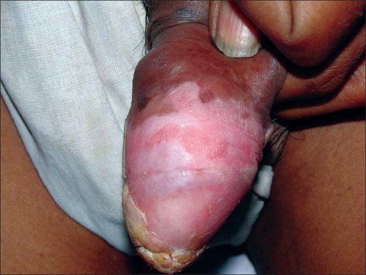
Clinical photograph of the glans penis in an uncircumcised male, showing a significant dermatological lesion on the dorsal surface. The primary finding is a large, well-demarcated erythematous plaque involving a substantial portion of the glans. Centrally and superiorly, the lesion displays areas of hyperkeratosis with white, mica-like scaling and a thickened, verrucous texture. The surrounding base is intensely erythematous. A finger is visible retracting the foreskin to expose the extent of the pathology. This presentation is characteristic of pseudoepitheliomatous keratotic and micaceous balanitis (PKMB), often clinically resembling penile psoriasis or Bowen's disease but distinguished by its aggressive, verrucous growth and thick scaling. The image serves as an educational tool for identifying premalignant or hypertrophic penile skin conditions in urology and dermatology.
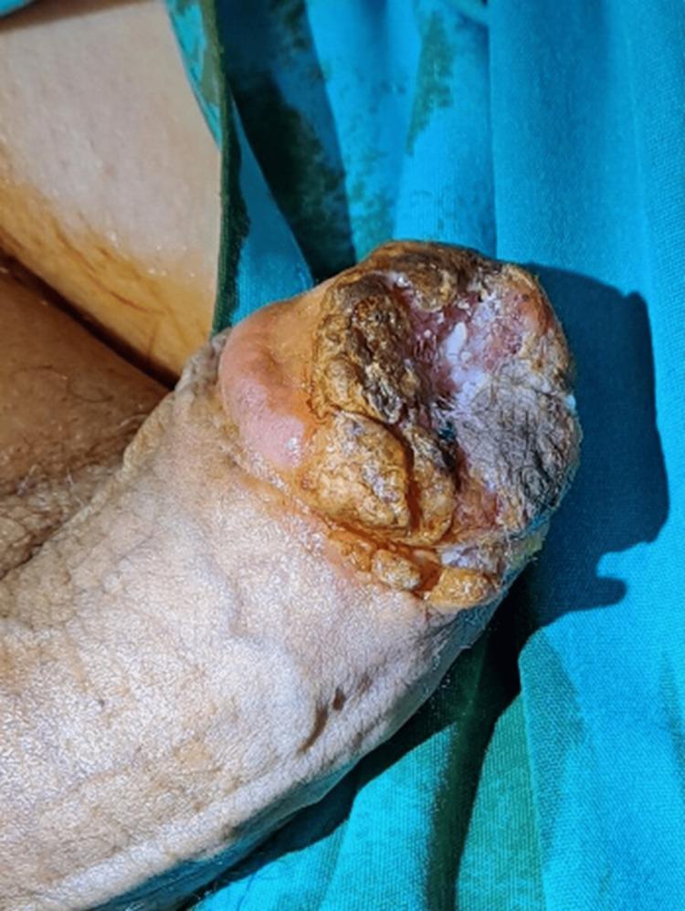
This clinical photograph displays a primary skin lesion involving the glans penis in a circumcised adult male. The lesion is characterized by a significant exophytic and verrucous (wart-like) growth, primarily located on the distal aspect of the glans. It exhibits a highly heterogeneous surface with areas of hyperkeratosis, thick yellowish-brown crusting, and focal gray-white scaling. The central portion of the lesion appears irregular and erosive, with mixed fleshy pink and white plaque-like features. The margins are asymmetrical and poorly defined, showing infiltration into the glans tissue and potential extension toward the proximal penile shaft. The urethral meatus is obscured by the progressive proliferation. In the clinical context of Pseudoepitheliomatous Keratotic and Micaceous Balanitis (PKMB) progressing to verrucous carcinoma, this image illustrates high-risk features for malignancy, including its hard, nodular appearance and resistance to conservative therapy. The image serves as a teaching tool for dermatologic oncology and urology in identifying potentially malignant penile transformations.
Enlargement / Swelling of the Glans Penis - Differential Diagnosis
1. Balanitis / Balanoposthitis (Most Common)
- Presentation: Redness, swelling, tenderness, itching of the glans; pus or discharge may be present; in severe cases the glans is "red-raw"
- Causes:
- Infectious - Candida (most common in uncircumcised men with poor hygiene or diabetes), bacterial (mixed organisms, STIs like gonorrhea/chlamydia), viral
- Inflammatory - Reactive arthritis (Reiter syndrome), psoriasis, lichen sclerosis
- Poor hygiene / phimosis - foreskin cannot be retracted and cleaned
- Risk factors: Diabetes mellitus, immunosuppression, uncircumcised status
- Treatment: Broad-spectrum antibiotics or antifungals, local hygiene; recurrent episodes require circumcision
- (Textbook of Family Medicine 9e, p. 1198; Bailey & Love's Surgery 28e)
2. Paraphimosis (Urological Emergency)
- Foreskin is retracted but cannot be reduced - venous congestion causes acute, painful swelling and enlargement of the glans
- Glans becomes engorged, edematous, and progressively ischemic
- Requires urgent manual reduction or dorsal slit/emergency circumcision
- (Rosen's Emergency Medicine; Campbell-Walsh Urology)
3. Phimosis-Related Ballooning
- Tight foreskin causes obstruction to urine flow, with glans appearing enlarged or ballooned
- In adults, often caused by balanitis xerotica obliterans (BXO / lichen sclerosis)
4. Precancerous Lesions
| Condition | Features |
|---|---|
| Leukoplakia | White plaque at meatus; linked to diabetes; histology shows acanthosis + hyperkeratosis |
| Balanitis Xerotica Obliterans (BXO) | White patch on glans/prepuce, often involves meatus; middle-aged diabetics; collagen abnormalities on biopsy |
| Giant Condylomata Acuminata | Cauliflower-like HPV lesions on glans/prepuce; may be hard to distinguish from SCC |
| Erythroplasia of Queyrat (PeIN) | SCC in situ of the glans; appears as red, velvety plaque with ulcerations; linked to high-risk HPV |
| Bowen's Disease | SCC in situ of the shaft; similar histology to Erythroplasia of Queyrat |
5. Squamous Cell Carcinoma (SCC) of the Penis
- Most common penile malignancy (98% of cases); most frequently originates on the glans
- Presentation: Painless, indurated, non-healing ulcer or papillary/fungating growth on the glans; may be associated with phimosis and chronic balanitis
- Appearance is papillary or ulcerative; may present as a subtle nodule initially
- Spreads via lymphatics to inguinal nodes
- Verrucous carcinoma - a variant (5-16%), papillary, well-demarcated deep margins, slower growing
- Risk factors: HPV infection, phimosis, poor hygiene, uncircumcised status, lichen sclerosis
- (Smith & Tanagho's General Urology 19e; Robbins Pathology)
6. Zoon Balanitis (Balanitis Plasmacellularis)
- Benign chronic inflammatory lesion of the glans penis
- Represents ~7% of persistent genital lesions biopsied for diagnosis
- Histology: dense plasma cell-rich infiltrate in dermis
- Appears as a shiny, moist, well-defined erythematous plaque on the glans
- (Andrews' Diseases of the Skin)
7. Sexually Transmitted Infections (STI)
| Condition | Key Features |
|---|---|
| Primary Syphilis (Chancre) | Painless, indurated, clean-based ulcer; well-defined margins on glans |
| Herpes Simplex | Painful clustered vesicles → ulcers; recurrent |
| Chancroid | Painful, soft, ragged ulcer; tender inguinal lymphadenopathy |
| Condyloma Acuminata | HPV warts; soft, flesh-colored papillary growths |
8. Pseudoepitheliomatous Keratotic and Micaceous Balanitis (PKMB)
- Rare condition with verrucous, hyperkeratotic plaques on the glans with thick mica-like scaling
- Considered premalignant; can progress to verrucous carcinoma
- Resembles psoriasis or Bowen's disease clinically
Diagnostic Approach
- History: Duration, pain (painful = inflammatory/infectious; painless = carcinoma/syphilis), sexual history, hygiene, diabetes, circumcision status
- Examination: Character of the lesion - ulcer, plaque, warty growth, diffuse swelling; foreskin retractability; inguinal lymph nodes
- Investigations:
- Swab for culture/sensitivity, STI screen (gonorrhea, chlamydia, syphilis VDRL/TPHA, herpes PCR)
- Fasting blood glucose / HbA1c (diabetes screening)
- Biopsy - mandatory for any persistent, non-healing, or suspicious lesion to exclude carcinoma
- Ultrasound or MRI if deep invasion suspected
Red Flags (Warrant Urgent Biopsy/Referral)
- Painless, non-healing ulcer or mass
- Hard, indurated margins
- Inguinal lymphadenopathy
- Failure to respond to antibiotics/antifungals
- Age >40, uncircumcised, history of phimosis or chronic balanitis