
Hba1c biochemistry
Reading File
HbA1c (glycated hemoglobin) is formed by the non-enzymatic glycosylation of hemoglobin A, reflecting average blood glucose over the prior 2-3 months. It serves as the gold standard marker for long-term glycemic control in diabetes.
Searching sources...
Finding Sources
Finding Sources
Reading File
Reading File
Finding Sources
Finding Sources
Reading File
Reading File
Reading File
Finding Sources
Loading Image
Reading File
Loading Image
Here is a complete, textbook-grounded overview of HbA1c biochemistry.
HbA1c (Glycated Hemoglobin) - Biochemistry
1. What Is HbA1c?
HbA1c is a specific fraction of adult hemoglobin A (HbA) in which glucose has formed a stable, covalent adduct at the N-terminal valine of the beta (β) chain. It belongs to a broader family of glycated hemoglobins (HbA1), which also includes HbA1a1, HbA1a2, and HbA1b (see table below).
| Fraction | Adduct Attached |
|---|---|
| HbA1a1 | Fructose-1,6-diphosphate at β-chain N-terminal valine |
| HbA1a2 | Glucose-6-phosphate at β-chain N-terminal valine |
| HbA1b | Pyruvic acid at β-chain N-terminal valine |
| HbA1c | Glucose at β-chain N-terminal valine |
| Pre-HbA1c | Unstable Schiff base (labile intermediate) |
Source: Tietz Textbook of Laboratory Medicine, 7th Ed.
2. Mechanism of Formation (Glycation Reaction)
The formation of HbA1c is a non-enzymatic, irreversible two-step process - part of the broader Maillard/Amadori chemistry:
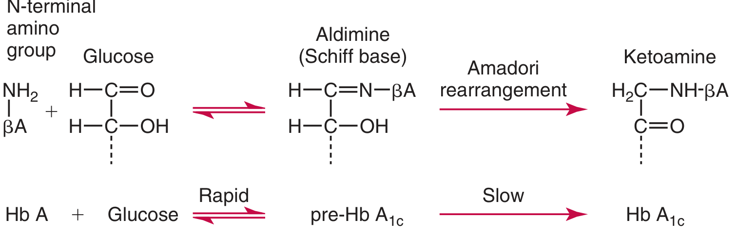
Step 1 - Rapid and Reversible:
The aldehyde group of glucose condenses with the free amino group (ε-amino of lysyl residues or the α-amino group of the N-terminal valine of β-chains) to form an unstable Schiff base (aldimine) called pre-HbA1c. This reaction is rapid and reversible.
Step 2 - Slow and Irreversible:
The Schiff base undergoes an Amadori rearrangement to yield a stable ketoamine - this is the true HbA1c. This second step is slow and essentially irreversible under physiological conditions.
Key point: Glycation (non-enzymatic) is distinct from glycosylation (enzyme-catalyzed addition of sugar moieties to proteins). - Harper's Illustrated Biochemistry, 32nd Ed.
3. Why Does It Reflect Long-Term Glucose Control?
- Erythrocyte lifespan is approximately 120 days
- HbA1c accumulates gradually over the RBC lifespan - the rate of formation is proportional to ambient blood glucose
- HbA1c concentration therefore reflects the mean blood glucose over the preceding 8-12 weeks
- The glycation rate is highest in the first 2 months, followed by more gradual change approaching steady state at ~3 months
- Normally, about 5% of hemoglobin is glycated in healthy individuals
4. Labile Fraction (Pre-HbA1c) - Clinical Pitfall
- Pre-HbA1c (the Schiff base intermediate) amounts to 5-8% of total HbA1 in healthy individuals and 8-30% in diabetics
- It changes rapidly with acute glucose fluctuations and does NOT reflect long-term control
- Some assay methods (especially older ion-exchange methods) inadvertently include the labile fraction, causing falsely elevated results
- The labile fraction can be removed by incubating washed red blood cells in saline (in the absence of glucose, it reverts back to glucose + HbA)
- Boronate affinity methods promote rapid dissociation of the Schiff base, minimizing this problem
5. Clinical Utility
Diagnosis of Diabetes
Since 2010, HbA1c has been accepted as a diagnostic criterion for diabetes:
- ≥6.5% = Diabetes mellitus
- 5.7% - 6.4% = Prediabetes
- <5.7% = Normal
Advantages of HbA1c over plasma glucose testing:
- Fasting is NOT required
- Very low biological variability
- Sample is stable
- Predicts risk of microvascular complications
Source: Robbins & Kumar Basic Pathology; Tietz Textbook of Laboratory Medicine, 7th Ed.
Monitoring Diabetes
- Target for most diabetics: <6.5 to 7% (48-53 mmol/mol) per major diabetes organizations
- Each 1% reduction in HbA1c is associated with:
- 37% reduction in risk of microvascular disease
- 21% reduction in diabetes-related death
- 14% reduction in myocardial infarction (UKPDS trial data)
- There is no HbA1c concentration below which microvascular risk is eliminated - risk varies exponentially
6. Measurement Methods
Over 250 methods have been described. They broadly fall into two categories:
Charge-based separation:
- Ion-exchange chromatography (HPLC) - most common; susceptible to labile fraction interference
- Electrophoresis
- Isoelectric focusing
- Capillary electrophoresis
Structure-based separation:
- Boronate affinity chromatography - least affected by Hb variants
- Immunoassay
Chemical/enzymatic analysis:
- Enzymatic assay (specifically measures HbA1c)
Results are expressed as a fraction (%) of total hemoglobin. The NGSP (National Glycohemoglobin Standardization Program) certifies assay methods traceable to the DCCT reference, ensuring comparability of results across labs. The IFCC reference method uses endoproteinase Glu-C digestion followed by HPLC-ESI-MS or capillary electrophoresis of the glycated N-terminal hexapeptide of the β-chain.
7. Test Limitations and Interferences
HbA1c is only valid when RBC lifespan is normal. False results occur in:
| Cause | Effect on HbA1c |
|---|---|
| Hemolytic anemia | Falsely LOW (shortened RBC lifespan) |
| Recent blood transfusion | Falsely LOW (dilution with non-glycated donor RBCs) |
| Recent significant blood loss | Falsely LOW (higher fraction of young erythrocytes) |
| Iron deficiency anemia | Falsely HIGH (older RBCs accumulate more glycation) |
| Hemoglobin variants (HbS, HbC, HbF) | Results can be spuriously increased or decreased - depends on assay method |
| Labile pre-HbA1c not removed | Falsely HIGH |
- Boronate affinity methods are minimally affected by Hb variants
- HPLC and capillary electrophoresis chromatograms can visually flag the presence of variant hemoglobins
- Point-of-care HbA1c devices should not be used for screening or diagnosis of diabetes
Summary
| Feature | Detail |
|---|---|
| Type of modification | Non-enzymatic glycation (not glycosylation) |
| Primary site | α-amino group of N-terminal valine, β-chains of HbA |
| Intermediate | Pre-HbA1c (Schiff base/aldimine) - labile |
| Stable product | HbA1c (ketoamine) via Amadori rearrangement |
| Reflects glucose over | 8-12 weeks (RBC lifespan ~120 days) |
| Normal fraction | ~5% of total hemoglobin |
| Diagnostic cut-off | ≥6.5% for diabetes; 5.7-6.4% for prediabetes |
| Gold standard assay | NGSP-certified HPLC |
Sources: Harper's Illustrated Biochemistry (32nd Ed.), Tietz Textbook of Laboratory Medicine (7th Ed.), Robbins & Kumar Basic Pathology
This is a shared conversation. Sign in to Orris to start your own chat.