
How to use AED in detailed
Reading File
An AED (Automated External Defibrillator) is used to restore normal heart rhythm during cardiac arrest caused by ventricular fibrillation or pulseless ventricular tachycardia. The basic steps are: turn it on, attach pads to bare chest, follow voice prompts, let it analyze, stand clear, and deliver shock if advised. Now searching textbooks and current guidelines for the full detailed protocol.
Searching sources...
Finding Sources
Searching the Web
AED automated external defibrillator step by step use 2024 guidelines
Finding Sources
Reading Web Page
https://www.mayoclinic.org/diseases-conditions/heart-arrhyth…
Reading File
Reading File
Reading File
Reading File
Now I have comprehensive information from multiple authoritative sources. Here is the full detailed guide:
How to Use an AED (Automated External Defibrillator) - Complete Guide
What Is an AED?
An AED is a portable device that analyzes the heart's electrical rhythm and delivers an electric shock (defibrillation) to restore a normal heartbeat. It is designed for use in sudden cardiac arrest (SCA) caused by ventricular fibrillation (VF) or pulseless ventricular tachycardia (VT). Two types exist:
- Semi-automatic AED - analyzes the rhythm and charges automatically, but requires the user to press a button to deliver the shock.
- Fully automatic AED - detects, charges, and delivers the shock without any button press - all bystanders must stay clear during charging.
Before You Begin - Recognize Cardiac Arrest
Watch for these signs:
- Sudden collapse and unresponsiveness
- No normal breathing (absent or only gasping)
- No pulse when checked for less than 10 seconds
- Loss of consciousness
Step-by-Step AED Use
Step 1 - Assess the Scene and the Person
- Make sure the environment is safe before approaching.
- Tap the person's shoulders firmly and shout, "Are you okay?"
- Check for normal breathing and pulse for no more than 10 seconds.
- If unresponsive with no breathing/no pulse, act immediately.
Step 2 - Call for Help
- Call 911 (or local emergency number) immediately, or direct a bystander to call while you stay with the victim.
- If others are present: one person performs CPR, another locates and retrieves the AED.
- If you are alone: call first, then get the AED if one is nearby.
Step 3 - Start CPR Immediately
Do not wait for the AED before starting CPR. Begin chest compressions right away:
- Place the heel of your hand on the center of the chest (lower half of the sternum).
- Push hard and fast - at least 2 inches deep, at a rate of 100-120 compressions per minute.
- Ratio: 30 compressions to 2 rescue breaths.
- Minimize interruptions - keep compressions going until the AED is ready.
Step 4 - Turn On the AED
- Open the AED case - many models turn on automatically when opened.
- If not automatic, press the power button.
- Follow the voice prompts - the AED guides you through every step with clear spoken instructions.
Step 5 - Prepare the Chest
- Remove all clothing from the chest - expose the bare skin completely.
- The chest must be dry. If wet, dry it quickly with a cloth.
- If the person has a hairy chest, the pads may not stick. Use the razor (usually in the AED kit) to shave the pad placement areas.
- Remove any medication patches or metallic objects from the pad zones.
Step 6 - Apply the Electrode Pads Correctly
There are two pads, each labeled or pictured with a placement diagram:
| Pad | Placement |
|---|---|
| Sternal pad (upper/right) | Below the right collarbone, to the right of the sternum |
| Apical pad (lower/left) | Midaxillary line at the 5th intercostal space (left side of chest, below the armpit) |
Important pad placement notes:
- Peel the backing off and press pads firmly to bare skin - no air pockets.
- If pads might touch each other (small child or small chest), place one pad in the center of the chest and the other on the back between the shoulder blades.
- If the patient has an implanted pacemaker or ICD (visible as a lump under the skin), place the pad at least 2 inches away from it.
- Plug the pad connector cable into the AED if it is not pre-connected.
Step 7 - Let the AED Analyze the Heart Rhythm
- The AED will announce: "Analyzing rhythm - do not touch the patient."
- Stop CPR and make sure nobody touches the person during analysis - even minor movement can interfere with rhythm detection.
- The AED determines automatically whether a shockable rhythm (VF or pulseless VT) is present.
Step 8 - Deliver the Shock (If Advised)
- If a shock is needed, the AED will announce "Shock advised" and will charge automatically to a preset energy level.
- During charging on a semi-automatic AED: resume CPR while it charges. Stand clear just before the shock.
- On a fully automatic AED: stand clear immediately during charging - it will deliver the shock on its own.
- Loudly call out "Stand clear!" and visually confirm no one is touching the patient or the surface they are lying on.
- On a semi-automatic: press the flashing SHOCK button to deliver the shock.
- The shock will cause a sudden muscle contraction in the patient.
Step 9 - Resume CPR Immediately After the Shock
- Do NOT pause to check the pulse or re-analyze the rhythm immediately after the shock.
- Start chest compressions again right away - 30 compressions to 2 ventilations.
- Continue for 5 cycles (approximately 2 minutes) of CPR.
- This is critical because the heart may be stunned after defibrillation and needs compression support to regain full pumping function.
Step 10 - Reassess and Repeat if Needed
- After 2 minutes of CPR, the AED will prompt you to stop and re-analyze.
- If another shock is needed, follow Steps 7-9 again.
- If no shock is advised, check for a pulse and signs of circulation.
- Continue CPR + AED cycles until:
- Emergency medical services (EMS) arrive and take over.
- The person shows clear signs of life (normal breathing, coughing, movement).
- You are physically unable to continue.
Special Situations
| Situation | What to Do |
|---|---|
| Child (1-8 years / under 55 lbs) | Use pediatric pads or a pediatric dose-attenuator if available. If only adult pads available, use them - one on chest, one on the back. |
| Infant (under 1 year) | Manual defibrillation by trained providers is preferred; AED is a last resort only. |
| Wet environment / near water | Move the person to a dry surface. Dry the chest before applying pads. |
| Implanted pacemaker/ICD | Place pads at least 2 inches away from the device. |
| Medication patch on chest | Remove the patch with a gloved hand and wipe the area before pad placement. |
| Pregnant person | Use the AED normally - it is safe and should not be withheld. |
| Hairy chest | Press pads firmly first; if no contact, quickly shave the area using the razor in the kit, then reapply pads. |
Energy Levels (For Healthcare Providers Using Manual Defibrillators)
AEDs set energy automatically, but for reference:
- Biphasic defibrillator (standard AED type): 120-200 J (manufacturer-specific default, typically ~200 J)
- Monophasic defibrillator: 360 J
For subsequent shocks, the same or higher energy may be used at the team leader's discretion, per Tintinalli's Emergency Medicine guidelines.
After the Event - AED Maintenance
- Replace the electrode pads after each use (single-use only).
- Replace batteries as needed (typically every 4 years or per manufacturer).
- AEDs perform self-checks automatically - if an alarm/beep is triggered, contact the manufacturer.
- Register your AED with the manufacturer to receive safety alerts and recall notices.
- Store it in a clearly marked, easily accessible location and ensure everyone nearby knows where it is.
Key Reminders
- Speed matters - every minute without defibrillation during VF reduces survival by 7-10%.
- CPR + AED together dramatically increases survival compared to either alone.
- You cannot harm someone with an AED - the device will only advise a shock if it detects a shockable rhythm. It will say "No shock advised" for any other rhythm.
- Modern AEDs also provide CPR feedback via metronome beats or real-time guidance on compression rate and depth, which improves CPR quality and the chance of successful defibrillation.
- Training is recommended but not required - the voice prompts are designed for untrained bystanders to follow successfully.
Sources: Roberts and Hedges' Clinical Procedures in Emergency Medicine (8th ed.), Tintinalli's Emergency Medicine (8th ed.), Mayo Clinic AED Guide, American Red Cross AED Steps, 2024 AHA/Red Cross First Aid Guidelines.
What is nstemi how do you diagnosis
Quick answer: NSTEMI (Non-ST-Elevation Myocardial Infarction) is a type of heart attack where part of the heart muscle is damaged due to partial blockage of a coronary artery. Unlike STEMI, the ECG does NOT show ST elevation, but cardiac biomarkers (troponin) are elevated, confirming myocardial necrosis. Diagnosis relies on symptoms + ECG changes (ST depression, T-wave inversions) + rising troponin levels.
Searching sources...
Reading File
Finding Sources
Searching the Web
NSTEMI diagnosis criteria 2024 ACC AHA guidelines troponin ECG
Finding Sources
Finding Sources
Reading File
Reading File
Reading File
Reading File
Reading File
Reading File
Reading File
Reading File
Loading Image
I now have comprehensive information from Harrison's, Tintinalli's, Washington Manual, and Fuster's Heart. Here is the full detailed answer:
NSTEMI - What It Is and How to Diagnose It
What Is NSTEMI?
NSTEMI stands for Non-ST-Elevation Myocardial Infarction - a heart attack in which part of the myocardium (heart muscle) is permanently damaged due to prolonged ischemia, but the ECG does not show the classic ST-segment elevation seen in STEMI.
It belongs to the broader category of Acute Coronary Syndromes (ACS), which are classified as:
| Condition | ECG | Troponin | Meaning |
|---|---|---|---|
| STEMI | ST elevation | Elevated | Full-thickness infarction, complete occlusion |
| NSTEMI | No ST elevation (ST depression / T-wave changes / normal) | Elevated | Partial-thickness infarction, partial/transient occlusion |
| Unstable Angina (UA) | No ST elevation | Normal | Ischemia without necrosis |
The key distinction between NSTEMI and UA is troponin elevation - if troponin rises above the 99th percentile, it is NSTEMI; if troponin is normal, it is UA.
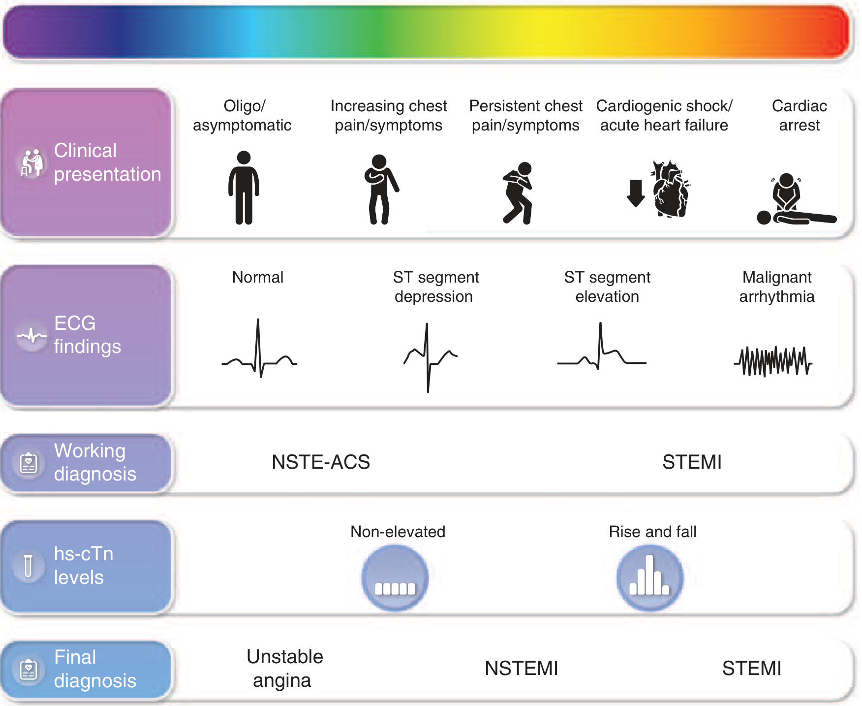
Pathophysiology (Why It Happens)
NSTEMI results from an imbalance between myocardial oxygen supply and demand, typically from coronary arterial thrombosis caused by one or more of these mechanisms:
- Plaque rupture with inflammation - thin-capped lipid-rich plaque fissures; inflammatory T-cell response drives thrombosis.
- Plaque rupture without inflammation - mechanical fissuring.
- Plaque erosion - present in at least one-third of ACS cases; the overlying endothelium erodes, exposing the subendothelium and triggering thrombus.
- Spasm or microvascular dysfunction - no thrombosis; epicardial or microvascular spasm, typically on a background of fixed obstruction.
The thrombus is non-occlusive (partial blockage) - this is why ST elevation does not appear (subendocardial ischemia pattern) but necrosis still occurs.
On coronary angiography, patients with NSTEMI show:
- Left main stenosis: <10%
- Three-vessel CAD: ~35%
- Two-vessel CAD: ~25%
- Single-vessel CAD: ~20%
- No apparent critical stenosis: ~10% (microvascular or spasm)
Clinical Presentation
Symptoms
Chest discomfort in NSTEMI typically has at least one of three features:
- Occurs at rest or with minimal exertion, lasting more than 10 minutes
- Recent onset (within the prior 2 weeks)
- Crescendo pattern - distinctly more severe, prolonged, or frequent than previous episodes
Character of chest pain:
- Usually substernal, severe, pressure-like or squeezing
- Radiates to the left arm, left shoulder, neck, or jaw
- Associated with diaphoresis, nausea, shortness of breath
Anginal equivalents (especially in women, elderly, and diabetics):
- Dyspnea alone
- Epigastric discomfort / nausea
- Unexplained fatigue or weakness
- Pain only in the jaw, shoulder, or arm
Physical examination may be entirely normal, but in large NSTEMI:
- Diaphoresis, pale/cool skin
- Sinus tachycardia
- S3 or S4 heart sounds
- Basilar lung crackles (rales)
- Hypotension, or in extreme cases, cardiogenic shock
Always exclude life-threatening mimics: pulmonary embolism, aortic dissection, cardiac tamponade.
Diagnosis of NSTEMI
Diagnosis is based on a triad: symptoms + ECG + cardiac biomarkers.
1. History and Risk Factor Assessment
Increase clinical suspicion with:
- Age ≥65 years
- Known CAD (stenosis ≥50%)
- Diabetes mellitus
- Hypertension, hyperlipidemia, smoking
- Family history of premature CAD (first-degree relative before age 50)
- Prior MI, PCI, or CABG
2. ECG (12-Lead)
Perform within 10 minutes of presentation and interpret immediately (2025 ACC/AHA guideline, Class I).
NSTEMI ECG findings (~50% of patients have significant changes):
| Finding | Description | Significance |
|---|---|---|
| ST-segment depression | ≥0.5 mm in V2-V3; ≥1 mm in all other leads; in ≥2 contiguous leads | Most specific for ischemia, especially if dynamic and symptomatic |
| T-wave inversions | New deep inversions ≥0.3 mV | Significant; less specific unless new and deep |
| Normal ECG | No ischemic changes | Does NOT rule out NSTEMI - diagnosis still requires troponin |
| Transient ST elevation | Brief, resolving <20 min | Can occur during ischemic episodes |
Important special patterns:
-
Wellens' Syndrome - deep symmetric T-wave inversions or biphasic T waves in V2-V3 (sometimes V1-V6), seen when pain-free, with normal/minimally elevated biomarkers. Indicates a critical proximal LAD stenosis - very high risk for imminent large MI. About 15% of unstable angina patients display this sign.
-
ST depression in multiple leads + ST elevation in aVR and/or V1 - suggests ischemia from left main or multivessel disease - very high risk.
-
Posterior STEMI pattern (ST depression in V1-V3 = mirror image) - always obtain posterior leads (V7-V9) if suspected.
Serial ECGs should be obtained if the initial ECG is non-diagnostic. Compare with prior ECGs to identify new changes.
The posterior circulation (circumflex artery) is poorly seen on standard ECG - always consider posterior leads or urgent echocardiography when posterior NSTEMI is suspected.
3. Cardiac Biomarkers (Troponin) - The Definitive Diagnostic Test
Troponin I (cTnI) or Troponin T (cTnT) is the standard and preferred biomarker.
The diagnostic rule:
NSTEMI = troponin value above the 99th percentile of the upper reference limit, with a rising and/or falling pattern (delta troponin), in the appropriate clinical context.
Testing Protocol (ACC/AHA 2025 / ESC Guidelines):
| Protocol | Draw Times |
|---|---|
| Standard | At presentation, then at 3-6 hours |
| High-sensitivity troponin (hsTn) rapid algorithm | 0 h and 1 hour (ESC 0h/1h protocol) |
| If initial and 3-6h draws are normal but suspicion remains | Draw again at 6+ hours after symptom onset |
High-sensitivity cardiac troponin (hs-cTn):
- Detects 90-100% of AMI at arrival
- Identifies small rises and falls (delta change) that older assays missed
- The 1-hour rapid rule-out algorithm (no abnormal elevation at 0 h or 1 h) has been endorsed by recent guidelines
- Has led to an increase in NSTEMI diagnoses (and corresponding decrease in UA) because smaller levels of necrosis are now detectable
Key biomarker facts:
- Troponin peaks at 12-24 hours after symptom onset and gradually decreases
- Elevations persist for up to 14 days (or longer)
- CK-MB is no longer recommended as a primary diagnostic marker for NSTEMI - it lacks cardiac specificity (also present in skeletal muscle)
- BNP/NT-proBNP elevation in ACS without known heart failure suggests large infarction - warrants urgent angiography
- Troponin elevation of any amount is always associated with worse outcomes - more elevation = worse prognosis
Elevated troponin with a 3-fold higher risk of death (1.6% vs. 5.3%) compared to non-elevated patients.
Causes of Troponin Elevation That Are NOT NSTEMI (must distinguish):
| Category | Examples |
|---|---|
| Reduced supply (non-ACS ischemia) | Coronary spasm, hypotension, severe anemia, tachyarrhythmia |
| Cardiac non-ischemic | Myocarditis, heart failure, cardiomyopathy, cardiac contusion, ablation |
| Systemic illness | Pulmonary embolism, sepsis, renal failure, stroke, rhabdomyolysis |
| Endocrine | Hypothyroidism, pheochromocytoma |
For NSTEMI, troponin must show an acute dynamic pattern (rise and/or fall) consistent with ischemic symptoms and ECG changes.
4. Additional Investigations
| Test | Purpose |
|---|---|
| Complete blood count | Anemia contributing to ischemia; thrombocytopenia altering antiplatelet management |
| Basic metabolic panel / electrolytes | Hyperkalemia can mimic ischemic ECG changes; renal function affects drug dosing |
| Fasting glucose + lipid panel | Cardiovascular risk factors |
| Chest X-ray | Assess for pulmonary edema, cardiac size, aortic widening (dissection) |
| Echocardiography | Wall motion abnormalities in the ischemic territory; LV function; excludes other diagnoses |
| Coronary angiography | Definitive for anatomy; combined with PCI for revascularization |
5. Risk Stratification Scores
Once NSTEMI is confirmed, quantify risk using validated scores:
TIMI Score (7 variables, each = 1 point):
- Age ≥65 years
- Known CAD (stenosis ≥50%)
- ST deviation >0.5 mm on ECG
- ≥2 anginal events in prior 24 hours
- Elevated cardiac markers
- Prior aspirin use (paradoxically indicates higher-risk patients)
- ≥3 CAD risk factors
| TIMI Score | 14-day risk of death/MI/urgent revascularization |
|---|---|
| 0-1 | 5% |
| 2 | 8% |
| 3 | 13% |
| 4 | 20% |
| 5 | 26% |
| 6-7 | 41% |
GRACE Score - uses 8 variables (age, heart rate, BP, creatinine, Killip class, cardiac arrest, ST deviation, troponin) - most validated for predicting in-hospital and 6-month mortality.
HEART Score - History, ECG, Age, Risk factors, Troponin - commonly used in emergency departments to guide early discharge vs. admission decisions.
6. Summary: Diagnostic Criteria for NSTEMI
A diagnosis of NSTEMI requires ALL of the following:
- Clinical symptoms consistent with ACS (chest pain/discomfort at rest, or crescendo pattern)
- No persistent ST-segment elevation on ECG (the absence of STEMI)
- Troponin above the 99th percentile of the upper reference limit with a dynamic rise and/or fall pattern
- No alternative explanation for the troponin elevation (e.g., not purely due to PE, sepsis, renal failure alone)
Sources: Harrison's Principles of Internal Medicine, 22e (2025); Tintinalli's Emergency Medicine, 8e; The Washington Manual of Medical Therapeutics; Fuster and Hurst's The Heart, 15th ed.; 2025 ACC/AHA ACS Guidelines; AHA/ACC 2014 NSTE-ACS Guideline
Uh, what is unstable angina? And, how do you diagnosis?
Unstable angina (UA) is chest pain or discomfort caused by reduced blood flow to the heart that occurs at rest, with minimal exertion, or in a crescendo pattern - without actual heart muscle death. It sits on the ACS spectrum between stable angina and NSTEMI. The key differentiator from NSTEMI is that troponin remains normal (no myocardial necrosis).
Searching sources...
Finding Sources
Searching the Web
unstable angina diagnosis criteria 2025 ACC AHA guidelines
Finding Sources
Finding Sources
Reading File
Reading File
Reading File
Reading File
Reading File
Your credits are exhausted. You can purchase a top-up or subscribe to a plan to continue using Orris. If you're already subscribed, your daily credits will reset tomorrow.
This is a shared conversation. Sign in to Orris to start your own chat.