
Why does albumin come in urine due to damage of which layer and does it come normally in urine being a low molecular weight
Reading File
Now let me search the medical library for the relevant content on glomerular filtration, albumin, and proteinuria.
Finding Sources
Finding Sources
Good results. Now let me read the key sections for detailed content.
Reading File
Reading File
Reading File
Excellent - I have very rich textbook content. Let me also fetch the glomerular filtration barrier diagram for visual clarity.
Loading Image
I now have everything needed for a thorough, well-illustrated answer. Here it is:
Albumin in Urine: The Glomerular Filtration Barrier
Does albumin normally appear in urine?
No, not in significant amounts. Although albumin is ~67 kDa - which by some measures could be considered moderate-to-large molecular weight - it is almost completely excluded from the glomerular filtrate under normal conditions. Only a tiny trace amount crosses the barrier (roughly 1 g of all plasma protein per 180 L of filtrate/day), and even that small amount is reabsorbed by proximal tubule cells. The final urine contains <150 mg/day of total protein, with albumin <30 mg/day (normal threshold).
Why isn't albumin normally filtered? (Two mechanisms)
Even though albumin (67 kDa, Stokes-Einstein radius ~3.5 nm) is smaller than some excluded molecules, it is kept out by two simultaneous selectivity mechanisms of the glomerular filtration barrier:
- Size selectivity - Molecules with radius >~2 nm face increasing restriction; albumin at 3.5 nm is near the exclusion cutoff (~5 nm), so size alone significantly limits its passage.
- Charge selectivity - This is the key one for albumin. Albumin carries a net negative charge at physiologic pH. The glomerular filtration barrier is also heavily negatively charged, due to heparan sulfate proteoglycans and other anionic components in the glomerular basement membrane (GBM) and the glycocalyx. Like charges repel, so albumin is electrostatically repelled from the barrier far more than a neutral molecule of the same size would be.
As stated in Medical Physiology (Boron & Boulpaep): "Because albumin is highly negatively charged, its clearance ratio is nearly zero... Glomerular diseases causing loss of negative charge in the glomerular barrier lead to the development of albuminuria." - [Medical Physiology, p. 1108]
The Three Layers of the Filtration Barrier
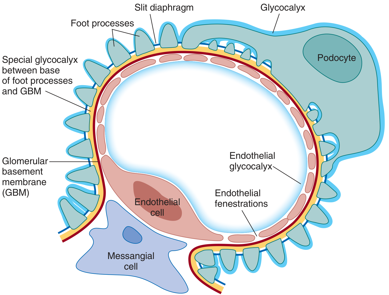
The glomerular filtration barrier (GFB) has three major layers. Damage to any one of them causes pathological proteinuria:
| Layer | Components | Main Function |
|---|---|---|
| 1. Fenestrated endothelium | Endothelial cells with large pores (~70-100 nm) covered by glycocalyx | Blocks blood cells; glycocalyx provides charge barrier |
| 2. Glomerular Basement Membrane (GBM) | Three sublayers (lamina rara interna, lamina densa, lamina rara externa) with heparan sulfate proteoglycans | Primary charge and size barrier for large proteins |
| 3. Podocytes + Slit Diaphragm | Visceral epithelial cells with foot processes; slit diaphragm proteins (nephrin, podocin) bridge 40-nm filtration slits | Last line of defense - ultimate size/charge barrier |
"Damage to any one of the three layers of the glomerular filtration barrier allows proteins through, resulting in abnormal, 'glomerular' proteinuria." - [Brenner & Rector's The Kidney, p. (Chapter 13 - Normal Physiology)]
Which Layer is the Most Clinically Important Cause of Albuminuria?
The podocyte/slit diaphragm is the most critical layer for albumin exclusion and is the primary site implicated in most proteinuric diseases:
- The slit diaphragm is formed by proteins nephrin and NEPH1 (from adjacent podocytes zipping together), along with podocin, CD2AP, and others.
- Loss, mutation, or redistribution of these proteins directly causes massive proteinuria.
- At least 26 podocyte-specific gene defects (including nephrin and podocin mutations) have been identified as hereditary causes of nephrotic syndrome.
- In minimal change disease, the podocytes lose their foot processes ("foot process effacement") - the slit diaphragm is destroyed, and massive albuminuria results.
- In diabetic nephropathy, the GBM thickens and loses its anionic charge, breaking both size and charge selectivity.
Summary Table
| Condition | Layer Damaged | Result |
|---|---|---|
| Minimal Change Disease | Podocyte foot processes / slit diaphragm | Massive albuminuria (nephrotic) |
| Focal Segmental Glomerulosclerosis | Podocytes | Heavy proteinuria |
| Diabetic Nephropathy | GBM (thickening + charge loss) | Progressive albuminuria |
| Glomerulonephritis (e.g., IgA, lupus) | Endothelium + GBM + podocytes | Mixed proteinuria + hematuria |
| Anti-GBM disease (Goodpasture) | GBM directly attacked | Severe proteinuria + nephritis |
What About Low Molecular Weight (LMW) Proteins?
This is a separate concept. True LMW proteins (<20,000 Da) - like beta-2-microglobulin, retinol-binding protein, alpha-1-microglobulin - are freely filtered across the GFB (because they're small enough to pass). They do NOT normally appear in urine because the proximal tubule reabsorbs them almost completely. When they do appear in urine (called "tubular proteinuria"), it indicates tubular damage (e.g., Fanconi syndrome, heavy metal toxicity), NOT glomerular damage.
Albumin is NOT a low molecular weight protein. At 67 kDa it is a high molecular weight protein, and its exclusion from urine is a glomerular (barrier) function, not a tubular reabsorption function.
- Brenner and Rector's The Kidney, 2-Volume Set - Chapter 3 (Permeability of the Glomerular Filtration Barrier)
- Brenner and Rector's The Kidney - Chapter 13 (Normal Physiology of proteinuria)
- Medical Physiology (Boron & Boulpaep) - Chapter 34 (Glomerular filtration and charge selectivity)
This is a shared conversation. Sign in to Orris to start your own chat.