
Hepatic encephalopathy management guidelines evidences
"hepatic encephalopathy"[MeSH Terms] AND management
hepatic encephalopathy management guidelines 2024 AASLD EASL
PMID: 35724930
PMID: 38439005
PMID: 34006606
PMID: 34600143
"hepatic encephalopathy" AND ("ACG" OR "American College of Gastroenterology") AND guideline
"hepatic encephalopathy" AND guideline
PMID: 33840331
PMID: 37127462
hepatic encephalopathy pathophysiology diagram ammonia
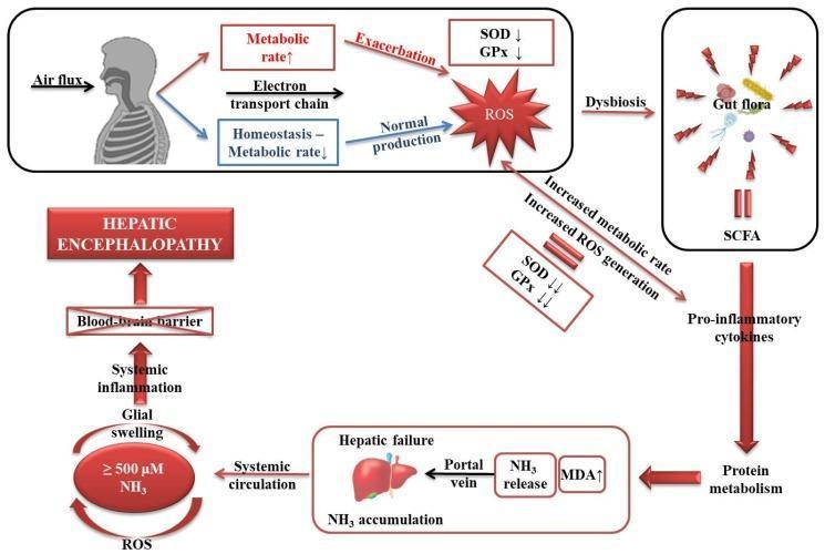
This pathophysiology diagram illustrates the multi-organ signaling pathways involved in the development of hepatic encephalopathy (HE) driven by oxidative stress and hyperammonemia. The process begins with systemic metabolic rate changes: an increased metabolic rate and electron transport chain activity lead to a depletion of antioxidant enzymes (SOD and GPx) and the exacerbation of Reactive Oxygen Species (ROS). Concurrently, gut dysbiosis and short-chain fatty acids (SCFA) trigger pro-inflammatory cytokines, affecting protein metabolism and contributing to hepatic failure. In the liver, hepatic failure results in NH3 (ammonia) accumulation and increased MDA (malondialdehyde) levels. High levels of NH3 (≥ 500 μM) enter systemic circulation and, together with ROS, induce glial swelling and systemic inflammation. The final stage of the pathway shows these systemic effects crossing the blood-brain barrier, leading to the clinical manifestation of hepatic encephalopathy. The diagram highlights the compensatory struggle between homeostasis and oxidative damage in the context of liver dysfunction.
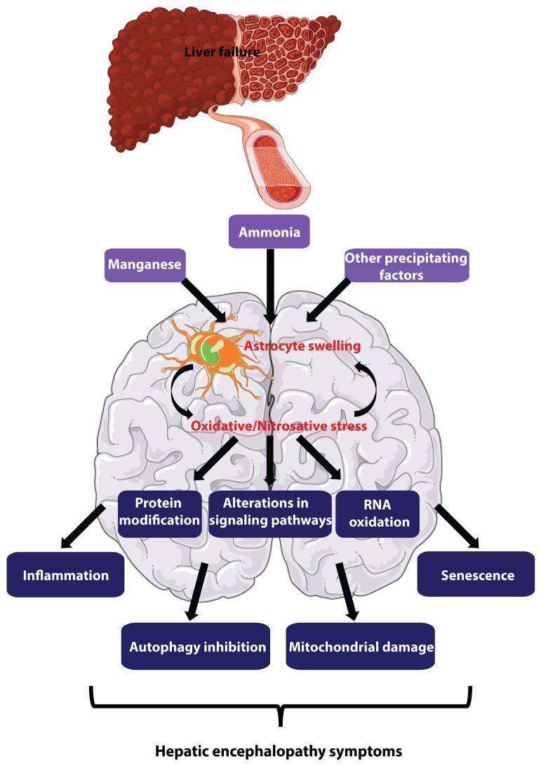
This pathophysiology diagram illustrates the major pathogenesis mechanisms of hepatic encephalopathy (HE). The flow begins with liver failure, shown as a cirrhotic liver, releasing metabolic toxins like ammonia and manganese into the systemic circulation via a blood vessel. These toxins, along with other precipitating factors, target the brain, specifically inducing astrocyte swelling. A central self-amplifying loop is depicted where astrocyte swelling and oxidative/nitrosative stress mutually reinforce each other. This cycle triggers a cascade of detrimental intracellular processes, including protein modification, RNA oxidation, and alterations in signaling pathways. Downstream effects featured include inflammation, autophagy inhibition, mitochondrial damage, and cellular senescence. These multi-factorial changes are linked via brackets to the ultimate manifestation of hepatic encephalopathy symptoms. The illustration uses a combination of anatomical representations (liver and brain) and text-based flow nodes to describe the transition from hepatic dysfunction to neurometabolic impairment, emphasizing the central role of astrocytes in the disease progression.
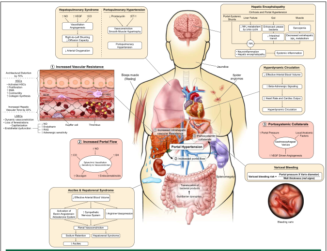
Summary : This is a comprehensive medical illustration summarizing the pathophysiology, clinical features, and complications of portal hypertension in liver disease, integrating vascular, hemodynamic, and systemic effects with annotated diagrams, flowcharts, and explanatory boxes.
Hepatic Encephalopathy: Management Guidelines & Evidence
Definition & Overview
Pathophysiology (Key Mechanisms)
- Colon bacteria metabolize proteins/nitrogen into ammonia; enterocytes produce ammonia from glutamine
- In cirrhosis, reduced hepatocyte function + portosystemic shunting = elevated circulating ammonia
- Ammonia crosses the blood-brain barrier → astrocyte swelling, cytotoxic brain edema, disrupted neurotransmission
- The GABA-benzodiazepine system is upregulated via neurosteroid production
- Other contributors: inflammatory cytokines, oxidative/nitrosative stress, gut dysbiosis, manganese toxicity
Classification & Grading
- Type A: Acute liver failure
- Type B: Portosystemic bypass/shunting
- Type C: Cirrhosis (most common)
| Grade | Consciousness | Symptoms | Signs |
|---|---|---|---|
| MHE | Normal | None (clinically) | Abnormal neuropsych testing only |
| 1 | Sleep-wake reversal, restlessness | Forgetfulness, mild confusion, irritability | Tremor, asterixis |
| 2 | Lethargy, slow responses | Disorientation, amnesia, inappropriate behavior | Asterixis, ataxia, dysarthria |
| 3 | Somnolence, confusion | Disorientation, aggressive behavior | Asterixis, hyperreflexia, Babinski sign |
| 4 | Coma | Unresponsive | Decerebration |
- Minimal HE (MHE) found in 60% of cirrhotic patients; overt HE in 30-45%. - Symptom to Diagnosis, 4th Ed., p. 330
Key Guidelines
1. EASL Clinical Practice Guidelines (2022) - PMID: 35724930
2. AASLD/EASL Joint Practice Guideline (2014) - Still widely referenced
3. ACG Clinical Guideline (2026) - Bajaj JS et al.
4. ISHEN Consensus (2020) - PMID: 32447806
Treatment: Stepwise Evidence-Based Approach
Step 1: Identify and Treat Precipitating Factors (Always First)
- GI bleeding (most common trigger)
- Infection / SBP - treat aggressively
- Constipation - laxative adjustment
- Medication noncompliance (lactulose)
- Electrolyte disturbances - hypokalemia, hyponatremia, azotemia
- Opioids, benzodiazepines, sedatives - minimize/discontinue
- Dehydration (diuretic overuse)
- TIPS and portosystemic shunts
"The mainstay of therapy is elimination of the precipitating event and excess ammonia." - Sleisenger and Fordtran's, p. 1815
Step 2: First-Line Pharmacotherapy - Lactulose
- Mechanism: Cathartic action removes ammonia; lowers colonic pH → inhibits ammonia absorption; traps ammonia as ammonium ions in the gut; reduces urease-producing bacterial population
- Dose: 15-45 mL PO (or via NGT) BID-QID; titrate to 3 soft stools/day
- In acute phase: start 30 mL every 1-2 hours, then taper as response occurs
- Enema option: 300 mL lactulose in 700 mL distilled water for patients unable to take orally or with aspiration risk
- Adverse effects: Flatulence, abdominal cramping, diarrhea, hypovolemia, hypernatremia - overuse must be avoided
- Improves symptoms vs. placebo but no clear mortality benefit. - Washington Manual, p. 747; Sleisenger and Fordtran's, p. 1816
Step 3: Second-Line / Combination - Rifaximin
- Approved by FDA (2010) for prevention of overt HE recurrence in advanced liver disease
- Used in combination with lactulose for patients not responding to lactulose alone, or as add-on therapy
- Superior tolerability vs. lactulose (fewer GI adverse effects), though higher cost
- Network meta-analysis (21 RCTs, 2024): Rifaximin significantly reduced:
- Primary HE prevention (OR 0.66; 95% CI 0.45-0.96)
- Secondary prevention / recurrence risk (OR 0.38; 95% CI 0.28-0.52)
- MHE progression to overt HE (OR 0.17; 95% CI 0.04-0.63)
- Improved clinical symptoms in MHE and overt HE (OR 3.76)
- Did NOT reduce mortality at any stage (OR 0.79; p=0.13)
- Optimal dosing: 400 mg TID for primary/secondary prevention; 600 mg BID for MHE treatment
- Fang et al., BMC Gastroenterol 2024 - PMID: 38439005
Step 4: Adjunctive Therapies (Evidence-Supported)
| Agent | Mechanism | Evidence Summary |
|---|---|---|
| L-Ornithine L-Aspartate (LOLA) | Activates urea cycle, enhances ammonia clearance | Cochrane review: possible benefit on mortality and HE; not available in USA |
| Probiotics | Modify gut flora, reduce ammonia generation | Cochrane review: probably improves recovery, overt HE, QoL, plasma ammonia; no mortality benefit |
| Zinc | Activates ornithine transcarbamylase (urea cycle); deficiency common in cirrhosis | Some improvement in HE; efficacy not clearly established |
| Acarbose | α-glucosidase inhibitor; increases saccharolytic vs. proteolytic bacteria | Shown to improve mild HE in cirrhosis + T2DM (RCT) |
| Albumin infusion | Reduces systemic inflammation, binds toxins | Meta-analysis (2021, PMID: 34600143): significant reduction in persistent HE (RR 0.60; p=0.03) and mortality (RR 0.54; p=0.02) |
| Sodium benzoate/phenylbutyrate | Enhance urinary ammonia excretion | FDA-approved for urea cycle disorders; efficacy in cirrhosis HE not well-established; high sodium load with benzoate |
| MARS (extracorporeal albumin dialysis) | Reduces blood ammonia levels | Benefit in severe HE with acute-on-chronic liver failure |
Step 5: Nutrition & Dietary Management (ACG Nutrition Guideline 2025)
- Do NOT restrict protein routinely - protein restriction worsens sarcopenia and outcomes
- Target 1.2-1.5 g/kg/day protein intake
- Prefer branched-chain amino acids (BCAA) and vegetable/dairy protein sources over meat protein (less ammoniagenic)
- Small, frequent meals; late-evening snack to reduce nocturnal fasting
- Nutritional support (enteral/parenteral if needed) for severely malnourished patients
- Correct zinc deficiency (common in cirrhosis)
Step 6: Fecal Microbiota Transplantation (FMT) - Emerging Evidence
- FMT improved neurocognitive test results in 4 human studies
- Donor microbiota persisted in recipients at 1 year post-FMT
- Lower readmission rates vs. standard of care
- Conclusion: FMT may improve neurocognitive function, but current evidence is limited to small trials. Large-scale RCTs are needed. - Madsen et al., Scand J Gastroenterol 2021
Step 7: Secondary Prophylaxis (Preventing Recurrence)
- Lactulose + rifaximin combination is the standard of care for ongoing secondary prophylaxis
- Continue indefinitely after an episode of overt HE
- Management of minimal HE (MHE) with treatment is evolving - not universally mandated but increasingly supported
Step 8: Liver Transplantation
- HE is an indication for liver transplant evaluation
- Transplantation generally reverses HE
- Post-transplant cognitive deficits can persist in some patients (systematic review 2021, PMID: 33790729 found persistent cognitive impairment after resolution)
Special Situations
TIPS-Induced HE
- TIPS (transjugular intrahepatic portosystemic shunt) placement precipitates HE in ~30% of patients
- Prevention: lactulose + rifaximin prophylaxis; consider TIPS reduction/closure if refractory
Acute Liver Failure (Type A HE)
- Cerebral edema is the primary concern (distinguishes from chronic HE)
- Management focuses on ICP monitoring, osmotherapy (mannitol, hypertonic saline), airway protection, lactulose/rifaximin as above
- Urgent liver transplant evaluation
Acute-on-Chronic Liver Failure (ACLF)
- More severe, rapid-onset HE
- MARS and extracorporeal support may be beneficial
- ICU-level management often required
Summary Algorithm
Overt HE episode
    ↓
1. Identify + treat precipitating factor
    ↓
2. Lactulose (titrate to 3 stools/day) ← First-line
    ↓ (if inadequate response)
3. Add rifaximin 550 mg BID ← Second-line
    ↓ (ongoing)
4. Secondary prophylaxis: lactulose + rifaximin indefinitely
    ↓
5. Adjuncts: probiotics, BCAA, zinc, albumin, LOLA per clinical judgment
    ↓
6. Assess for liver transplant candidacy
Key Evidence Summary
| Study/Guideline | Year | Key Finding | Evidence Level |
|---|---|---|---|
| EASL CPG on HE | 2022 (PMID: 35724930) | Comprehensive PICO-based management guidance | Practice Guideline |
| AASLD/EASL Joint Guideline | 2014 | Standardized nomenclature; lactulose + rifaximin backbone | Practice Guideline |
| ACG Clinical Guideline (Bajaj JS et al.) | 2026 | Latest North American guidance | Practice Guideline |
| Fang et al. (rifaximin network meta-analysis) | 2024 (PMID: 38439005) | Rifaximin prevents HE at all stages; 400 mg TID best for prevention | Level 1 (Meta-Analysis) |
| Hoilat et al. (PEG vs. lactulose) | 2021 (PMID: 34006606) | PEG faster HE resolution vs. lactulose | Level 1 (Meta-Analysis) |
| Bombassaro et al. (albumin) | 2021 (PMID: 34600143) | Albumin reduces HE persistence and mortality | Level 1 (Meta-Analysis) |
| Madsen et al. (FMT) | 2021 (PMID: 33840331) | FMT promising but insufficient RCT evidence | Level 1 (Systematic Review) |