
Ganz dislocation of hip joint
Ganz surgical hip dislocation trochanteric osteotomy
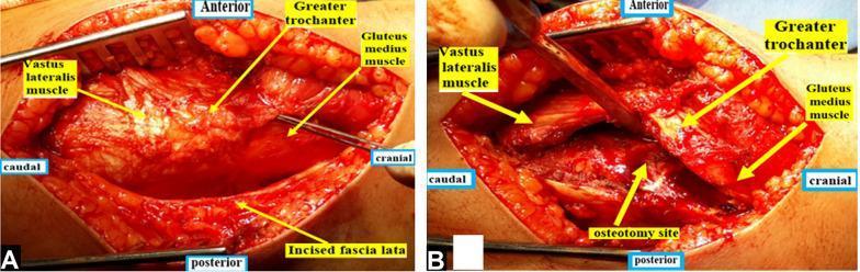
Two intraoperative clinical photographs (A and B) demonstrating a modified Ganz surgical hip dislocation approach. Image A shows a straight lateral skin incision with superficial dissection, exposing the underlying musculoskeletal anatomy. Key structures are labeled, including the incised fascia lata, the vastus lateralis muscle (caudal-anterior), the greater trochanter (cranial-anterior), and the gluteus medius muscle (cranial). Image B depicts the subsequent greater trochanteric osteotomy. An oscillating saw has been used to create a bone cut, labeled as the osteotomy site, located posterior to the vastus lateralis ridge. This procedure allows for the mobilization of the greater trochanter with its attached gluteus medius to preserve the blood supply to the femoral head. Both images include anatomical orientation markers (anterior, posterior, cranial, and caudal) to clarify the surgical perspective. This visual material is intended for orthopedic education, specifically regarding surgical techniques for treating slipped capital femoral epiphysis (SCFE) or other hip-preserving surgeries.
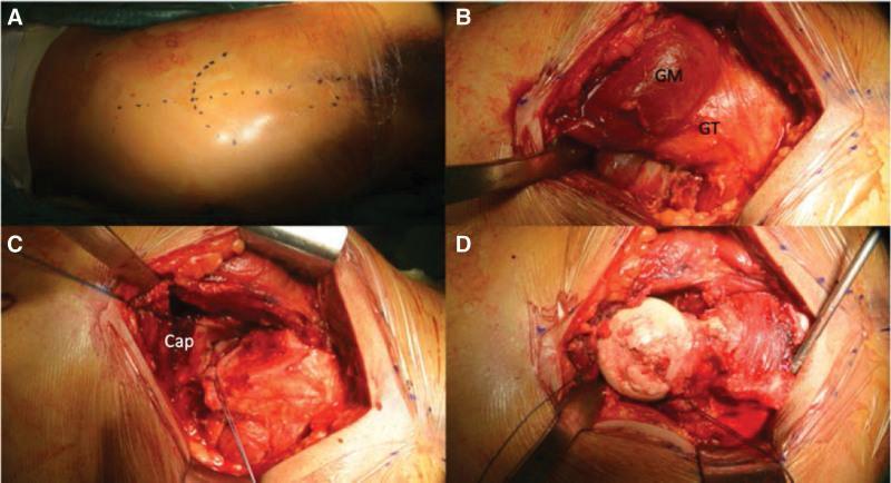
A series of four clinical photographs (A–D) documenting a surgical hip dislocation procedure following Ganz's technique for cartilage repair. (A) Preoperative skin marking on the lateral right thigh shows a longitudinal incision line approximately 10 cm in length, centered over the greater trochanter (GT). (B) Intraoperative view showing surgical exposure of the gluteus medius (GM) and the posterior aspect of the greater trochanter (GT) prior to trochanteric osteotomy. (C) Deep surgical dissection illustrating the disrupted hip joint capsule (Cap) following anterior and superior capsulotomy. (D) View of the dislocated femoral head, which exhibits a large, irregular chondral defect on the superior-lateral weight-bearing surface, measuring approximately 35 mm × 27 mm. The surrounding healthy cartilage and the exposed subchondral bone are visible within the operative field. This surgical approach provides full 360-degree access to the femoral head while preserving its vascularity for subsequent procedures such as mosaicplasty.
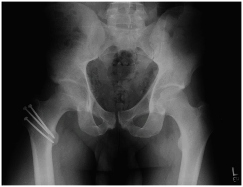
**Modality:** Anterior-posterior (AP) pelvic radiograph. **Anatomy:** Pelvis and proximal femora, including the acetabular-femoral joints, iliac wings, and pubic symphysis. **Observation:** The image demonstrates a postoperative state of the right hip. There is evidence of a completed trochanteric osteotomy. The osteotomy fragment is anatomically reduced and fixated using three cannulated metallic compression screws. The screws are oriented from the lateral aspect of the greater trochanter, extending medially and inferiorly toward the femoral neck/metaphysis. **Pathology/Condition:** Postoperative surgical hip dislocation (Ganz approach). The right femoral head is reduced within the acetabulum with a preserved joint space. No acute fracture or hardware failure is visualized. The contralateral (left) hip joint appears unremarkable with normal joint space and osseous morphology. **Differentiating Features:** Characteristic triple-screw fixation of the greater trochanter, commonly utilized for trochanteric flip osteotomy to provide surgical access to the acetabulum and femoral head while preserving vascularity. Overall pelvic alignment and pubic symphysis are within normal limits.
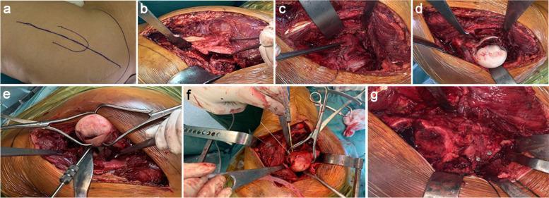
A multi-panel series of clinical photographs documenting the surgical steps of open reduction and internal fixation (ORIF) for a Pipkin type femoral head fracture via a safe surgical hip dislocation and Ganz trochanteric flip osteotomy. Panel (a) shows preoperative skin markings for the Gibson approach. Panels (b-c) demonstrate deep dissection, including the Z-shaped trochanteric osteotomy and subsequent Z-shaped capsulotomy to preserve the blood supply to the femoral head. Panel (d) reveals the exposed femoral head fracture post-dislocation. Panel (e) depicts the reduction of fracture fragments using Weber pointed reduction clamps. Panel (f) illustrates the definitive internal fixation of the femoral head using cannulated Herbert screws. The final panel (g) shows the anatomic reduction and synthesis of the greater trochanter with cortical screws. This series serves as an educational guide for orthopedic surgeons on the technique of Ganz surgical dislocation, emphasizing exposure, fragment manipulation, and stabilization in complex proximal femur trauma.
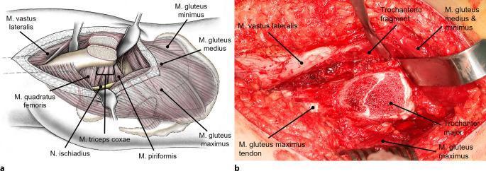
This composite educational resource demonstrates a trochanteric flip osteotomy for surgical hip dislocation. Panel A is a labeled anatomical diagram illustrating the surgical site after the osteotomy. Key structures shown include the mobilized trochanteric fragment with the vastus lateralis, gluteus medius, and gluteus minimus muscles attached. Deep structures visible include the piriformis, triceps coxae, quadratus femoris, and the sciatic nerve (N. ischiadicus). Panel B is a corresponding intraoperative clinical photograph providing a real-world perspective of the same surgical stage. It highlights the separated trochanteric fragment held by a surgical retractor, the exposed greater trochanter (trochanter major), and the mobilized gluteus medius and minimus muscles. The image serves to teach the surgical approach and relevant musculoskeletal anatomy necessary to preserve femoral head perfusion during hip reconstruction, emphasizing the anterior mobilization of the trochanteric fragment to expose the hip joint capsule while maintaining the integrity of the attached musculature.
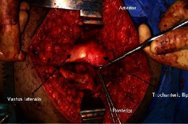
This clinical photograph captures a surgical procedure involving a trochanteric osteotomy of the hip, commonly utilized in surgical hip dislocation. The image shows a deep lateral incision centered over the greater trochanter, with soft tissue retracted to expose the musculoskeletal structures. Key anatomical landmarks are annotated: the 'Anterior' and 'Posterior' orientations are marked to provide spatial context. A 'Trochanteric flip' is indicated by an arrow, showing the mobile osteotomized fragment of the greater trochanter being displaced anteriorly. The 'Vastus lateralis' is also labeled, illustrating its relationship to the surgical site and its mobilization as part of the digastric trochanteric flap. Surgical retractors and elevators are visible within the field, reflecting active tissue manipulation to expose the underlying joint capsule. This visual demonstrates the surgical approach used for conditions such as Slipped Capital Femoral Epiphysis (SCFE) or femoroacetabular impingement, highlighting the preservation of the blood supply to the femoral head via the retinacular vessels.
"surgical hip dislocation" AND Ganz
Ganz Surgical Hip Dislocation (SHD)
Overview
Vascular Anatomy - The Critical Basis
- Keeping the piriformis tendon insertion intact on the proximal femur (not included in the trochanteric fragment)
- Performing an anterior Z-capsulotomy rather than a posterior one
- Carefully preserving the posterior capsule and retinacular vessels
The Trochanteric Flip Osteotomy (Digastric Osteotomy)
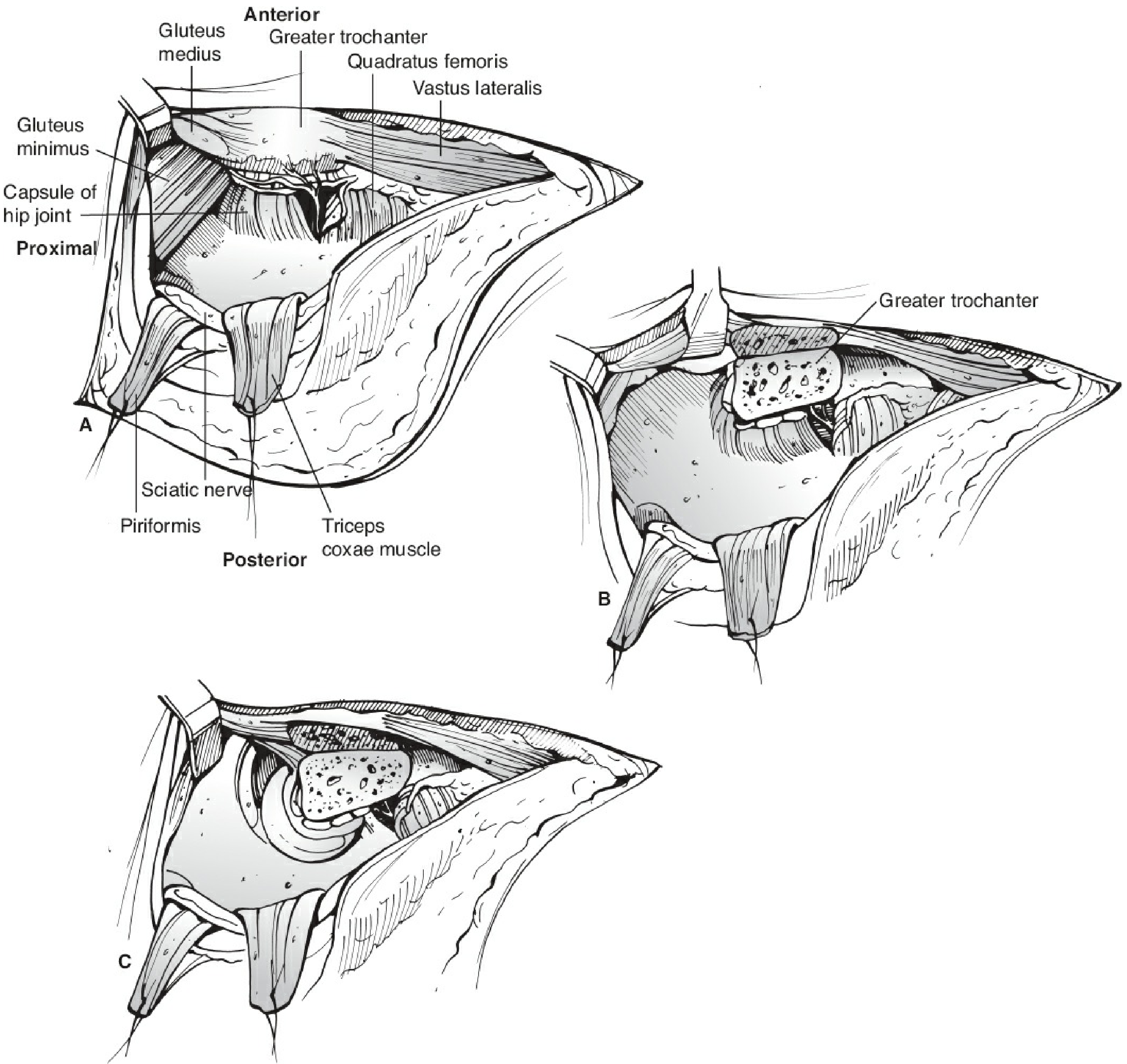
- Splits the posterior ridge of the greater trochanter, exiting anteriorly just medial to the gluteus medius/minimus insertions
- Creates a "digastric" flap because both the abductor muscles (gluteus medius + minimus) and the vastus lateralis remain attached to the trochanteric fragment
- The piriformis tendon stays on the intact proximal femur - this is the key vascular-protective step
- The trochanteric fragment is then retracted anteriorly, opening the joint
Surgical Steps (Ganz Technique - Campbell's Technique 34.4)
- Patient positioned in lateral decubitus
- Modified Gibson (straight lateral) or Kocher-Langenbeck approach - develop the interval between gluteus maximus and tensor fascia lata, or split gluteus maximus
- Identify and protect the sciatic nerve
- Perform trochanteric flip osteotomy - cut posterior to the vastus lateralis ridge, preserving piriformis
- Retract the trochanteric fragment anteriorly
- Perform anterior Z-shaped capsulotomy (Z-capsulotomy) - this protects the posterior retinacular vessels
- Dislocate the femoral head anteriorly by flexion, external rotation, and adduction
- Perform the necessary intra-articular procedure (osteochondroplasty, labral repair, fracture fixation, etc.)
- Reduce the femoral head
- Repair the capsule
- Reattach the trochanteric fragment with 3 cortical screws (triple-screw fixation)
Indications
| Indication | Notes |
|---|---|
| Femoroacetabular impingement (FAI) | Cam, pincer, or combined - when severe or arthroscopy is insufficient |
| Femoral head fractures (Pipkin) | Especially Pipkin type I, II, and IV; allows anatomic ORIF |
| Acetabular fractures | Combined femoral head + posterior wall fractures (Pipkin IV) |
| Legg-Calvé-Perthes disease | Osteochondroplasty for hinge abduction and FAI sequelae |
| Slipped capital femoral epiphysis (SCFE) | Subcapital realignment |
| Osteochondral defects | Full 360° access for mosaicplasty or OCA |
| Labral pathology | Repair of complex labral tears |
| Loose bodies | Removal of intra-articular loose bodies |
| Multiple hereditary exostoses | Excision of osteochondromas |
| Hip resurfacing | In select cases as an alternative to anterolateral approach |
Advantages
- 360-degree visualization of the entire femoral head and acetabulum
- No risk of AVN when performed correctly - the most feared complication of hip dislocation is effectively eliminated
- Permits treatment of severe deformities not accessible arthroscopically
- Allows soft tissue repair (labrum, chondral flaps)
- Can assess and correct dynamic impingement intraoperatively
Complications
| Complication | Rate |
|---|---|
| Osteonecrosis (AVN) | 0% in 334 hips (when technique is correct) |
| Trochanteric nonunion | ~1.8% |
| Heterotopic ossification | Uncommon |
| Overall complication rate | <5% |
Comparison: SHD vs. Hip Arthroscopy for FAI
| Feature | Surgical Hip Dislocation | Hip Arthroscopy |
|---|---|---|
| Exposure | 360° complete | Limited zones |
| Severe deformities | Excellent | Less effective |
| Invasiveness | Higher | Lower |
| Recovery | Longer | Shorter |
| Soft tissue repair | Better access | Adequate for most |
Intraoperative Images

Sources
- Miller's Review of Orthopaedics, 9th Ed - Treatment of FAI, p. 2897-2921
- Campbell's Operative Orthopaedics, 15th Ed (2026) - Technique 34.4 (Ganz), pp. 1486-1487; Pipkin fracture management, pp. 3473-3474
- Rockwood and Green's Fractures in Adults, 10th Ed (2025) - Trochanteric Flip Osteotomy, pp. 2506-2507; Hip Dislocation, p. 2568