
Minimal indentation of cisternal segment of left 3rd cranial neve by aberrant loop of P1 segment of left PCA. Essentially normal study of brain parenchyma. Orbits: Essentially normal study of orbits. Result of MRI brain and orbits visual prhway protocol.
MRI cisternal segment 3rd nerve compression P1 PCA indentation clinical significance
MRI cisternal segment oculomotor nerve third nerve PCA compression neurovascular conflict
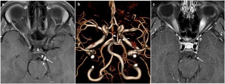
This composite of diagnostic images illustrates neurovascular conflict of the oculomotor nerve in a patient with ophthalmoplegic migraine. Panel (a) shows an axial post-contrast T1 TSE fat-saturated MRI demonstrating focal enhancement of the left oculomotor nerve at its exit zone from the cerebral peduncle. A nearby flow void corresponding to the left posterior cerebral artery (PCA) is seen compressing the nerve against the midbrain. Panel (b) provides a 3D volume-rendered time-of-flight (TOF) MRA, revealing a prominent sharp loop in the proximal P2 segment of the left PCA, just distal to the junction with the posterior communicating artery, which anatomically correlates to the site of nerve compression. Panel (c) is a follow-up axial T1 TSE fat-saturated image showing a significant reduction in nerve enhancement and swelling after symptom resolution. These findings highlight the role of vascular anomalies in the pathophysiology of recurrent cranial nerve palsies and migraine-like clinical presentations.
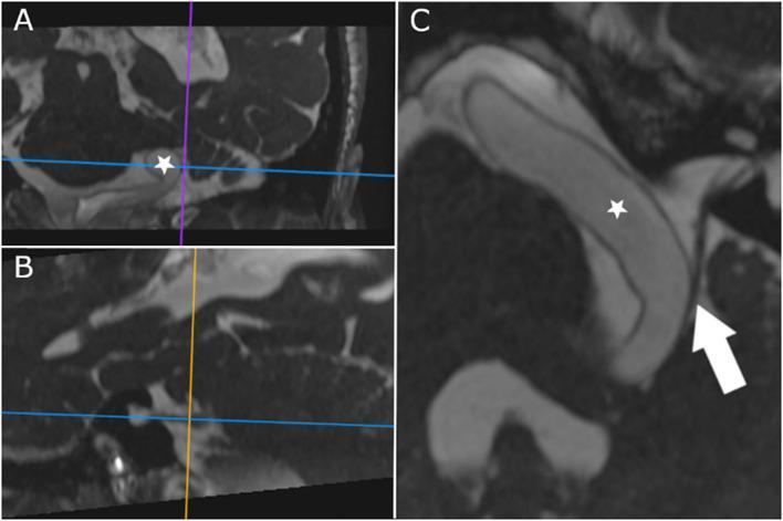
Diagnostic Magnetic Resonance Imaging (MRI) using Constructive Interference in Steady State (CISS) sequences, displayed in three panels (A, B, and C) to demonstrate a neurovascular conflict in the posterior fossa. Panel A and B show multiplanar reconstructions with reference lines centered on the cisternal segment of the cranial nerves. Panel C is an oblique axis reconstruction oriented along the cisternal course of the facial (CN VII) and vestibulocochlear (CN VIII) nerves. A prominent, dilated, and elongated basilar artery (marked with a star) is visualized within the prepontine and cerebellopontine angle cisterns. In the oblique view (C), the tortuous artery makes direct contact with and causes mechanical displacement of the nerve bundle (indicated by a white arrow). This imaging demonstrates the anatomical basis for neurovascular compression syndromes, where a dolichoectatic basilar artery indents the vestibulocochlear and facial nerves, potentially leading to symptoms such as hemifacial spasm or vestibular dysfunction. The high-resolution CISS sequence provides excellent contrast between the cerebrospinal fluid, vascular structures, and fine cranial nerve fibers.
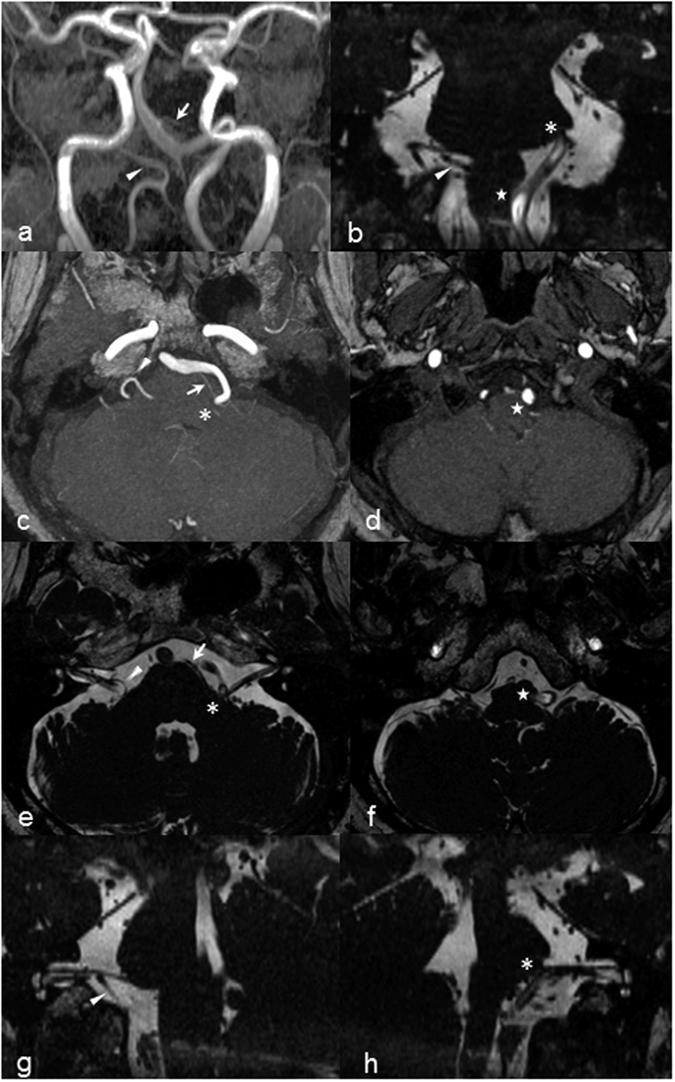
A multi-panel Brain MRI and MRA study demonstrating neurovascular conflict in a patient with left hemifacial spasm. Panel (a) shows a coronal MRA Maximum Intensity Projection (MIP) of dolichoectatic vertebrobasilar arteries. Panels (b), (e), (f), (g), and (h) utilize Constructive Interference at Steady State (CISS) sequences to provide high-resolution anatomical detail. The images highlight vascular compression of the left facial nerve root exit zone (REZ) and the left ventrolateral medulla (VLM). Specifically, the tortuous left vertebral artery (VA) and anterior inferior cerebellar artery (AICA) are seen indenting and displacing the brainstem and cranial nerve origin. Comparative oblique sagittal reconstructions show a normal facial nerve path on the right (panel g) versus pontine distortion and nerve elevation on the left (panel h). Markers identify the left AICA (arrow), right PICA (arrowhead), facial nerve REZ (asterisk), and VLM compression (star). This series illustrates the typical imaging findings of neurovascular compression syndromes where ectatic vessels impinge upon the cisternal segment of cranial nerves.
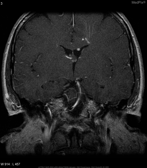
**Imaging Modality:** Magnetic Resonance Imaging (MRI), Coronal view. **Anatomical Region:** Brainstem and posterior fossa, specifically the prepontine cistern and the cerebellopontine angle (CPA). **Observed Pathology:** The image demonstrates a neurovascular contact or conflict involving the right trigeminal nerve (CN V). A prominent, tortuous vascular structure, likely the superior cerebellar artery (SCA) or an elongated basilar artery, is seen in close proximity to the right trigeminal nerve root entry zone. **Characteristic Visual Features:** - **Vascular contact:** A hypointense flow void or enhancing vessel is positioned immediately adjacent to the cisternal segment of the right trigeminal nerve. - **Asymmetry:** Compared to the contralateral (left) side, the right prepontine cistern shows crowding due to the vascular loop. - **Landmarks:** Visible structures include the pons, third ventricle, and the petrous temporal bone. **Clinical Context:** These radiologic findings are highly suggestive of trigeminal neuralgia (tic douloureux) caused by vascular compression of the nerve. **Diagnostic Features:** The key differentiating feature is the direct contact between a vascular loop and the trigeminal nerve at its point of exit from the pons, a common etiology for facial pain syndromes.
MRI Brain & Orbits - Visual Pathway Protocol: Report Explanation
What the Report Found
Key Anatomy
- After leaving the midbrain, CN III travels through the subarachnoid space (cisterns) before entering the cavernous sinus. This free-floating portion in the cerebrospinal fluid is called the cisternal segment.
- It is the most vulnerable part of the nerve to vascular compression because it is surrounded by CSF with no bony protection.
- The nerve controls: eyelid elevation (levator palpebrae), most eye movements, and pupil constriction.
- The P1 segment is the first part of the PCA, running from the basilar artery tip to the posterior communicating artery junction.
- Normally, this artery runs close to but does not contact CN III.
- An aberrant (abnormal/anomalous) loop means this artery has an unusual course - it loops closer to or directly against the cisternal nerve.
- When a blood vessel contacts and indents a cranial nerve in the cistern, this is called neurovascular conflict. The pulsatile pressure from arterial flow can irritate or compress the nerve.
- Well-known examples include trigeminal neuralgia (CN V compressed by SCA) and hemifacial spasm (CN VII compressed by AICA/PICA). PCA compression of CN III is a rarer but documented variant.
Clinical Significance of This Finding
| Severity | Description | Clinical Implication |
|---|---|---|
| Minimal indentation (as in this report) | Artery touches or barely dents the nerve | Usually asymptomatic / incidental |
| Moderate contact with displacement | Artery displaces the nerve | May cause partial CN III symptoms |
| Severe compression | Distinct groove on nerve | More likely to cause CN III palsy |
- This is a mild, likely incidental finding in the context of a normal brain parenchyma study.
- It does NOT by itself confirm a diagnosis or require immediate treatment.
- Its clinical relevance depends entirely on whether the patient has symptoms referable to CN III dysfunction.
Possible CN III Symptoms to Watch For
- Ptosis (drooping eyelid)
- Diplopia (double vision) - typically oblique/diagonal
- Eye deviated "down and out" (exotropia + hypotropia)
- Dilated, non-reactive pupil (pupil-involving palsy = serious, rule out aneurysm)
- Intermittent or episodic eye symptoms - could suggest NVC
Important note: A pupil-involving CN III palsy always requires urgent CTA/DSA to exclude a posterior communicating artery or basilar artery aneurysm - this MRI finding alone does not substitute for that workup if the pupil is involved.
Why Was This Imaged on a "Visual Pathway Protocol"?
- Brain parenchyma: essentially normal - no infarct, tumor, demyelination, or structural lesion.
- Orbits: essentially normal - no optic nerve pathology, orbital mass, or proptosis.
Relevant Image - Neurovascular Conflict of CN III by PCA
Summary
| Point | Detail |
|---|---|
| Finding | Minimal vascular contact/indentation of left CN III cisternal segment by aberrant P1-PCA loop |
| Nature | Neurovascular contact - a vascular anatomical variant |
| Brain & orbits | Normal |
| Clinical relevance | Depends on symptoms - likely incidental if patient is asymptomatic |
| Action needed | Correlate with clinical symptoms (ptosis, diplopia, pupil size); if asymptomatic, MRI follow-up or observation; if pupil involved, urgent vascular imaging |