
Contact granuloma
I'll look this up in the medical library first.
Reading File
Now let me search the medical library and PubMed in parallel.
Finding Sources
Searching PubMed
"contact granuloma" larynx vocal cord
Two excellent textbook sources found. Let me read both sections in parallel.
Reading File
Reading File
Let me get the rest of the Cummings section which was truncated.
Reading File
Loading Image
I have comprehensive information. Let me now compile the full answer.
Contact Granuloma (Vocal Fold Granuloma / Contact Ulcer)
A contact granuloma is a reactive, inflammatory lesion arising on the vocal process of the arytenoid cartilage -- the posterior, cartilaginous part of the glottis. It is also called a contact ulcer when it presents as an excavation rather than a heaped-up mass.
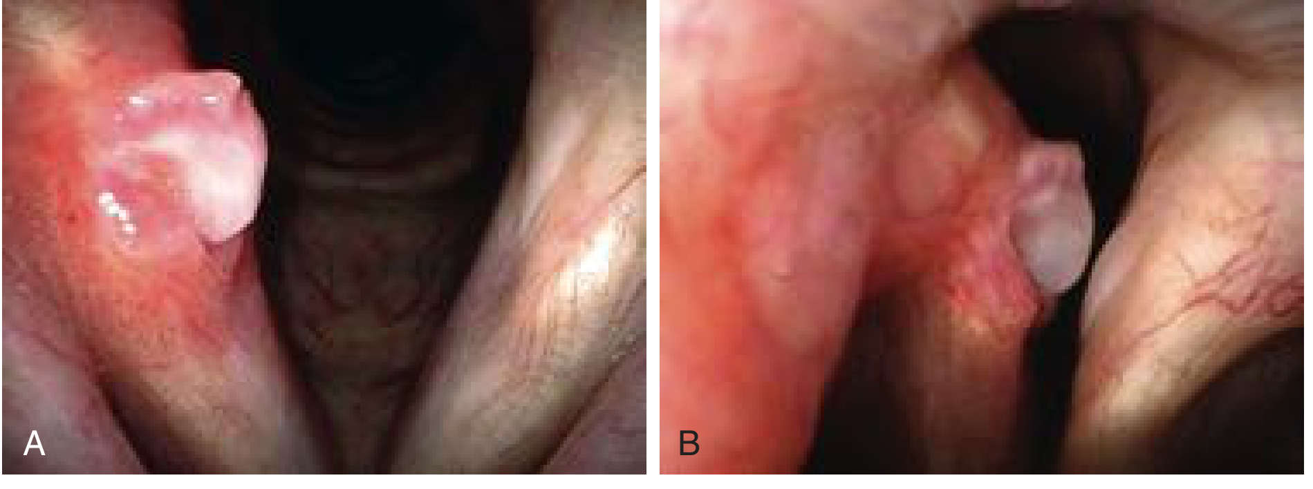
Fig. A: Contact granuloma on the right posterior vocal fold. Note the bilobed appearance and surrounding inflammation. At phonatory contact (B), the medial surface of the left arytenoid fits into the cleft of the bilobed granuloma.
Epidemiology
- Predominantly affects men
- Associated with vocally demanding occupations, hard glottal attack speakers, chronic throat clearers, and patients with acid reflux
- Psychological stress has also been implicated
Causes / Etiology
| Cause | Notes |
|---|---|
| Vocal abuse / misuse | Forceful glottal stroke, habitual low pitch, hard attack |
| Throat clearing / chronic coughing | Repetitive traumatic apposition of arytenoids |
| Laryngopharyngeal reflux (LPR) / GERD | Acid inflames the vulnerable posterior laryngeal mucosa |
| Endotracheal intubation | Pressure necrosis from the ETT tip on the vocal process |
| Glottal incompetence | Vocal fold atrophy causes compensatory hyperfunction |
Pathophysiology
The thin mucosa and perichondrium overlying the cartilaginous glottis are vulnerable to:
- Overly forceful apposition (slamming) of the arytenoids at glottal onset
- Chronic coughing/throat clearing
- Acid-mediated inflammation of the vocal process region
The traumatized area either ulcerates (contact ulcer) or forms a heaped-up granuloma through a reparative process. Bilobed morphology is characteristic -- when the contralateral arytenoid closes, it fits into the cleft between the two lobes.
Clinical Features
Symptoms:
- Unilateral discomfort over the midthyroid cartilage area, often with referred pain to the ipsilateral ear (otalgia)
- Foreign body sensation / globus
- Hoarseness (only when lesion is large)
- Frequent throat clearing
- History of intubation or chronic reflux
Voice characteristics:
- Speaking voice may sound normal or only slightly husky
- Habitual use of an overly low fundamental frequency
- Held-back, constrained vocal quality
- Low, monotone speech
Laryngeal Examination
- A depressed, ulcerated area with whitish exudate OR a bilobed, heaped-up lesion at the vocal process of the arytenoid
- Erythema on the vocal process extending up the medial arytenoid surface
- At phonatory closure, the contralateral vocal process fits into the granuloma cleft
- A maturing granuloma becomes pedunculated and may flip above and below the vocal fold plane
- Detection of early ulcers may require a rigid telescope or distal-chip camera
- Stroboscopy can reveal glottal incompetency as a contributing factor
Management
1. Anti-reflux Therapy (First-line)
- Empiric PPIs even without overt GERD symptoms (LPR is often clinically silent)
- Lifestyle and diet changes: avoid caffeine, alcohol, late-night eating
- Effective even in patients without classic GERD symptoms
2. Voice Therapy
- Vocal hygiene instruction
- Abolish habitual throat clearing
- Raise average speaking pitch
- Reduce hard glottal attack
3. Corticosteroid Injection
- Depot corticosteroid (e.g., triamcinolone) injected directly into the lesion and its base via videoendoscopy
- Inhaled triamcinolone or mometasone can be used to avoid systemic effects
- Re-assess at 6-8 week intervals; continue if size is reducing
4. Botulinum Toxin Injection
- Injection into the thyroarytenoid-lateral cricoarytenoid muscle complex
- Reduces forceful vocal process apposition
- Reserved for refractory cases or as an adjunct to surgery
- Causes significant temporary dysphonia -- patient must be counseled
5. Surgery (Last Resort)
- Indicated only for symptomatic lesions not responding to medical therapy, or when malignancy must be excluded
- Spontaneous maturation and detachment occurs in 3-6 months with conservative treatment -- surgery should wait
- When performed, removal should be conservative, leaving the base/pedicle undisturbed to minimize recurrence
- Recurrence rate is very high; recurrent lesions are often more recalcitrant than the original
Intubation Granuloma (Special Subtype)
- More common in females (unlike phonotraumatic granuloma, which favors males)
- Occurs after brief or prolonged intubation
- Mechanism: pressure necrosis from the ETT at the posterior commissure and vocal process
- Natural history: often mature and spontaneously detach without intervention (see management above)
- Characteristic "divots" at the posterior commissure may become visible after detachment
Key Points to Remember
- Location: always at the vocal process of the arytenoid (posterior, cartilaginous glottis), NOT the membranous vocal fold
- Contact ulcer = excavated form; granuloma = heaped-up reparative form -- they are a spectrum
- Referred otalgia is a characteristic symptom (via Arnold's nerve, a branch of CN X)
- Spontaneous resolution is the rule with medical management (3-6 months)
- Surgery is a last resort due to high recurrence
- Botulinum toxin is an important tool for refractory or recurrent cases
Sources: Cummings Otolaryngology Head and Neck Surgery, p. 1125-1126; K.J. Lee's Essential Otolaryngology, p. 923
A recent 2026 review in J Voice (PMID 37985285) confirms these management principles, with updates on botulinum toxin and multimodal treatment approaches for refractory cases.
Convert to a pdf format
Reading File
Running Command
ls /home/daytona/workspace/ 2>/dev/null || echo "workspace empty or missing"
Running Command
mkdir -p /home/daytona/workspace/contact-granuloma && python3 -c "from reportlab.lib.pagesizes import A4; print('reportlab ok')" && python3 -c "import markdown2; print('markdown2 ok')" 2>&1
I'll use ReportLab directly to build a well-formatted PDF.
Writing File
~/contact-granuloma/generate_pdf.py
from reportlab.lib.pagesizes import A4
from reportlab.lib.styles import getSampleStyleSheet, ParagraphStyle
from reportlab.lib.units import cm
from reportlab.lib import colors
from reportlab.platypus import (
    SimpleDocTemplate, Paragraph, Spacer, Table, TableStyle,
    HRFlowable, ListFlowable, ListItem, KeepTogether
)
from reportlab.lib.enums import TA_LEFT, TA_CENTER, TA_JUSTIFY
from reportlab.platypus import PageBreak
import io, urllib.request

OUTPUT = "/home/daytona/workspace/contact-granuloma/Contact_Granuloma.pdf"

# ── Styles ─────────────────────────────────────────────────────────────────
styles = getSampleStyleSheet()

BRAND_BLUE   = colors.HexColor("#1a4a7a")
BRAND_TEAL   = colors.HexColor("#0f7b8c")
LIGHT_BLUE   = colors.HexColor("#e8f4f8")
LIGHT_GRAY   = colors.HexColor("#f5f5f5")
MED_GRAY     = colors.HexColor("#555555")
TABLE_HEADER = colors.HexColor("#1a4a7a")
TABLE_ALT    = colors.HexColor("#eaf4fb")

title_style = ParagraphStyle(
    "DocTitle", parent=styles["Title"],
    fontSize=22, textColor=colors.white,
    spaceAfter=4, leading=28, alignment=TA_LEFT,
    fontName="Helvetica-Bold"
)
subtitle_style = ParagraphStyle(
    "DocSubtitle", parent=styles["Normal"],
    fontSize=11, textColor=colors.HexColor("#d0eaf5"),
    spaceAfter=0, leading=14, alignment=TA_LEFT,
    fontName="Helvetica"
)
h1_style = ParagraphStyle(
    "H1", parent=styles["Heading1"],
    fontSize=14, textColor=BRAND_BLUE,
    spaceBefore=14, spaceAfter=4, leading=18,
    fontName="Helvetica-Bold", borderPad=2,
    borderColor=BRAND_TEAL, borderWidth=0,
)
h2_style = ParagraphStyle(
    "H2", parent=styles["Heading2"],
    fontSize=11, textColor=BRAND_TEAL,
    spaceBefore=10, spaceAfter=3, leading=14,
    fontName="Helvetica-Bold",
)
body_style = ParagraphStyle(
    "Body", parent=styles["Normal"],
    fontSize=10, textColor=colors.HexColor("#222222"),
    spaceAfter=5, leading=15, alignment=TA_JUSTIFY,
    fontName="Helvetica"
)
bullet_style = ParagraphStyle(
    "Bullet", parent=body_style,
    leftIndent=14, spaceAfter=3,
)
caption_style = ParagraphStyle(
    "Caption", parent=styles["Normal"],
    fontSize=8.5, textColor=MED_GRAY,
    spaceAfter=8, leading=12, alignment=TA_CENTER,
    fontName="Helvetica-Oblique"
)
source_style = ParagraphStyle(
    "Source", parent=styles["Normal"],
    fontSize=8, textColor=MED_GRAY,
    spaceAfter=3, leading=11,
    fontName="Helvetica-Oblique"
)
keypoint_style = ParagraphStyle(
    "KeyPoint", parent=body_style,
    leftIndent=10, textColor=BRAND_BLUE,
    fontName="Helvetica-Bold", fontSize=10,
)

def hr():
    return HRFlowable(width="100%", thickness=1, color=BRAND_TEAL, spaceAfter=6, spaceBefore=4)

def h1(text):
    return Paragraph(text, h1_style)

def h2(text):
    return Paragraph(text, h2_style)

def body(text):
    return Paragraph(text, body_style)

def bullet(items):
    elems = []
    for item in items:
        elems.append(ListItem(Paragraph(item, bullet_style), bulletColor=BRAND_TEAL, value='bullet'))
    return ListFlowable(elems, bulletType='bullet', leftIndent=18, bulletFontSize=8)

def spacer(h=6):
    return Spacer(1, h)

# ── Document ────────────────────────────────────────────────────────────────
doc = SimpleDocTemplate(
    OUTPUT,
    pagesize=A4,
    leftMargin=2.2*cm, rightMargin=2.2*cm,
    topMargin=2*cm, bottomMargin=2*cm,
    title="Contact Granuloma",
    author="Orris Medical Library",
    subject="Otolaryngology – Vocal Fold Granuloma"
)

story = []

# ── Header Banner ────────────────────────────────────────────────────────────
banner_data = [[
    Paragraph("Contact Granuloma", title_style),
]]
banner_table = Table(banner_data, colWidths=[doc.width])
banner_table.setStyle(TableStyle([
    ("BACKGROUND", (0,0), (-1,-1), BRAND_BLUE),
    ("TOPPADDING",    (0,0), (-1,-1), 16),
    ("BOTTOMPADDING", (0,0), (-1,-1), 12),
    ("LEFTPADDING",   (0,0), (-1,-1), 14),
    ("RIGHTPADDING",  (0,0), (-1,-1), 14),
    ("ROUNDEDCORNERS", [6]),
]))
story.append(banner_table)

subtitle_data = [[
    Paragraph("Vocal Fold Granuloma / Contact Ulcer  |  Otolaryngology Reference", subtitle_style)
]]
sub_table = Table(subtitle_data, colWidths=[doc.width])
sub_table.setStyle(TableStyle([
    ("BACKGROUND", (0,0), (-1,-1), BRAND_TEAL),
    ("TOPPADDING",    (0,0), (-1,-1), 6),
    ("BOTTOMPADDING", (0,0), (-1,-1), 6),
    ("LEFTPADDING",   (0,0), (-1,-1), 14),
    ("RIGHTPADDING",  (0,0), (-1,-1), 14),
]))
story.append(sub_table)
story.append(spacer(12))

# ── Definition ───────────────────────────────────────────────────────────────
story.append(h1("Definition"))
story.append(hr())
story.append(body(
    "A contact granuloma is a reactive, inflammatory lesion arising on the <b>vocal process of the "
    "arytenoid cartilage</b> — the posterior, cartilaginous part of the glottis. When the lesion "
    "presents as an excavation rather than a heaped-up mass it is termed a <b>contact ulcer</b>. "
    "The two forms represent a spectrum of the same pathological process."
))
story.append(spacer(4))

# ── Epidemiology ─────────────────────────────────────────────────────────────
story.append(h1("Epidemiology"))
story.append(hr())
story.append(bullet([
    "Predominantly affects <b>men</b>",
    "Associated with vocally demanding occupations and hard glottal attack speakers",
    "Chronic throat clearers and acid reflux patients are at elevated risk",
    "Psychological stress has also been implicated as a contributing factor",
]))
story.append(spacer(4))

# ── Etiology ─────────────────────────────────────────────────────────────────
story.append(h1("Causes / Etiology"))
story.append(hr())

etio_data = [
    ["Cause", "Notes"],
    ["Vocal abuse / misuse", "Forceful glottal stroke, habitual low pitch, hard attack"],
    ["Throat clearing / chronic coughing", "Repetitive traumatic apposition of arytenoids"],
    ["LPR / GERD", "Acid inflames the vulnerable posterior laryngeal mucosa"],
    ["Endotracheal intubation", "Pressure necrosis from ETT tip on the vocal process"],
    ["Glottal incompetence", "Vocal fold atrophy causes compensatory hyperfunction"],
]
etio_table = Table(etio_data, colWidths=[6.5*cm, doc.width - 6.5*cm])
etio_table.setStyle(TableStyle([
    ("BACKGROUND",    (0,0), (-1,0),  TABLE_HEADER),
    ("TEXTCOLOR",     (0,0), (-1,0),  colors.white),
    ("FONTNAME",      (0,0), (-1,0),  "Helvetica-Bold"),
    ("FONTSIZE",      (0,0), (-1,-1), 9.5),
    ("BOTTOMPADDING", (0,0), (-1,0),  7),
    ("TOPPADDING",    (0,0), (-1,0),  7),
    ("ROWBACKGROUNDS",(0,1), (-1,-1), [colors.white, TABLE_ALT]),
    ("ALIGN",         (0,0), (-1,-1), "LEFT"),
    ("VALIGN",        (0,0), (-1,-1), "MIDDLE"),
    ("TOPPADDING",    (0,1), (-1,-1), 5),
    ("BOTTOMPADDING", (0,1), (-1,-1), 5),
    ("LEFTPADDING",   (0,0), (-1,-1), 8),
    ("RIGHTPADDING",  (0,0), (-1,-1), 8),
    ("GRID",          (0,0), (-1,-1), 0.4, colors.HexColor("#cccccc")),
    ("ROUNDEDCORNERS",[4]),
]))
story.append(etio_table)
story.append(spacer(8))

# ── Pathophysiology ───────────────────────────────────────────────────────────
story.append(h1("Pathophysiology"))
story.append(hr())
story.append(body(
    "The thin mucosa and perichondrium overlying the <b>cartilaginous glottis</b> are vulnerable to:"
))
story.append(bullet([
    "Overly forceful apposition (slamming) of the arytenoids at glottal onset",
    "Chronic coughing and repetitive throat clearing",
    "Acid-mediated inflammation of the vocal process region via LPR",
]))
story.append(body(
    "The traumatized area either <b>ulcerates</b> (contact ulcer) or forms a <b>heaped-up granuloma</b> "
    "through reparative granulation tissue formation. The characteristic <b>bilobed morphology</b> arises "
    "because the contralateral arytenoid contacts the lesion at its midpoint during phonation — at closure, "
    "the opposing vocal process fits exactly into the cleft between the two lobes."
))
story.append(spacer(4))

# ── Clinical Features ─────────────────────────────────────────────────────────
story.append(h1("Clinical Features"))
story.append(hr())
story.append(h2("Symptoms"))
story.append(bullet([
    "Unilateral discomfort over the midthyroid cartilage area",
    "<b>Referred otalgia</b> to the ipsilateral ear (via Arnold's nerve, a branch of CN X) — characteristic",
    "Foreign body sensation / globus pharyngeus",
    "Hoarseness (only when the lesion is large)",
    "Frequent throat clearing",
    "History of intubation, chronic reflux, or heavy voice use",
]))
story.append(spacer(4))
story.append(h2("Voice Characteristics"))
story.append(bullet([
    "May sound normal or only slightly husky",
    "Habitual use of an overly <b>low fundamental frequency</b>",
    "Held-back, constrained vocal quality",
    "Low, monotone speech pattern",
]))
story.append(spacer(8))

# ── Laryngeal Examination ─────────────────────────────────────────────────────
story.append(h1("Laryngeal Examination"))
story.append(hr())
story.append(bullet([
    "Depressed, ulcerated area with whitish exudate <b>or</b> a bilobed, heaped-up lesion at the <b>vocal process of the arytenoid</b>",
    "Erythema on the vocal process extending up the medial arytenoid surface",
    "At phonatory closure, the contralateral vocal process fits into the granuloma cleft",
    "Maturing granuloma becomes <b>pedunculated</b> and may flip above/below the vocal fold plane",
    "Detection of early ulcers may require a rigid telescope or distal-chip camera",
    "<b>Stroboscopy</b> can reveal glottal incompetency as a contributing factor",
]))
story.append(spacer(8))

# ── Management ────────────────────────────────────────────────────────────────
story.append(h1("Management"))
story.append(hr())

mgmt_data = [
    ["Step", "Treatment", "Details"],
    ["1st line", "Anti-reflux therapy",
     "Empiric PPIs even without overt GERD symptoms. Lifestyle changes: avoid caffeine, alcohol, late-night eating."],
    ["1st line", "Voice therapy",
     "Vocal hygiene, abolish throat clearing, raise average speaking pitch, reduce hard glottal attack."],
    ["2nd line", "Corticosteroid injection",
     "Depot steroid (triamcinolone) injected into lesion + base via videoendoscopy. Inhaled triamcinolone or mometasone also used. Re-assess at 6-8 week intervals."],
    ["Refractory", "Botulinum toxin",
     "Inject TA-LCA muscle complex to reduce forceful arytenoid apposition. Causes significant temporary dysphonia — counsel patient."],
    ["Last resort", "Surgery",
     "Only for lesions not responding to medical therapy or if malignancy must be excluded. Wait for spontaneous maturation (3-6 months). Leave base/pedicle undisturbed. High recurrence rate."],
]

col_w = [2.5*cm, 3.8*cm, doc.width - 6.3*cm]
mgmt_table = Table(mgmt_data, colWidths=col_w)
mgmt_table.setStyle(TableStyle([
    ("BACKGROUND",    (0,0), (-1,0),  TABLE_HEADER),
    ("TEXTCOLOR",     (0,0), (-1,0),  colors.white),
    ("FONTNAME",      (0,0), (-1,0),  "Helvetica-Bold"),
    ("FONTSIZE",      (0,0), (-1,-1), 9),
    ("BOTTOMPADDING", (0,0), (-1,0),  7),
    ("TOPPADDING",    (0,0), (-1,0),  7),
    ("ROWBACKGROUNDS",(0,1), (-1,-1), [colors.white, TABLE_ALT]),
    ("ALIGN",         (0,0), (-1,-1), "LEFT"),
    ("VALIGN",        (0,0), (-1,-1), "TOP"),
    ("TOPPADDING",    (0,1), (-1,-1), 5),
    ("BOTTOMPADDING", (0,1), (-1,-1), 5),
    ("LEFTPADDING",   (0,0), (-1,-1), 7),
    ("RIGHTPADDING",  (0,0), (-1,-1), 7),
    ("GRID",          (0,0), (-1,-1), 0.4, colors.HexColor("#cccccc")),
]))
story.append(mgmt_table)
story.append(spacer(10))

# ── Intubation Granuloma ──────────────────────────────────────────────────────
story.append(h1("Intubation Granuloma (Special Subtype)"))
story.append(hr())
story.append(bullet([
    "More common in <b>females</b> (unlike phonotraumatic granuloma, which favors males)",
    "Occurs after brief or prolonged endotracheal intubation",
    "Mechanism: pressure necrosis from the ETT at the posterior commissure and vocal process",
    "Natural history: often matures and spontaneously detaches without intervention",
    'Characteristic "divots" at the posterior commissure may become visible after detachment',
]))
story.append(spacer(8))

# ── Key Points ────────────────────────────────────────────────────────────────
story.append(h1("Key Points"))
story.append(hr())

keys = [
    ("Location:", "Always at the vocal process of the arytenoid (posterior, cartilaginous glottis) — NOT the membranous vocal fold."),
    ("Spectrum:", "Contact ulcer = excavated form; granuloma = heaped-up reparative form. They are the same entity."),
    ("Otalgia:", "Referred pain to the ipsilateral ear is characteristic (Arnold's nerve, branch of CN X)."),
    ("Natural history:", "Spontaneous resolution is the rule with medical management (3-6 months)."),
    ("Surgery:", "Last resort due to very high recurrence rate."),
    ("Botulinum toxin:", "Important tool for refractory or recurrent cases — reduces forceful arytenoid apposition."),
    ("Intubation type:", "More common in females; phonotraumatic type more common in males."),
]

kp_data = [[Paragraph(f"<b>{k}</b>", body_style), Paragraph(v, body_style)] for k, v in keys]
kp_table = Table(kp_data, colWidths=[4*cm, doc.width - 4*cm])
kp_table.setStyle(TableStyle([
    ("BACKGROUND",    (0,0), (-1,-1), LIGHT_BLUE),
    ("ALIGN",         (0,0), (-1,-1), "LEFT"),
    ("VALIGN",        (0,0), (-1,-1), "TOP"),
    ("TOPPADDING",    (0,0), (-1,-1), 5),
    ("BOTTOMPADDING", (0,0), (-1,-1), 5),
    ("LEFTPADDING",   (0,0), (-1,-1), 8),
    ("RIGHTPADDING",  (0,0), (-1,-1), 8),
    ("LINEBELOW",     (0,0), (-1,-2), 0.3, colors.HexColor("#b0c8d8")),
]))
story.append(kp_table)
story.append(spacer(14))

# ── Sources ───────────────────────────────────────────────────────────────────
story.append(hr())
story.append(Paragraph("<b>Sources</b>", source_style))
story.append(Paragraph(
    "Cummings Otolaryngology Head and Neck Surgery, pp. 1125-1126",
    source_style
))
story.append(Paragraph(
    "K.J. Lee's Essential Otolaryngology, p. 923",
    source_style
))
story.append(Paragraph(
    "Teng TZJ et al. Vocal Fold Granuloma: Updates and Advancements in Treatment. J Voice. 2026 Mar. [PMID: 37985285]",
    source_style
))

# ── Build ─────────────────────────────────────────────────────────────────────
doc.build(story)
print(f"PDF written to: {OUTPUT}")
Running Command
python3 /home/daytona/workspace/contact-granuloma/generate_pdf.py 2>&1
Your PDF is ready. Download it here:
Contact Granuloma
PDF Document · PDF
The document includes all sections from the previous answer - definition, epidemiology, etiology table, pathophysiology, clinical features, management table, intubation granuloma subtype, key points summary, and source citations - formatted with a clean blue header banner, color-coded tables, and structured headings.
Sculpted incus
Finding Sources
Searching PubMed
"sculpted incus" ossiculoplasty
Finding Sources
Finding Sources
Excellent - rich results from Shambaugh, Cummings, Scott-Brown's, and KJ Lee. Let me read the key sections in parallel.
Reading File
Reading File
Reading File
Let me now fetch the surgical illustration and read the Shambaugh section on results.
Loading Image
Reading File
Loading Image
I have comprehensive information from three major textbooks with excellent surgical illustrations. Here is the full answer:
Sculpted Incus (Incus Interposition Graft)
A sculpted incus (also called a "fitted incus" or "incus interposition graft") is a type A ossiculoplasty technique in which the patient's own incus is removed, reshaped with an otologic drill, and reinterposed as a strut between the malleus handle and the stapes capitulum to restore the ossicular chain. It is the preferred method when the lenticular process of the incus is eroded but the stapes superstructure and malleus handle remain intact and favorably aligned.
Indication
Type A defect (malleus present [M+], stapes superstructure present [S+]) with:
- Erosion of the lenticular process (incudostapedial joint)
- Manubrium of the malleus in close proximity to the stapes capitulum (favorable spatial relationship)
- Absent or minimal incudomalleolar contact requiring bridging
If the manubrium is positioned far anterior to the stapes (unfavorable relationship), a sculpted incus is unstable and a PORP is preferred instead.
The most common indication is chronic otitis media with isolated lenticular process erosion.
Surgical Technique
Step 1 - Incus Extraction
The incus is removed from the middle ear and held in the Sheehy ossicular holder for shaping.
Step 2 - Sculpting (Reshaping) with the Drill
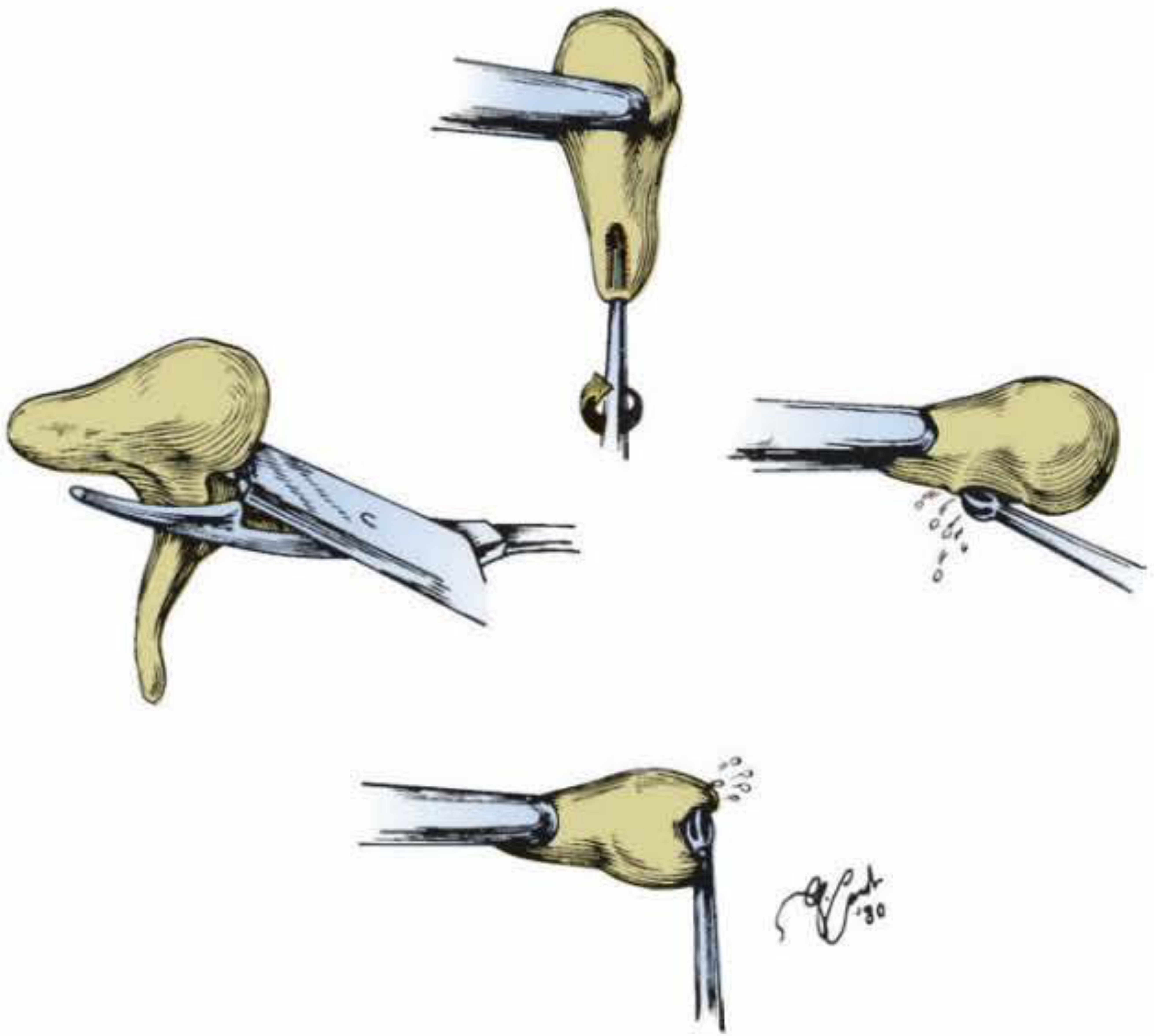
Fig. 29-1: Fashioning a sculpted incus interposition graft (Shambaugh Surgery of the Ear)
Key sculpting steps using a light drilling technique:
- Remove the residual long process - to minimize mass and reduce risk of fixation to the fallopian canal or promontory
- Create a groove on the malleus-contact surface using a 1.5-mm diamond burr, roughly following the natural groove of the incudomalleolar joint
- Drill an acetabulum (cup) into the short process using a 0.5- or 0.7-mm diamond burr - hollowed out as much as possible in an oval shape without gouging the walls (prevents the prosthesis from slipping off the capitulum)
- Trim the short process to desired length - roughly measured by holding a 2-mm round knife adjacent to the gap between the capitulum and manubrium
- The completed sculpted incus must slightly exceed the gap size (~2.4 mm) to create appropriate tension
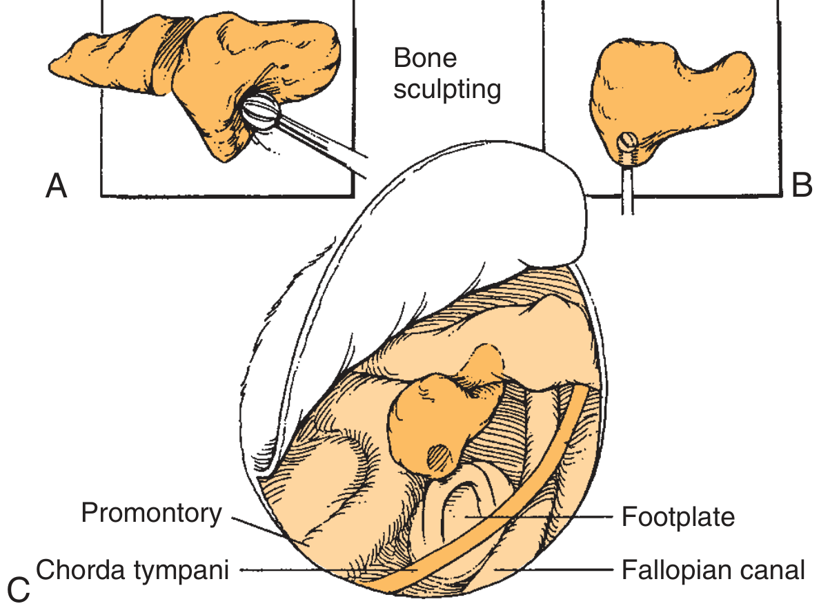
Fig. 145.10: (A) Bone sculpting of the incus. (B) Sculpted incus. (C) Modified incus positioned to restore ossicular continuity. (Cummings Otolaryngology)
Step 3 - Placement
- The sculpted incus is set to engage the malleus handle in its groove
- Both ossicles are elevated laterally with a curved pick in the acetabulum
- The incus is rotated onto the stapes capitulum and pressure slowly released
- Care is taken that the incus does not exert excessive pressure on the stapes
- The incus is slipped superiorly along the malleus until it contacts the tensor tympani tendon - this maximizes stability and hearing optimization
- The posterior scutum is curetted to prevent contact between the graft and the canal wall
Critical Tension Principle
- Too long: risks fixation (ankylosis to promontory or facial ridge)
- Too short: risks displacement
- Correct tension: prosthesis holds firmly without needing Gelfoam packing
Silastic sheeting or hyaluronic acid sheets can be placed in the middle ear to prevent fixation of the graft to the promontory or facial ridge.
Wehrs Technique (Modification)
If the interposed incus does not bridge the vertical distance between the capitulum and the malleus handle after initial sculpting, the incus can be resculpted according to the Wehrs technique to achieve adequate length.
Postoperative Appearance
Once healed, the sculpted incus is usually visible on otomicroscopy as a shadow or bulge just posterior to the malleus.
Results
| Metric | Result |
|---|---|
| Air-bone gap closure within 20 dB | ~66-68% of patients |
| Hearing stability | Good long-term stability in experienced hands |
| Preferred graft material | Autologous incus (best biocompatibility) |
Advantages of Autologous Sculpted Incus
- Biocompatibility - autologous bone is the ideal material; no foreign body reaction
- Low cost - no prosthesis required
- Tailored fit - can be customized intraoperatively to exact dimensions
- No extrusion risk (unlike alloplastic prostheses)
Alternatives When Sculpted Incus is Not Suitable
| Situation | Alternative |
|---|---|
| Incus unavailable or unusable | PORP (partial ossicular replacement prosthesis) |
| Unfavorable malleus-stapes geometry | PORP with cartilage cap |
| Stapes superstructure also absent | TORP (total ossicular replacement prosthesis) |
| Homograft available | Homograft incus (long process resting on footplate) |
| Cartilage / cortical bone | Can be sculpted as substitute, but more prone to resorption or ankylosis |
Sources: Shambaugh Surgery of the Ear, pp. 517-518; Cummings Otolaryngology Head & Neck Surgery, pp. 604-605; K.J. Lee's Essential Otolaryngology, p. 923; Scott-Brown's Otorhinolaryngology Head & Neck Surgery Vol. 2
Tuning fork tests for malingering
Searching sources...
Finding Sources
Finding Sources
Finding Sources
Finding Sources
Excellent leads from multiple textbooks. Let me read all relevant sections in parallel.
Reading File
Reading File
Reading File
Now let me search specifically for the tuning fork tests used in malingering detection.
Finding Sources
Finding Sources
Finding Sources
Finding Sources
Finding Sources
Reading File
Reading File
I now have comprehensive content. Let me compile the full answer, drawing also on my own knowledge for the classic tuning fork-specific tests.
Tuning Fork Tests for Malingering (Pseudohypacusis / Functional Hearing Loss)
Terminology
Before the tests, note the terminology hierarchy:
| Term | Meaning |
|---|---|
| Pseudohypacusis | Umbrella: hearing behaviors discrepant with true audiologic function (deliberate or not) |
| Malingering | Deliberate feigning for external gain (compensation, litigation, avoiding duty) |
| Factitious disorder | Deliberate feigning for internal gain (sick role, attention) - more common in children |
| Conversion disorder | Unintentional, no organic basis, follows psychological distress |
| Functional overlay | Real organic loss + willful exaggeration on top |
Psychological labels (especially "malingering") should be applied cautiously - a battery approach is always advisable.
Tuning Fork Tests Specifically for Malingering
1. Stenger Test (Tuning Fork Version) - Most Important
Principle (Stenger effect): When two tones of the same frequency are presented simultaneously to both ears, the listener perceives the sound only in the ear where it is louder. The subject cannot selectively suppress the louder sound.
Application in unilateral or asymmetric claimed hearing loss (>30 dB difference between ears):
| Step | Action |
|---|---|
| 1 | Strike a tuning fork |
| 2 | Hold it closer to the "deaf" ear (louder side) |
| 3 | Simultaneously hold an identical fork (or the same fork) further from the good ear (softer side - just above its threshold) |
| 4 | Observe response |
Interpretation:
- Negative test (genuine loss): Patient responds - they heard it in the good ear, as expected
- Positive test (malingering): Patient fails to respond - the louder tone dominates in the "deaf" ear, the patient perceives it there and chooses not to acknowledge it, unaware they should still hear it in the good ear. This unmasking is pathognomonic of pseudohypacusis.
The audiometric version uses pure tones: tone at 5 dB above threshold in the good ear + identical tone at 5 dB below the claimed threshold in the poor ear simultaneously. Failure to respond = positive Stenger = pseudohypacusis.
2. Weber Test - Simple Lateralization Clue
Principle: In a true unilateral sensorineural hearing loss (SNHL), the Weber lateralizes to the better ear. In a true unilateral conductive loss, it lateralizes to the worse ear.
Malingering clue:
- A malingerer claiming complete unilateral deafness (anacusis) will expect the Weber to go to the good side.
- However, if the claimed "deaf" side is actually hearing normally, a 256 or 512 Hz tuning fork placed on the forehead or vertex will lateralize to neither side (perceived in midline) - contradicting the claimed anacusis.
- If the Weber lateralizes to the allegedly deaf side, this contradicts a claimed SNHL on that side and raises suspicion of organic conductive pathology OR fabrication.
Trap for the malingerer (Chimani-Moos test variant): Occlude the good ear - if hearing is truly absent on the "bad" side, no Weber lateralization should occur to either side with the good ear occluded. If the malingerer still reports hearing in the good (occluded) side, inconsistency is exposed.
3. Rinne Test - Inconsistency Detection
Normal principle: Air conduction (AC) > Bone conduction (BC) in a normal ear or SNHL (positive Rinne). BC > AC in conductive loss (negative Rinne).
In claimed unilateral deafness:
- Place a vibrating fork on the mastoid of the claimed deaf ear
- A true deaf ear should transmit no sound via BC to that cochlea (if the cochlea is dead)
- However, due to transcranial transmission, bone conduction on either mastoid stimulates both cochleae (crossover occurs at approximately 0 dB for BC)
- Therefore, a malingerer claiming total unilateral deafness who denies hearing a BC fork placed on the "deaf" mastoid is being inconsistent - the vibration will reach the contralateral (good) cochlea by bone conduction regardless
This is the basis of the "false negative Rinne" concept:
- If the malingerer claims BC > AC on the "deaf" side (i.e., reports hearing the fork on the mastoid of the claimed deaf ear but NOT when moved to in front of it), this is physiologically impossible unless there is a genuine conductive component
- True anacusis cannot produce a negative Rinne - any response by BC must be cochlear crossover to the good ear
4. Bing Test / Occlusion Effect - Malingering Clue
Principle: When the external canal of a normal ear (or SNHL ear) is occluded, BC thresholds improve by ~10-15 dB (occlusion effect) because low-frequency sound energy is trapped.
Malingering application:
- If a patient claims no hearing in one ear, occlude that ear and have them report if the tuning fork (on vertex/forehead) becomes louder
- A genuine totally deaf ear cannot contribute to this occlusion effect
- If the patient reports an occlusion effect on the "deaf" side, it implies the ear does have some cochlear function
5. Absolute Bone Conduction (ABC) Comparison / Schwabach Test
A vibrating fork placed on the mastoid of the "deaf" ear should eventually go silent for the patient if hearing is genuinely absent. Normally, the examiner (assumed to have normal hearing) and patient both hear the fork for the same duration by bone conduction. If a patient claims deafness yet hears the fork for as long as the normal-hearing examiner, this is inconsistent.
6. Lombard Test (Voice + Noise - not a tuning fork test but a bedside test)
Principle (Lombard effect): In the presence of loud background noise, a person with normal hearing involuntarily raises their voice (to overcome auditory self-monitoring interference).
How it works:
- Patient reads aloud; masking noise introduced via headphones
- True hearing loss: voice volume does not change (patient doesn't hear the noise)
- Pseudohypacusis: patient's voice becomes louder involuntarily
Sensitivity is only fair - rarely used when OAEs/ABR are available, but useful in field settings.
Summary Table
| Test | Principle | Positive (Malingering) Finding |
|---|---|---|
| Stenger (TF version) | Louder tone dominates perception in bilateral simultaneous presentation | Fails to respond when louder fork is at "deaf" ear |
| Weber | Lateralization in asymmetric loss | Fails to lateralize correctly; contradictions with claimed anacusis |
| Rinne | AC vs BC comparison | "False negative Rinne" on claimed deaf side; BC crossover inconsistency |
| Bing/Occlusion | Occlusion effect on BC | Reports occlusion effect on allegedly deaf ear |
| Schwabach/ABC | Duration of BC perception | Hears BC fork as long as normal examiner despite claiming deafness |
| Lombard | Involuntary voice raise in noise | Raises voice when masking noise introduced |
Objective Tests That Supplement Tuning Fork Tests
| Test | What It Detects |
|---|---|
| OAEs (TEOAEs/DPOAEs) | Cochlear (OHC) function - presence implies hearing not worse than ~30-40 dB HL |
| ABR / ASSR | Objective threshold estimation - does not require patient cooperation |
| Acoustic reflex | Reflex at 5 dB or less above voluntary threshold = strong suspicion of pseudohypacusis |
| PTA-SRT mismatch | Discrepancy >10 dB between pure-tone average and speech reception threshold |
Classic Indicators of Pseudohypacusis on Routine Testing
- Test-retest variability >5 dB (without inattention as explanation)
- PTA-SRT gap >10 dB
- Good word recognition scores at or near claimed SRT (SRT + 10 dB)
- Acoustic reflexes present despite claimed severe loss
- Absence of shadow curve in claimed unilateral anacusis (crossover should occur by ~65 dB for AC)
- Behavioral observations: leaning toward stimulus, straining, questioning intonation, half-word responses
Sources: K.J. Lee's Essential Otolaryngology, pp. 320-322; Shambaugh Surgery of the Ear, p. 217; Cummings Otolaryngology Head & Neck Surgery
This is a shared conversation. Sign in to Orris to start your own chat.