
adrenal glands
Reading File
Now let me search the medical library for adrenal gland content in parallel.
Finding Sources
Finding Sources
Good results. Let me now read the most comprehensive sections in parallel - the physiology and histology sections:
Reading File
Reading File
Reading File
I have rich content. Let me fetch the key diagram images and read more detail on cortisol/aldosterone regulation and the medulla:
Loading Image
Reading File
Reading File
I now have comprehensive content from multiple authoritative textbooks. Let me compose a thorough answer.
Adrenal Glands
Overview
The paired adrenal (suprarenal) glands sit atop the superior pole of each kidney in the retroperitoneal space. Each weighs approximately 4 g. Despite their small size, they receive one of the highest rates of blood flow per gram of any organ in the body. Each gland is functionally two distinct organs - an outer cortex and an inner medulla - with different embryological origins, structures, and hormonal products.
- Medical Physiology (Barrett), p. 50-1
Embryology
| Region | Embryological Origin | Key Point |
|---|---|---|
| Cortex | Mesoderm (coelomic epithelium) | Differentiates by gestational week 8; fetal cortex involutes after birth |
| Medulla | Neural crest cells | Migrate into developing cortex; differentiate into chromaffin cells |
The fetal adrenal cortex is distinct from the adult cortex and is responsible for steroid production throughout intrauterine life. It involutes soon after birth and is replaced by the three-layered adult cortex.
- Langman's Medical Embryology; Costanzo Physiology 7e, p. 431
Gross Anatomy & Blood Supply
The adrenal gland has no hilum. Blood enters via three suprarenal arteries arising from larger abdominal vessels, which penetrate the capsule independently and immediately branch into a subcapsular arterial plexus. From there, separate arterioles supply the cortex and medulla. Notably, the medulla receives a dual blood supply:
- Direct arterial blood from medullary arterioles
- Venous blood draining from cortical capillaries (meaning medullary chromaffin cells are exposed to high concentrations of cortical steroids)
Venous drainage is via the suprarenal veins.
- Junqueira's Basic Histology 17e, p. 1020
Adrenal Cortex (80% of gland mass)
The cortex has three concentric zones, each producing distinct hormones:
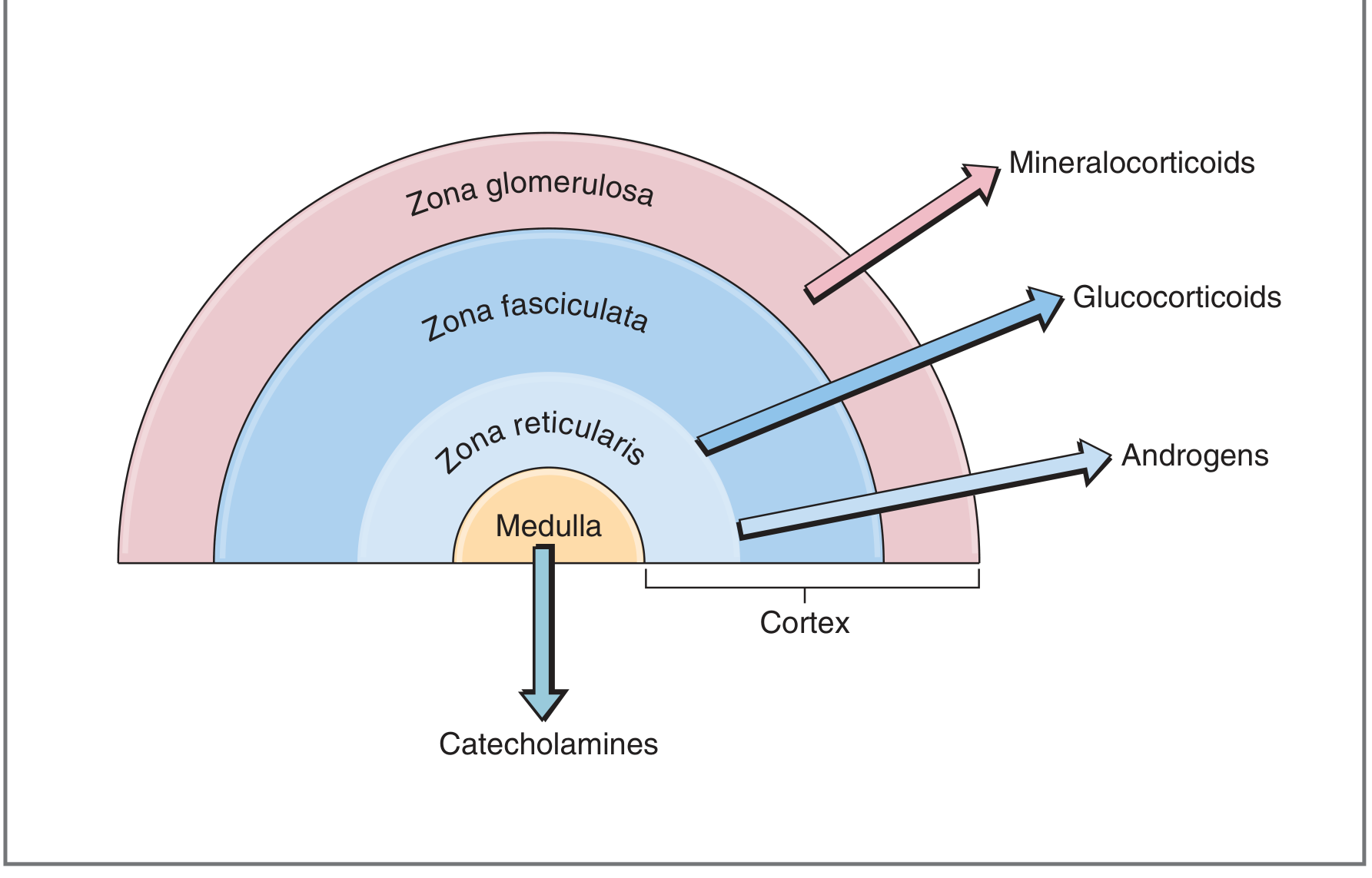
The Three Zones (GFR mnemonic - outside to inside)
| Zone | Location | Hormone | Class |
|---|---|---|---|
| Zona Glomerulosa | Outermost | Aldosterone | Mineralocorticoid |
| Zona Fasciculata | Middle (widest) | Cortisol | Glucocorticoid |
| Zona Reticularis | Innermost | DHEA, DHEAS, Androstenedione | Androgens |
All cortical steroids are synthesized from cholesterol as the precursor.
Histology of Cortical Cells
Cortical cells (adrenocortical cells) share features typical of all steroid-secreting cells:
-
Acidophilic cytoplasm rich in lipid droplets (cholesterol esters)
-
Abundant smooth ER (SER) - contains enzymes for cholesterol synthesis and steroid conversion
-
Mitochondria with tubular/vesicular cristae (not the usual shelf-like cristae) - contain enzymes for cholesterol → pregnenolone conversion and other steroidogenic steps
-
Steroids are not stored in granules - as lipid-soluble molecules, they diffuse freely across the plasma membrane
-
Junqueira's Basic Histology 17e, p. 1021
Cortisol (Glucocorticoid)
Synthesis: Zona fasciculata and (to a lesser degree) zona reticularis.
Key actions across body tissues:
- Liver: Activates gluconeogenesis; promotes glycogen synthesis
- Muscle/adipose: Mobilizes amino acids and fatty acids (catabolic)
- Immune system: Anti-inflammatory; suppresses lymphocyte and eosinophil activity; stabilizes mast cell membranes
- Bone: Inhibits osteoblasts; chronic excess causes osteoporosis
- CNS: Affects mood, cognition, appetite
- Cardiovascular: Supports vascular responsiveness to catecholamines
Regulation - HPA Axis:
Hypothalamus → CRH (41-amino acid peptide, from paraventricular nuclei) → Anterior pituitary corticotrophs → ACTH → Adrenal cortex → Cortisol
- Cortisol exerts negative feedback at three points: (1) directly inhibits hypothalamic CRH, (2) via hippocampal neurons that synapse on the hypothalamus, (3) inhibits CRH action on the anterior pituitary
Diurnal rhythm: Cortisol secretion is pulsatile (~10 bursts/day). Levels are lowest around midnight and peak just before awakening (~8 AM). The pre-awakening burst accounts for ~50% of total daily cortisol output. This pattern is driven by ACTH, which in turn is driven by CRH. The diurnal rhythm is abolished by coma, blindness, or constant light/dark exposure.
- Costanzo Physiology 7e, pp. 435-436
Aldosterone (Mineralocorticoid)
Synthesis: Zona glomerulosa exclusively.
Actions: Promotes Na⁺ reabsorption and K⁺ excretion in the renal collecting tubule; increases water retention; raises blood pressure.
Regulation: Primarily by the renin-angiotensin-aldosterone system (RAAS) and plasma K⁺ levels. ACTH plays only a minor role (unlike with cortisol).
Structurally, aldosterone differs from cortisol by lacking the -OH at C17 and having an aldehyde group at C18 - a seemingly minor difference that completely eliminates glucocorticoid activity at physiological concentrations.
Adrenal Androgens
Synthesis: Zona reticularis.
Key hormones: Dehydroepiandrosterone (DHEA), DHEAS, and androstenedione - all weak androgens (far weaker than testosterone or DHT).
- DHEAS circulates at the highest concentration of any adrenal steroid and shows no diurnal variation
- Plasma DHEA follows the same diurnal pattern as cortisol
- In women, adrenal androgens are a significant source of androgen activity
Adrenal Medulla (20% of gland mass)
Origin: Neuroectoderm (neural crest). The medulla is essentially a modified sympathetic ganglion.
Cell type: Chromaffin cells (named for their staining with chromium salts), innervated by preganglionic sympathetic neurons.
Hormones: Catecholamines
- Epinephrine (adrenaline): ~80% of output; synthesized from norepinephrine via PNMT enzyme (induced by high local cortisol from cortical venous blood)
- Norepinephrine: ~20% of output; also produced at sympathetic nerve terminals elsewhere
Catecholamines are stored in granular complexes with chromogranins (49 kDa proteins), Ca²⁺, and ATP, and released by exocytosis in response to preganglionic stimulation.
Fight-or-flight effects:
-
Epinephrine: increases heart rate, dilates bronchioles, dilates skeletal/cardiac muscle arteries, elevates blood glucose (glycogenolysis)
-
Norepinephrine: constricts splanchnic and skin vessels, redirects blood to heart, muscles, and brain
-
Both: promote glycogenolysis and lipolysis
-
Junqueira's Basic Histology 17e, p. 1027
Clinical Summary: Major Disorders
| Disorder | Pathology | Key Features |
|---|---|---|
| Cushing's syndrome | Cortisol excess | Central obesity, moon face, striae, hypertension, hyperglycemia, osteoporosis |
| Addison's disease | Primary adrenocortical insufficiency | Fatigue, hypotension, hyponatremia, hyperkalemia, hyperpigmentation (↑ ACTH/MSH) |
| Primary hyperaldosteronism (Conn's) | Aldosterone excess | Hypertension, hypokalemia, metabolic alkalosis |
| Pheochromocytoma | Catecholamine-secreting medullary tumor | Paroxysmal hypertension, headache, sweating, palpitations; "10% tumor" |
| Congenital adrenal hyperplasia (CAH) | Enzyme defect in steroidogenesis (usually 21-hydroxylase) | Virilization, salt-wasting, ↑ ACTH driving adrenal hyperplasia |
Summary Table
| Feature | Cortex | Medulla |
|---|---|---|
| Origin | Mesoderm | Neural crest (neuroectoderm) |
| % of gland | ~80% | ~20% |
| Hormones | Cortisol, aldosterone, DHEA | Epinephrine, norepinephrine |
| Hormone class | Steroids | Catecholamines (amine) |
| Storage | Not stored; diffuse freely | Stored in chromaffin granules |
| Regulation | HPA axis (cortisol/androgens); RAAS + K⁺ (aldosterone) | Sympathetic innervation |
This is a shared conversation. Sign in to Orris to start your own chat.