
vesicouterine fistula
vesicouterine fistula
vesicouterine fistula Youssef syndrome
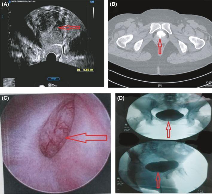
A four-panel figure illustrating diagnostic findings of a vesicouterine fistula (VUF). (A) Transvaginal ultrasound (TVUS) showing an interruption in the continuity of the anterior cervical wall at the internal os level (red arrow), indicating communication between the cervical canal and the bladder. (B) Axial pelvic CT (enhanced urography) demonstrating speckled high-density contrast media within the uterus, situated posterior/inferior to the bladder, confirming fistulous passage. (C) Cystoscopic view revealing a well-defined mucosal defect (approximately 3x2 cm) located superior to the trigone of the bladder. (D) Radiographic fluoroscopy during a methylene blue and contrast medium instillation test, depicting the bladder filled with contrast and demonstrating slight leakage into the vaginal vault. This series serves as a multimodal diagnostic comparison for detecting abnormal pelvic communications, typically presenting with Youssef's syndrome (cyclic hematuria and menouria).
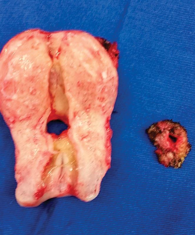
This clinical photograph displays a macroscopic view of surgical specimens from a hysterectomy and fistulectomy, illustrating a vesicouterine fistula (Youssef syndrome). The larger specimen is a mid-sagittal section of the uterus, showing a pale, fibromuscular myometrium. A distinct, rounded fistulous orifice is located in the lower uterine segment, just superior to the cervical canal. The surrounding tissue near the orifice exhibits yellowish-white discoloration, characteristic of chronic inflammation or scarring. To the right, a smaller, irregular excised segment of the bladder wall is shown, featuring a corresponding patent orifice with dark, hemorrhagic edges. This pathological specimen demonstrates the direct communication between the bladder and the uterus, which clinically manifests as cyclic menouria (presence of menstrual blood in urine) and the absence of normal vaginal menstruation. The image is an important educational resource for understanding the gross anatomy of iatrogenic urogenital fistulas following cesarean sections.
Vesicouterine Fistula (VUF)
Etiology
- Cesarean section is the overwhelming cause - the fistula develops when a bladder injury during a low-segment cesarean (or an occult bladder injury incorporated into the uterine closure) goes unrecognized. Repeat cesareans and VBAC also carry risk.
- Other causes: uterine rupture during obstructed labor (bladder tears along the rupture line), placenta percreta (chorionic villi invading through the uterine serosa into the bladder), IUD erosion, uterine artery embolization, pelvic brachytherapy, traumatic catheterization, endometrial ablation, induced abortion, D&C, and hysterectomy.
- Anatomically, the tract most often runs along the posterior bladder wall in the midline, communicating with the uterus just above the internal cervical os.
Clinical presentation - Youssef syndrome
- Menouria (menstrual blood passing via the urine)
- Cyclic hematuria with apparent amenorrhea (no vaginal bleeding)
- Urinary continence
- Often associated with infertility
Diagnosis
- Cystoscopy: reveals a midline lesion on the posterior bladder wall.
- Urine cytology: may show endometrial cells.
- Cystogram: contrast instilled into the bladder outlines the uterine cavity.
- Hysterosalpingogram: contrast in the uterus fills the bladder.
- MRI, CT, and ultrasonography are all used for diagnosis and evaluation.
- IVU or contrast-enhanced CT helps exclude a concomitant ureteral injury.
Management
- Conservative: prolonged bladder catheterization or fulguration of the tract with drainage can succeed in small, immature fistulae. Hormonal induction of menopause (to cause involution of the puerperal uterus) combined with continued bladder drainage has also been used successfully (Jozwik and Jozwik reported success in 8 of 9 patients).
- Surgical (definitive therapy):
- Fertility-preserving/uterine-sparing repair (an O'Conor-type approach) if the patient wants to retain fertility - successful pregnancy after repair is possible.
- Hysterectomy with bladder repair if fertility is no longer desired.
- Vaginal repair is feasible in selected cases, but most require an abdominal (or robotic-assisted) approach.
- Most common cause: low-segment cesarean section
- Continence is often preserved (unlike other urogenital fistulae)
- Nonsurgical (hormonal) management is a viable option in select patients
- Management decisions hinge on the patient's desire for future fertility