
Karyotyping
Reading File
Karyotyping is a lab technique used to visualize and analyze chromosomes. Let me search the medical library for comprehensive coverage.
Searching sources...
Finding Sources
Finding Sources
Good results. Let me read the most comprehensive sources in parallel.
Reading File
Reading File
Reading File
Reading File
Excellent content from multiple sources. Let me read more of the Henry's Clinical Pathology text (which has detailed lab method coverage) and fetch the karyogram image.
Reading File
Reading File
Loading Image
I now have comprehensive content from multiple authoritative textbooks. Let me also fetch the chromosome structure diagram and read the section on FISH and advanced techniques.
Loading Image
Reading File
I have excellent comprehensive content from multiple authoritative textbooks. Now I can compile a thorough answer.
Karyotyping
Definition
A karyotype refers to the full chromosome complement of an individual, arranged in a standardized format. The word has three uses: the standard chromosome set of an individual, the photomicrograph of those chromosomes arranged in pairs, and the process of preparing that figure ("to karyotype"). The ordered image itself is technically called a karyogram.
The normal human karyotype consists of 46 chromosomes arranged in 23 pairs - 22 pairs of autosomes and one pair of sex chromosomes (XX in females, XY in males).
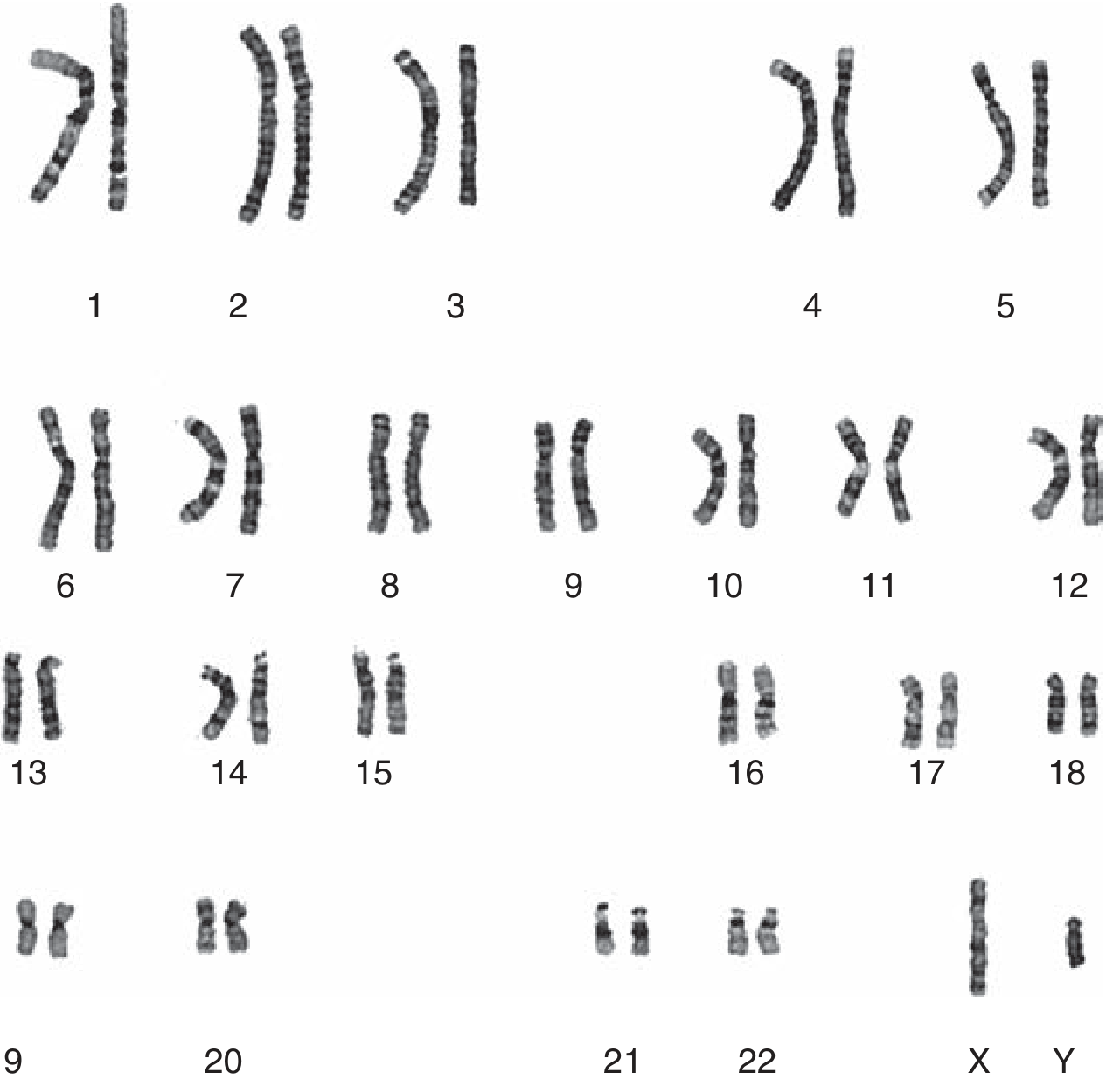
Normal male karyogram - 46 chromosomes ordered in pairs (1-22, X, Y). - Henry's Clinical Diagnosis and Management by Laboratory Methods, Fig. 71.1
Chromosome Structure
Each metaphase chromosome consists of two chromatids held together at the centromere (primary constriction). The centromere divides the chromosome into:
- p arm (short arm, from petit)
- q arm (long arm)
The ends are capped by telomeres - repeated TTAGGG sequences that stabilize chromosomes. Telomeres shorten with each division, and total loss has been linked to chromosomal instability, carcinogenesis, and aging.
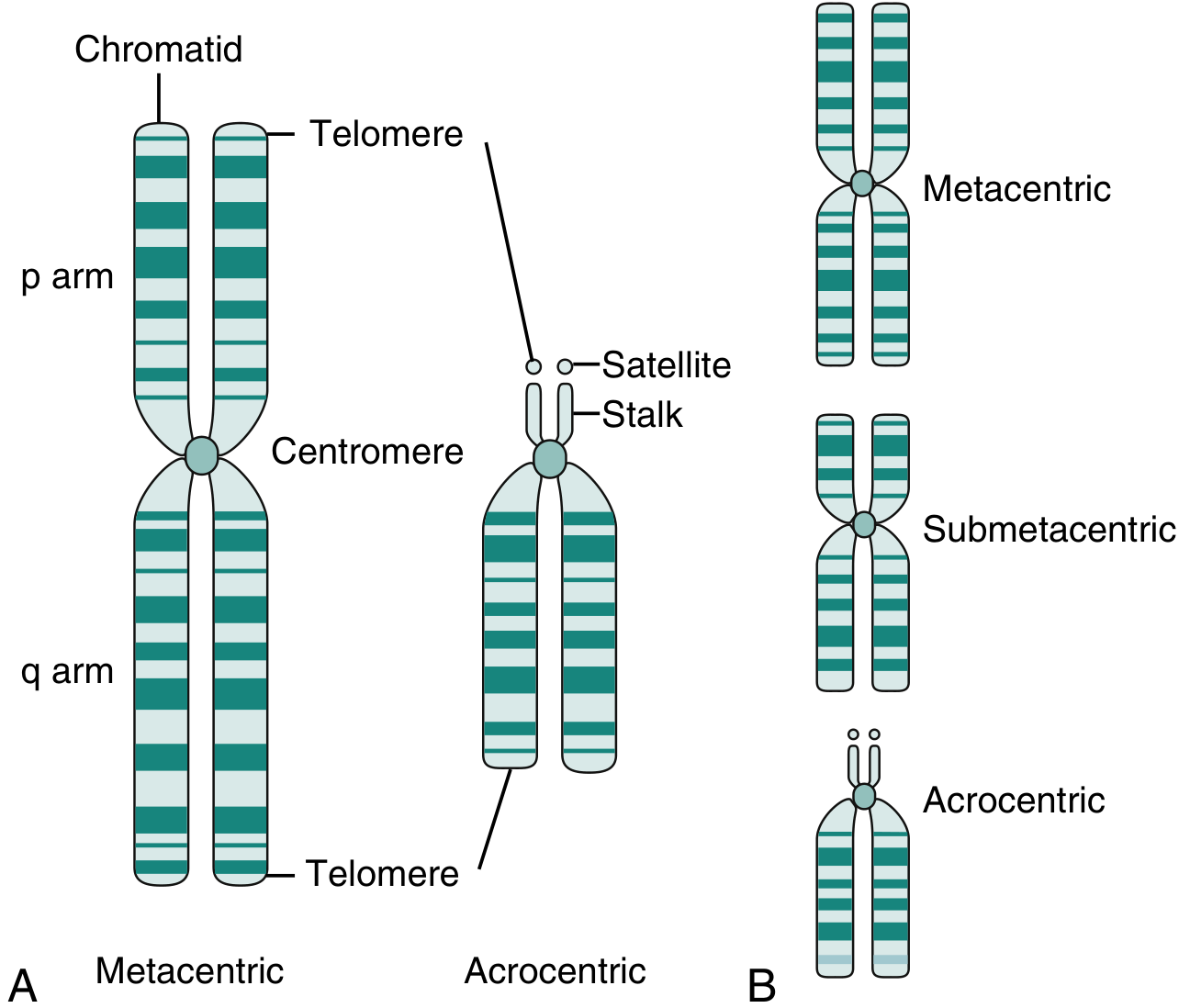
Chromosome anatomy - Henry's Clinical Diagnosis and Management, Fig. 71.2
Classification by centromere position
| Type | Description |
|---|---|
| Metacentric | Centromere roughly equidistant from both ends; arms approximately equal |
| Submetacentric | Centromere closer to one end; q arm longer than p arm |
| Acrocentric | Centromere very close to one end; minimal p arm with stalks and satellites encoding rRNA genes; common sites of Robertsonian translocations |
Chromosomes are numbered 1-22 by length (chromosome 1 is longest). Note: chromosome 21 is actually shorter than chromosome 22 - an historical naming error.
Step-by-Step Process of Karyotyping
1. Cell Source (Specimen Collection)
Any viable, nucleated, dividing cell can be used:
- Peripheral blood (heparinized): most common for routine analysis; lymphocytes used
- Bone marrow: preferred in hematologic malignancies
- Amniotic fluid (amniocentesis at 16-18 weeks): most common prenatal source; ~0.2-0.3% fetal loss risk
- Chorionic villus sampling (CVS) (10-13 weeks): earlier prenatal diagnosis; ~1% fetal loss risk
- Cordocentesis/PUBS: umbilical cord blood sampling
- Skin fibroblasts, products of conception, solid tumors
2. Cell Culture
Lymphocytes in peripheral blood do not normally divide. The sample is added to nutrient medium with phytohemagglutinin (PHA), which stimulates T-lymphocytes to divide. Cultured at 37°C for approximately 3 days.
3. Metaphase Arrest
Colchicine (or its derivative colcemid) is added to arrest dividing cells in metaphase - the stage when chromosomes are maximally condensed and most visible. Colchicine inhibits spindle formation.
4. Hypotonic Treatment
Hypotonic saline is added, causing cells to swell and lyse. This spreads the chromosomes so they do not overlap.
5. Fixation and Slide Preparation
Cells are fixed, spread on glass slides, and allowed to dry.
6. Staining (Banding)
The chromosomes are stained to produce characteristic banding patterns:
| Technique | Stain | Details |
|---|---|---|
| G-banding | Giemsa after mild trypsin | Most widely used; produces alternating dark (AT-rich) and light (GC-rich) bands; permanent preparation |
| Q-banding | Quinacrine (fluorescent) | Historical; now mainly used for rapid Y chromosome identification (bright Y heterochromatin) |
| R-banding | Reverse banding | Light/dark pattern reversed from G; better for detecting telomeric deletions |
| C-banding | Centromere banding | Highlights constitutive heterochromatin; used to detect dicentric chromosomes |
Up to 1000 bands can be resolved across all chromosomes. Each cytogenetic band contains ~50 or more genes.
7. Analysis
A cytogeneticist counts chromosomes (46 centromeres = normal diploid), identifies each pair by band pattern, and arranges them in the standard karyogram. Automated imaging software assists modern labs.
Chromosomal Nomenclature
Chromosome locations are written in the format:
[chromosome][arm][region][band].[subband]
Example: 18q21.3 = chromosome 18, long arm (q), region 2, band 1, subband 3 - this is where the BCL2 gene (involved in follicular lymphoma) resides.
Regions are numbered outward from the centromere (region 1 is closest).
Advanced Cytogenetic Techniques
Beyond conventional G-banding, modern labs use:
| Technique | Principle | Use |
|---|---|---|
| FISH (Fluorescence in situ hybridization) | Fluorescent DNA probes hybridize to specific chromosomal sequences | Detects specific deletions, amplifications, translocations; works in interphase cells |
| SKY (Spectral karyotyping) | Combinatorial FISH using multiple fluorescent probes | Paints all 46 chromosomes in different colors simultaneously; identifies complex rearrangements |
| Array CGH (Comparative genomic hybridization) | Patient vs. control DNA on microarray | Detects copy number variants genome-wide; higher resolution than conventional karyotyping |
| Whole genome sequencing | Next-generation sequencing | Highest resolution; increasingly used in cancer cytogenomics |
Clinical Indications for Karyotyping
- Prenatal diagnosis: maternal age >35, abnormal ultrasound findings, abnormal serum screening
- Congenital anomalies: multiple birth defects, dysmorphic features
- Developmental delay / intellectual disability
- Ambiguous genitalia / sex determination disorders
- Recurrent pregnancy loss
- Primary amenorrhea or infertility
- Hematologic malignancies: leukemia, lymphoma (bone marrow karyotype guides prognosis and treatment)
- Solid tumors: certain chromosomal rearrangements are diagnostic (e.g., t(9;22) Philadelphia chromosome in CML)
Common Chromosomal Abnormalities Detected
| Abnormality | Karyotype | Condition |
|---|---|---|
| Trisomy 21 | 47,XX or XY,+21 | Down syndrome |
| Trisomy 18 | 47,XX or XY,+18 | Edwards syndrome |
| Trisomy 13 | 47,XX or XY,+13 | Patau syndrome |
| Monosomy X | 45,X | Turner syndrome |
| 47,XXY | 47,XXY | Klinefelter syndrome |
| Robertsonian translocation | 46,XX,rob(14;21) | Familial Down syndrome |
| Philadelphia chromosome | 46,XY,t(9;22)(q34;q11) | CML |
Sources:
- Henry's Clinical Diagnosis and Management by Laboratory Methods, Chapter 71
- Thompson & Thompson Genetics and Genomics in Medicine, 9th ed., Chapter 2
- Emery's Elements of Medical Genetics and Genomics, Chapter 3
- Quick Compendium of Clinical Pathology, 5th ed., Section 7.2.1
This is a shared conversation. Sign in to Orris to start your own chat.