
Q,2 A 58-year-old woman with poorly controlled Type 2 diabetes mellitus presents to the Emergency Department with fever, severe left flank pain, vomiting, and progressive drowsiness for 2 days and oliguria. She completed a course of oral antibiotics for a urinary tract infection one week ago. A non-contrast CT KUB demonstrates gas within the left renal parenchyma extending into the perinephric space with an obstructing proximal ureteric calculus. What is the most likely diagnosis? Mention four important differential diagnoses. (3 Marks) Assessment and immediate stabilization priorities in the first hour. (4 Marks), current guidelines for evidence-based antimicrobial management. (4 Marks) Role of imaging and classify this disease using an appropriate radiological classification. How does the classification influence management? (3 Marks) Compare the available source-control options in this patient. (4 Marks) Name two validated prognostic/scoring systems applicable to this patient and mention five predictors of poor outcome. (2 Marks)
emphysematous pyelonephritis management classification
emphysematous pyelonephritis CT classification Huang Tseng management guidelines 2024
PMID: 35802159
https://www.ncbi.nlm.nih.gov/books/NBK599505
emphysematous pyelonephritis CT scan gas renal parenchyma perinephric
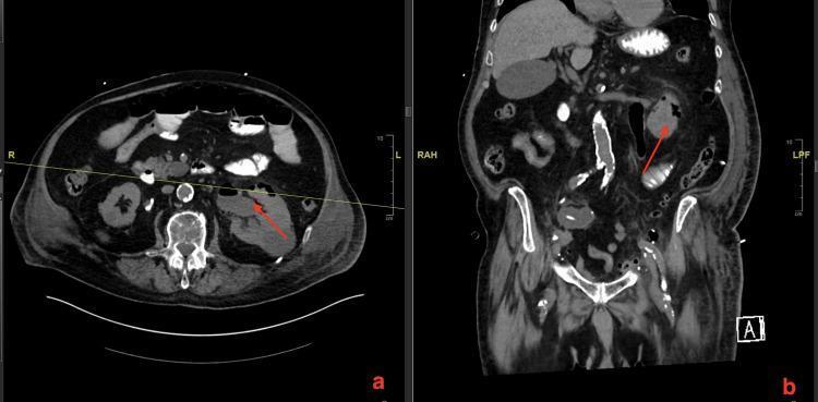
Non-contrast CT scan of the abdomen and pelvis presenting axial (a) and coronal (b) reconstructions of the kidneys. The images demonstrate classic features of Emphysematous Pyelonephritis (EPN) within the left kidney. Red arrows point to pathognomonic gas collections within the renal parenchyma and extending into the surrounding perinephric space. This gas appears as low-attenuation (black) pockets or bubbles, indicating necrotizing infection by gas-forming organisms. The axial section (a) shows air-fluid levels and intra-parenchymal destruction, while the coronal view (b) highlights the vertical extent of the gas tracking through the renal tissue. No significant gas is seen in the contralateral right kidney. The imaging serves as a critical diagnostic tool for identifying severe renal infection, particularly in patients with predisposing factors like diabetes or urinary tract obstruction.
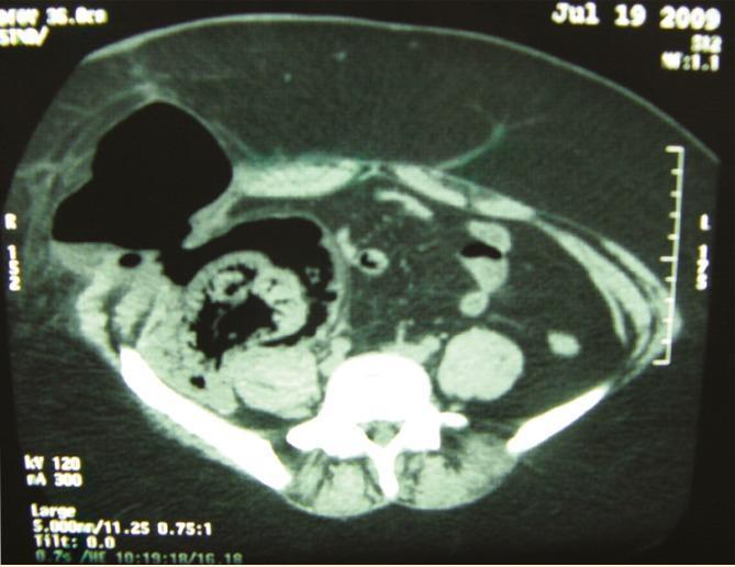
This axial non-contrast computerized tomography (CT) scan of the abdomen demonstrates severe emphysematous pyelonephritis involving a transplant kidney in the right iliac fossa. The image shows extensive, confluent gas collections (hypodense regions) within the renal parenchyma, indicating significant tissue destruction. There is prominent extension of gas into the extraperitoneal perinephric and paranephric spaces. A large, well-defined collection of air is visible in the right lower quadrant, extending anteriorly and disrupting the subcutaneous tissue planes of the abdominal wall. Normal anatomical landmarks are severely distorted by the massive retroperitoneal air and associated inflammation. The native kidneys appear small and atrophic, consistent with chronic renal failure. This imaging is characteristic of a life-threatening necrotizing infection of the renal system, showing both parenchymal gas and distant extraperitoneal migration of air.
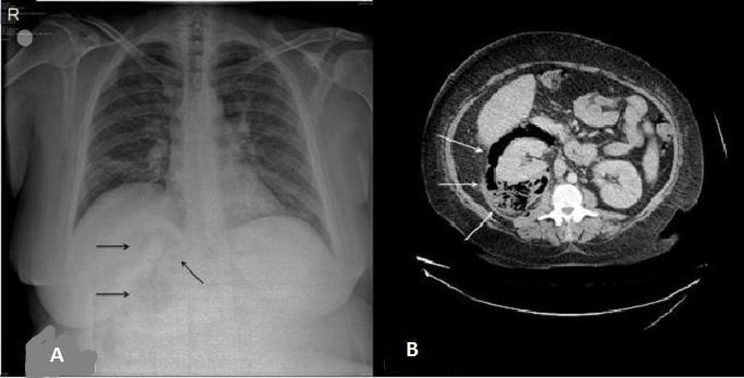
This diagnostic comparison illustrates emphysematous pyelonephritis through two modalities: a supine abdominal X-ray (A) and an axial non-contrast computed tomography (CT) scan (B). Image A displays subtle, ill-defined radiolucent (darker) areas in the right upper quadrant, marked by black arrows, which suggest gas within the renal parenchyma or collecting system. Image B provides a definitive view of the same pathology, showing the right kidney replaced by extensive, irregular hypoattenuating (dark) foci. These foci represent gas collections within the renal parenchyma and the perinephric space, characteristic of a severe necrotizing infection. The white arrows in the CT scan highlight the distribution of gas, which disrupts the normal homogeneous renal architecture compared to the contralateral kidney. This comparison highlights the superior sensitivity of CT over plain radiography in detecting and characterizing renal gas, which is critical for the diagnosis and staging of life-threatening urological emergencies like emphysematous pyelonephritis.
Emphysematous Pyelonephritis (EPN) - Comprehensive Clinical Analysis
PART 1 - Diagnosis and Differential Diagnoses (3 Marks)
Most Likely Diagnosis
Four Important Differential Diagnoses
| Differential | Key Distinguishing Features |
|---|---|
| 1. Emphysematous pyelitis | Gas confined to the collecting system only (no parenchymal involvement); much better prognosis; usually responds to antibiotics alone; less likely to occur in diabetics |
| 2. Xanthogranulomatous pyelonephritis (XGP) | Chronic destructive renal infection; CT shows "bear paw" sign with lipid-laden macrophages replacing parenchyma; rarely gas-forming; associated with staghorn calculi and Proteus |
| 3. Perinephric / renal abscess | Fluid collection with or without gas; CT shows ring-enhancing lesion; typically from haematogenous spread or ascending infection; no diffuse parenchymal gas |
| 4. Obstructive uropathy with infected hydronephrosis (pyonephrosis) | Dilated, infected collecting system; fever and flank pain but CT typically shows hydronephrosis with debris, not gas within parenchyma |
PART 2 - Assessment and Immediate Stabilisation in the First Hour (4 Marks)
A - Airway and Breathing
- Assess GCS; with progressive drowsiness, protect airway if GCS falls below 8
- High-flow O2 to maintain SpO2 >94%
- ABG: assess lactate, pH, pCO2 (metabolic acidosis expected)
B - Haemodynamic Resuscitation
- Two large-bore IV cannulae; begin crystalloid bolus 30 mL/kg IV in first 3 hours (Surviving Sepsis Campaign)
- Vasopressors (noradrenaline) if MAP <65 mmHg persists after fluid resuscitation
- Continuous cardiac monitoring; urinary catheter for hourly urine output monitoring
C - Investigations (draw before antibiotics)
- Blood: FBC (thrombocytopenia is a critical prognostic marker), U&E/creatinine, glucose, LFT, coagulation, blood cultures x2, serum lactate, CRP, procalcitonin
- Urine: MSU for MC&S (before antibiotics if possible)
- Serum HbA1c: confirms degree of glycaemic dyscontrol
- ABG/VBG with point-of-care lactate
D - Glycaemic Control
- Initiate insulin infusion for urgent blood glucose control (high glucose serves as a fermentable substrate for gas-forming bacteria - the direct driver of EPN)
- Target glucose 7-10 mmol/L; avoid hypoglycaemia
E - Imaging Confirmation
- CT KUB (non-contrast) has already been done here and is the gold standard - confirms diagnosis and allows classification
- Review urgently with urology, radiology, and intensive care
F - Urgent Specialist Referral
- Urology (source control within hours), ICU/HDU admission, nephrology input for AKI management
- Antibiotic administration within 1 hour of recognition of sepsis
PART 3 - Evidence-Based Antimicrobial Management (4 Marks)
Principles (Surviving Sepsis Campaign + IDSA/EAU Guidelines)
Empiric Regimen (Urosepsis with Source Control Required)
- Piperacillin-tazobactam 4.5 g IV q8h (extended infusion 4h preferred)
OR - Meropenem 1 g IV q8h OR Imipenem-cilastatin 500 mg IV q6h
(Carbapenems preferred given: prior antibiotic exposure 1 week ago raising resistance risk, severely ill, diabetic, obstructed)
- Ceftriaxone 2 g IV OD or Cefepime 2 g IV q8h
- Gentamicin or amikacin as synergistic combination if haemodynamic instability/bacteraemia suspected (aminoglycoside + beta-lactam for 48-72 hours)
- Cover Enterococcus with ampicillin or vancomycin if Gram-positive cocci on urine Gram stain
De-escalation
- Review at 48-72 hours with culture and sensitivity results
- Narrow spectrum based on sensitivity: E. coli - trimethoprim-sulfamethoxazole or fluoroquinolone if susceptible; Klebsiella - 3rd-gen cephalosporin if non-ESBL
- Duration: 3-4 weeks of parenteral antibiotics are typically required for EPN, transitioning to oral when the patient is clinically improving and can tolerate oral intake - Campbell-Walsh-Wein, p. 55
Special Considerations
- ESBL-producing organisms: Carbapenems mandatory; consider local antibiogram (prior antibiotic use within 7 days is a key risk factor for ESBL/resistance)
- MRSA: Unlikely in UTI but add vancomycin if Gram-positive cocci confirmed
- Antifungal: Add fluconazole if Candida isolated in immunocompromised diabetic patient
- Therapeutic drug monitoring (TDM): Especially for aminoglycosides in AKI (this patient has oliguria - dose-adjust all renally cleared drugs)
PART 4 - Imaging, Radiological Classification, and Management Influence (3 Marks)
Role of Imaging
- Detects gas in parenchyma/perinephric space with near 100% sensitivity (plain X-ray detects gas in only ~50% of cases)
- Characterises extent: parenchymal destruction, fluid collections, perinephric extension, bilateral involvement
- Identifies the precipitating cause (obstructing calculus in this case)
- Enables classification to guide management
- Guides percutaneous drainage (PCD)
Huang and Tseng CT Classification (2000) - Currently Widely Used
| Class | CT Finding | Mortality | Recommended Management |
|---|---|---|---|
| Class 1 | Gas in collecting system only (emphysematous pyelitis) | Low (<5%) | IV antibiotics + glycaemic control ± relief of obstruction |
| Class 2 | Gas in renal parenchyma only; no extension outside kidney | ~10-20% | Antibiotics + PCD + relieve obstruction |
| Class 3A | Gas or abscess extending to perinephric space | ~25-50% | Antibiotics + PCD; if ≥2 risk factors - nephrectomy |
| Class 3B | Gas or abscess extending beyond Gerota's fascia / bilateral disease | ~50-75% | Antibiotics + aggressive drainage; if ≥2 risk factors - emergency nephrectomy |
| Class 4 | Bilateral EPN or EPN in a solitary functioning kidney | Highest | IV antibiotics + PCD; nephrectomy with extreme caution (risk of dialysis-dependence) |
How Classification Influences Management
- Class 1-2: Medical management alone or with PCD; 66% success rate with conservative approach
- Class 3A/3B with 0-1 risk factors: aggressive medical management + PCD can be attempted first
- Class 3A/3B with ≥2 of the following risk factors: emergency nephrectomy recommended:
- Thrombocytopenia
- Acute renal failure (serum creatinine >1.4 mg/dL)
- Disturbance of consciousness
- Shock
- This patient has at least 3-4 risk factors (drowsiness, oliguria/AKI, uncontrolled diabetes, prior failed antibiotics) - pointing strongly toward nephrectomy after initial resuscitation
PART 5 - Source Control Options Compared (4 Marks)
1. Medical Management Alone (Antibiotics + Supportive Care)
- Indication: Class 1, selected Class 2 patients with no obstruction and no risk factors
- Technique: IV broad-spectrum antibiotics, fluid resuscitation, glycaemic control, correction of electrolytes
- Advantages: Non-invasive; avoids surgical risk in high-risk patients
- Disadvantages: Mortality rate ~50% when used alone (vs. 13.5% with PCD); unacceptable as sole therapy in Class 3/4 or multi-risk-factor patients
- Not appropriate as sole management in this patient given Class 3B disease and multiple risk factors - Brenner and Rector's The Kidney, p. 1644
2. Percutaneous Catheter Drainage (PCD) + Ureteric Stenting
- Indication: Class 2, Class 3 (0-1 risk factors), Class 4 (solitary kidney), as a bridge in Class 3B before definitive nephrectomy
- Technique: Ultrasound- or CT-guided percutaneous nephrostomy (PCN) to drain the collecting system; ureteric stenting (retrograde or antegrade) to relieve the obstructing calculus
- Advantages: Minimally invasive; preserves renal function; success rate up to 66%; avoids GA in critically ill; the 2022 meta-analysis (PMID 35802159) confirmed minimally invasive intervention (PCN/ureteric stent) was associated with significantly decreased mortality (OR 0.47)
- Disadvantages: May not achieve adequate drainage if extensive parenchymal necrosis; requires repeat procedures; failure necessitates nephrectomy anyway; risk of septic shower on manipulation
- Role in this patient: PCN for urgent decompression of the obstructed infected system should be performed as an emergency alongside resuscitation. The obstructing calculus can be managed by ureteric stent or PCN. This may serve as a bridge to delayed elective nephrectomy if the patient stabilises.
3. Nephrectomy (Emergency or Elective)
- Indication: Class 3A/3B/4 with ≥2 risk factors; failure of PCD; rapidly deteriorating patient; destroyed non-functioning kidney; bilateral disease where one kidney is unsalvageable
- Technique: Open or laparoscopic nephrectomy; laparoscopic approach associated with less morbidity in stable patients; open preferred in emergency setting or when perinephric spread is extensive
- Advantages: Definitive source control; removes all infected tissue; mortality with emergency nephrectomy ~25% vs. 50% with medical management alone
- Disadvantages: Major surgery in a critically ill, septic, poorly controlled diabetic; risk of haemorrhage, prolonged ICU stay, and permanent loss of renal function; AKI from contralateral kidney impairment is a major concern
- Timing: If PCD fails or patient deteriorates within 24-48 hours of PCD, proceed to nephrectomy without further delay
Summary Recommendation for This Patient
| Priority | Action |
|---|---|
| Immediate | Resuscitation (fluids, vasopressors, O2) + IV carbapenems within 1 hour |
| Emergency (same admission) | Percutaneous nephrostomy (PCN) to drain the obstructed left kidney + ureteric stent for the calculus |
| 48-72 hours | Reassess response; if no improvement or ≥2 risk factors persist - proceed to emergency left nephrectomy |
| Delayed | Manage ureteric calculus (ESWL/ureteroscopy) once infection resolved |
PART 6 - Prognostic/Scoring Systems and Predictors of Poor Outcome (2 Marks)
Two Validated Scoring Systems
-
Huang and Tseng Risk Factor Score (2000) - EPN-specific: counts the number of the following poor prognostic markers present: thrombocytopenia, acute renal failure, altered consciousness, shock. ≥2 risk factors = emergency nephrectomy recommended. Shown to correlate with mortality outcomes in EPN.
-
SOFA Score / qSOFA (Sepsis-3 Definitions, 2016) - applicable to all septic patients including urosepsis. qSOFA (Respiratory rate ≥22, altered mentation, systolic BP <100) serves as a rapid bedside predictor of poor outcomes; full SOFA score guides ICU admission and prognosis. This patient scores high on both (drowsiness + oliguria suggesting organ dysfunction).
Five Predictors of Poor Outcome in EPN
- Septic shock (systolic BP <90 mmHg) - OR 15.57 for mortality
- Disturbance of consciousness (altered mental status, drowsiness, confusion) - OR 12.11
- Thrombocytopenia (platelet count <60,000/μL or <100,000/μL) - OR 7.85
- Acute renal failure (rising creatinine, oliguria/anuria) - OR 5.41
- Huang-Tseng Class III-IV disease (bilateral or extensive extrarenal gas) - OR 2.4; also correlated: medical management alone (OR 2.04) - underscoring the necessity of procedural intervention
This patient has at least 3-4 of these poor prognostic factors at presentation (drowsiness, AKI/oliguria, prior failed antibiotics, Class 3B disease), placing her in the highest risk stratum and mandating aggressive combined medical-surgical management without delay.
Key References
- Huang JJ, Tseng CC. Emphysematous pyelonephritis: clinicoradiological classification, management, prognosis, and pathogenesis. Arch Intern Med. 2000;160:797-805
- Ngo XT et al. Prevalence and Risk Factors of Mortality in Emphysematous Pyelonephritis Patients: A Meta-Analysis. World J Surg. 2022 [PMID: 35802159]
- Wu SY et al. Emphysematous pyelonephritis: classification, management, and prognosis. Tzu Chi Med J. 2022 [PMID: 35912050]
- Campbell-Walsh-Wein Urology, 12th ed., Chapter 55
- Smith and Tanagho's General Urology, 19th ed., Chapter 14
- Brenner and Rector's The Kidney, Chapter on Emphysematous Pyelonephritis