
Takayaso artiritis
I'll look up Takayasu arteritis in the medical library for you right away.
Searching sources...
Reading File
Finding Sources
Searching PubMed
Takayasu arteritis
Good results. Now let me read the most comprehensive sources - Braunwald's Heart Disease and Andrews' Dermatology in parallel.
Reading File
Reading File
Now let me read more from Braunwald's (the most comprehensive cardiovascular source) for diagnosis, classification, and management details.
Reading File
Loading Image
Now let me read the Braunwald's treatment section for TA specifically and also the Brenner/Rector kidney text.
Reading File
Reading File
I now have comprehensive information from multiple textbooks. Let me also search for the ACR/EULAR diagnostic criteria paragraph and get the classification criteria details.
Finding Sources
Reading File
I now have a thorough picture from multiple authoritative sources. Let me compile the full answer.
Takayasu Arteritis
Definition and Overview
Takayasu arteritis (TA) is a granulomatous panarteritis of unknown etiology that affects the aorta and its major branches (and sometimes the pulmonary arteries). It is classified as a large-vessel vasculitis, the same category as giant cell arteritis (GCA), and the two diseases are now considered to lie on a spectrum of the same disease process.
Key epidemiological features:
- Predominantly affects young women (female:male ratio up to 10:1)
- Onset typically before age 40 (most often in the 2nd-3rd decade)
- More common in Japan, Southeast Asia, India, and South America
- Often called "pulseless disease" or "aortic arch syndrome"
Pathogenesis
Arteritic lesions show:
- Adventitial thickening with leukocyte accumulation in the media
- Infiltration by dendritic cells, T and B lymphocytes, macrophages, and multinucleated giant cells
- In TA specifically, the majority of lymphocytes are perforin-secreting killer cells (cytotoxic T cells and NK cells) - oligoclonal T cell receptors suggest a response to a specific but unknown antigen
- Growth factor-driven mesenchymal proliferation leads to intimal hyperplasia and fibrosis, causing stenosis or occlusion
- Local matrix metalloproteinase (MMP) synthesis may predispose to aneurysmal dilation
- Late phase: intimal proliferation, medial necrosis with scarring, adventitial fibrosis
Clinical Phases
Phase 1 - Systemic/Pre-pulseless phase:
- Fever, night sweats, arthralgia, malaise, profound fatigue, lethargy
- Weight loss
Phase 2 - Vascular/Occlusive phase:
- Upper limb claudication
- Carotidynia (tenderness over carotids) in up to 25% of patients
- Absent or diminished radial and carotid pulses
- Bruits over stenotic vessels
- BP difference between arms ≥20 mmHg
Vessels most commonly affected:
- Subclavian and common carotid arteries (most frequent)
- Aorta (can be involved throughout its length)
- Pulmonary arteries (up to 50% of patients)
- Renal arteries (causes renovascular hypertension in 40-60%)
Lesion types:
-
90% have stenotic/occlusive lesions
- ~25% have aneurysms
- Aortic valve regurgitation and coronary arteritis may occur
Imaging Findings
The figure below from Braunwald's Heart Disease shows the key imaging modalities:
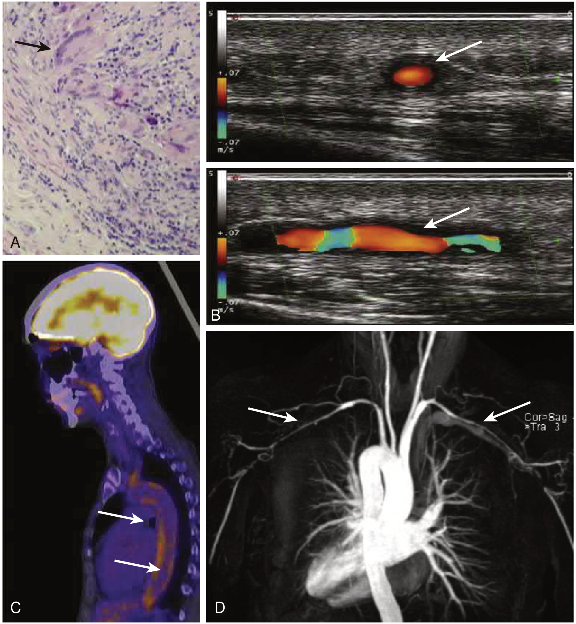
Panel A: Histology with granulomatous inflammation. Panel B: Color Doppler ultrasound showing vessel wall thickening. Panel C: 18F-FDG-PET-CT showing active arteritis uptake in the thoracic aorta. Panel D: MR angiogram showing bilateral subclavian/axillary artery stenoses.
Imaging options:
- Conventional arteriography - gold standard for luminal assessment
- CT angiography (CTA) - detects wall thickening and luminal changes
- MR angiography (MRA) - preferred for follow-up (no radiation); can detect mural inflammation
- 18F-FDG-PET-CT - best for detecting active inflammation
- Doppler ultrasound - useful for accessible vessels (carotid, subclavian)
Laboratory Findings
- Elevated ESR and CRP (markers of active inflammation)
- Mild anemia
- Elevated gamma-globulins
- Some patients: anti-endothelial cell antibodies, elevated pentraxin-3
- ANA, VDRL, complement levels are normal
- Hypertension in 40-60% (renal artery stenosis, decreased aortic elasticity)
Classification Criteria
ACR 1990 Criteria (≥3 of 6 = sensitivity 91%, specificity 98%):
- Age at onset ≤40 years
- Claudication of extremities
- Decreased brachial artery pulse
- BP difference between arms ≥10 mmHg
- Bruit over subclavian arteries or aorta
- Arteriographic abnormality (narrowing/occlusion of aorta, its primary branches, or large arteries in proximal extremities)
Newer ACR/EULAR Scoring (≥5 points required):
| Feature | Points |
|---|---|
| Female sex | +1 |
| Angina or ischemic cardiac pain | +2 |
| Arm or leg claudication | +2 |
| Vascular bruit | +2 |
| Reduced pulse in upper extremity | +2 |
| Carotid artery abnormality | +2 |
| Systolic BP difference in arms ≥20 mmHg | +1 |
| 1 arterial territory on imaging | +1 |
| 2 arterial territories | +2 |
| ≥3 arterial territories | +3 |
| Symmetric involvement of paired arteries | +1 |
| Abdominal aorta with renal/mesenteric involvement | +3 |
Renal Involvement (from Brenner & Rector)
- Renal disease is usually due to obliterative arteritis of the main renal artery or narrowing of renal ostia by abdominal aortitis
- Causes renovascular hypertension (40-60% of patients)
- May rarely cause mesangial proliferative glomerulonephritis, IgA nephropathy-like lesions, renal amyloidosis, or crescentic GN
- Progressive renal failure is uncommon
Cutaneous Manifestations (from Andrews' Dermatology)
- Loss of hair and skin atrophy due to ischemia
- Occasional necrotizing or granulomatous vasculitis of small vessels
- Erythema nodosum-like or erythema induratum-like nodules
- Sweet syndrome association
- Pyoderma gangrenosum-like ulcerations (especially in Japan; lesions precede TA diagnosis by ~3 years on average)
Treatment
First-line:
- Prednisone 1 mg/kg/day, tapered over 8-12 weeks to ≤20 mg/day
- Relapse is common when steroids are tapered
Steroid-sparing agents (most widely used):
- Methotrexate
- Mycophenolate mofetil (MMF)
- Azathioprine
Refractory/life-threatening disease (e.g., coronary arteritis, myocarditis):
- IV pulsed cyclophosphamide
Biologic therapies for refractory cases:
- TNF-α antagonists (infliximab, etanercept): complete remission in 37%, partial in 53.5%
- Tocilizumab (anti-IL-6R): initial trial showed beneficial effect; now increasingly used - note that tocilizumab suppresses CRP synthesis, complicating disease monitoring
- Rituximab, abatacept, and ustekinumab have also been used
Surgical/interventional indications:
- Aneurysmal enlargement with rupture risk
- Severe aortic regurgitation or coarctation
- Critical limb ischemia or severe cerebrovascular disease
- Uncontrolled hypertension from renal artery stenosis
- Surgery should be delayed until immunosuppression achieves remission
Prognosis
- Survival at 15 years: >95% in most Western/Japanese cohorts
- Survival falls to ~67% in patients with serious complications or progressive disease course
- 74% report compromised daily activities; 23% unable to work
- Follow-up should include angiographic monitoring with MRI (preferred over CT to avoid cumulative radiation)
Recent evidence: A 2025 systematic review (PMID 40982119) examined prevalence and clinical severity of Takayasu arteritis by angiographic type, providing updated data on type distribution and outcomes.
Sources: Braunwald's Heart Disease (2 Vol Set), Andrews' Diseases of the Skin, Brenner & Rector's The Kidney, Firestein & Kelley's Textbook of Rheumatology
Cellulitis treatment
Searching sources...
Reading File
Finding Sources
Searching PubMed
cellulitis treatment antibiotics
Finding Sources
Good. Now let me read the most relevant sources in parallel - Tintinalli (EM treatment table), Rosen's EM, Andrews' Dermatology, and Goldman-Cecil.
Reading File
Reading File
Reading File
Excellent. Now let me read more from Tintinalli (which has the detailed treatment section) and Goldman-Cecil for the non-purulent cellulitis treatment.
Reading File
Reading File
Now let me read the detailed non-purulent cellulitis treatment table from Tintinalli and the Goldman-Cecil cellulitis chapter.
Reading File
Reading File
Let me also get the non-purulent cellulitis treatment table from Tintinalli and fetch the clinical image.
Finding Sources
Loading Image
I have comprehensive information from multiple textbooks. Let me now compile the full answer.
Cellulitis - Treatment
Overview
Cellulitis is an infection of the subcutaneous tissues characterised by localized erythema, swelling, warmth, and tenderness. The key initial step in management is to distinguish purulent from non-purulent cellulitis and assess severity, as these drive the treatment choice.
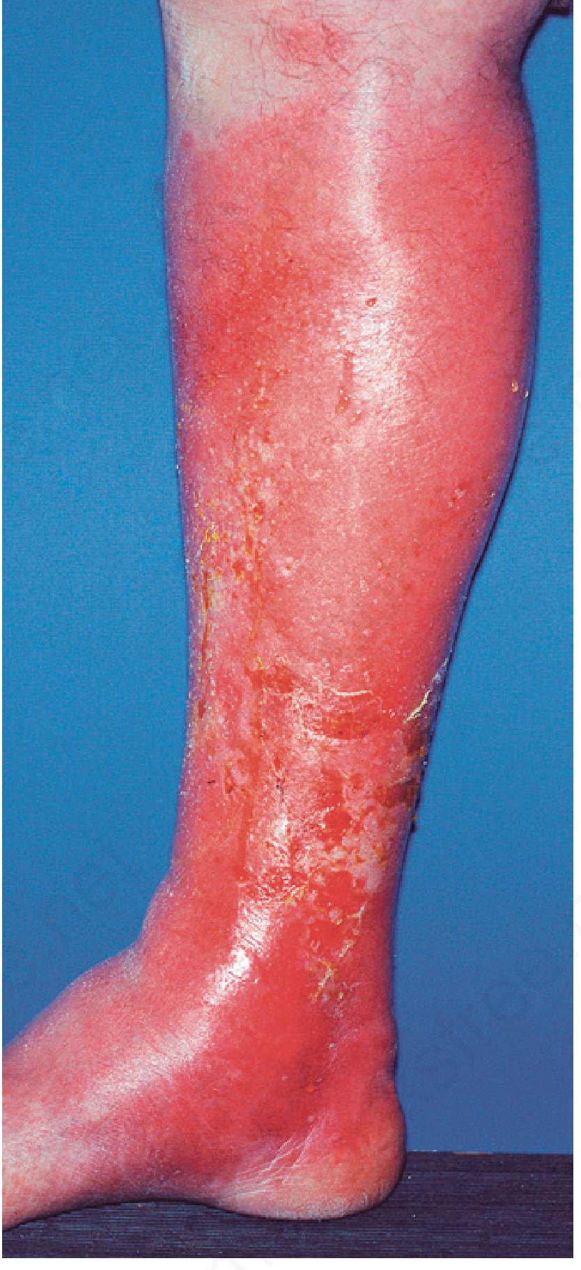
Step 1 - Distinguish Purulent vs. Non-Purulent
| Feature | Non-Purulent | Purulent |
|---|---|---|
| Discharge/pus | Absent | Present (abscess, exudate) |
| Predominant pathogen | Group A Streptococcus (~75%) | S. aureus (incl. MRSA) |
| Antibiotic target | Beta-lactam / penicillin-class | MRSA-active agent if risk factors present |
- Use ultrasound if abscess is clinically uncertain - abscess appears as a fluid-filled cavity; cellulitis shows "cobblestoning" with hypoechoic reticular stranding
- Mark the skin perimeter with an indelible marker to track spread at follow-up
Step 2 - Assess Severity
Mild: No systemic signs; outpatient management appropriate
Moderate: Systemic signs present (fever, tachycardia) but not septic; may need IV
Severe: Meets sepsis criteria (temp >39°C, HR >90, RR >24, WBC <4K or >12K, or immunocompromised)
Non-Purulent Cellulitis Treatment
Caused predominantly by Group A Streptococcus; MRSA coverage generally NOT required
Mild (Outpatient - Oral):
- Cephalexin 500 mg four times daily × 10 days (first choice)
- Dicloxacillin 500 mg four times daily × 10 days
- Penicillin VK 500 mg four times daily × 10 days (streptococcal)
- Clindamycin 300-450 mg four times daily × 10 days
5-day regimens have been successful where follow-up at day 5 is feasible
Moderate (IV Required):
- Penicillin G IV
- Ceftriaxone 1 g IV every 24 hours
- Cefazolin IV
- Clindamycin IV
Severe / Unclear Pathogen / Sepsis:
- Vancomycin (15-20 mg/kg IV q12h) + imipenem 500 mg IV q6h
- Or vancomycin + piperacillin-tazobactam 4.5 g IV q6h
- Or vancomycin + meropenem 500-1000 mg IV q8h
- Transition to oral once clinical response achieved
Purulent Cellulitis Treatment (incl. Abscess)
Target: MRSA-active agents
Mild (Drainable abscess, no systemic signs):
- Incision and drainage (I&D) alone - no antibiotics needed in immunocompetent patients if drainage is complete
- Wound culture recommended if antibiotics are given
Moderate (Purulent cellulitis without systemic signs, or abscess with mild-moderate systemic signs) - Oral:
- TMP-SMX DS (trimethoprim-sulfamethoxazole) 1-2 tablets PO twice daily × 7-10 days (preferred for MRSA)
- Doxycycline 100 mg PO twice daily × 7-10 days
- Clindamycin 300-450 mg PO four times daily × 7-10 days
Severe (Systemic signs, sepsis, or immunocompromised) - IV:
- Vancomycin 15 mg/kg IV q12h (MRSA coverage)
- Linezolid 600 mg IV q12h
- Daptomycin 4 mg/kg IV q24h
- Telavancin 10 mg/kg IV q24h
- Clindamycin 600 mg IV q8h
If sepsis / unclear etiology - add broad coverage:
- Piperacillin-tazobactam 4.5 g IV q6h, OR
- Meropenem 500-1000 mg IV q8h, OR
- Imipenem-cilastatin IV
Special Situations
Freshwater exposure (Aeromonas sp.):
- Doxycycline 100 mg IV q12h + Ceftriaxone 1 g IV q24h
Saltwater exposure (Vibrio sp.):
- Doxycycline 100 mg IV q12h + Ceftriaxone 1 g IV q24h
Penicillin allergy:
- Non-IgE-mediated: first-generation cephalosporin (cephalexin) is acceptable
- IgE-mediated anaphylaxis: use clindamycin, linezolid, or vancomycin
Admission Criteria
Admit patients who have:
- Systemic toxicity (fever, tachycardia, hypotension)
- Comorbidities: diabetes, alcoholism, immunosuppression
- Failed outpatient oral antibiotics
- Unable to tolerate oral medication
- Sepsis - consider ICU admission
Healthy patients without systemic toxicity can be discharged with close follow-up in 2-3 days.
Secondary Prevention (Recurrent Cellulitis)
Recurrence is driven by predisposing factors: lymphedema, venous insufficiency, obesity, tinea pedis. Management:
- Prophylactic Penicillin V 250 mg orally twice daily - effective for patients with chronic lymphedema and ≥3 episodes per year
- Treat tinea pedis aggressively (common portal of entry on the leg)
- Compression therapy to reduce lymphedema
- A 2026 systematic review (PMID 41275489) confirms compression and antibiotic prophylaxis as the main strategies for secondary prevention
Response Monitoring
- Most cases improve within 4-6 days
- Mark skin margin with pen at baseline - if erythema spreads beyond the mark within 24-48 hours of therapy, reassess for necrotizing fasciitis, deeper infection, or treatment failure
- Do NOT mistake bilateral lower leg stasis dermatitis, contact dermatitis, or eosinophilic cellulitis (insect bite reaction) for cellulitis - these mimics are common causes of over-diagnosis
Recent evidence: A 2024 network meta-analysis (PMID 39240378) comparing first- and second-line antibiotics for cellulitis/erysipelas supports beta-lactams as first-line for non-purulent cases, with TMP-SMX and doxycycline for MRSA-associated purulent disease.
Sources: Tintinalli's Emergency Medicine, Rosen's Emergency Medicine, Goldman-Cecil Medicine, Andrews' Diseases of the Skin
This is a shared conversation. Sign in to Orris to start your own chat.