
Dengue symptoms
dengue fever symptoms rash clinical presentation
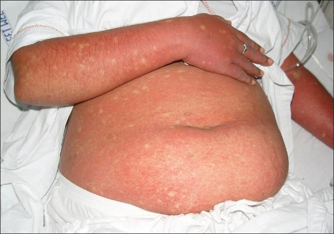
This clinical photograph displays a patient's abdomen and arm presenting a generalized, confluent erythematous rash, a characteristic cutaneous manifestation of Dengue Fever. The exanthem is marked by widespread, intense redness resulting from capillary dilatation and coalesced maculopapular lesions. A diagnostic hallmark visible here is the presence of 'islands of sparing'—small, rounded, or oval-shaped patches of normal-colored skin scattered throughout the erythematous background, often described as 'white islands in a sea of red'. These spared areas are randomly distributed across the affected trunk and limbs. The image serves as a key educational reference for recognizing the secondary rash phase of Dengue viral infection, typically occurring 3 to 6 days after the onset of fever. The visual presentation is essential for clinical diagnosis and differentiation from other viral exanthems, highlighting the specific immunologic response patterns seen in Dengue Fever (DF) and Dengue Hemorrhagic Fever (DHF).
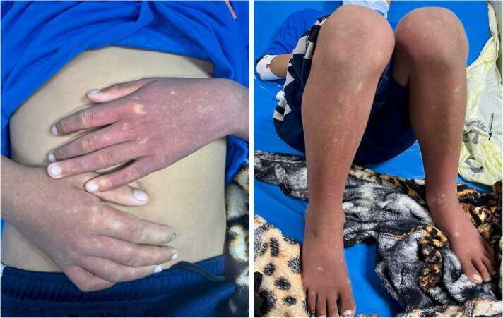
This clinical photograph displays a pediatric patient with a classic dermatological presentation of the recovery phase of Dengue Fever. Two side-by-side images show the dorsal surfaces of the hands, forearms, lower legs, and feet. The primary finding is a diffuse, confluent, erythematous rash covering the extremities. This background erythema is punctuated by multiple small, irregular macules and patches of pale, normal-appearing skin. This characteristic morphological pattern is classically described in clinical medicine as 'white islands in a sea of red.' The distribution is symmetric across the upper and lower limbs. Clinically, this convalescent rash typically appears after the defervescence of fever in dengue virus infection and is a key diagnostic physical sign indicating the recovery phase of the disease. The image is a valuable educational resource for infectious disease and pediatric dermatology, illustrating the morphological distinction between confluent erythematous lesions and spared areas of skin.
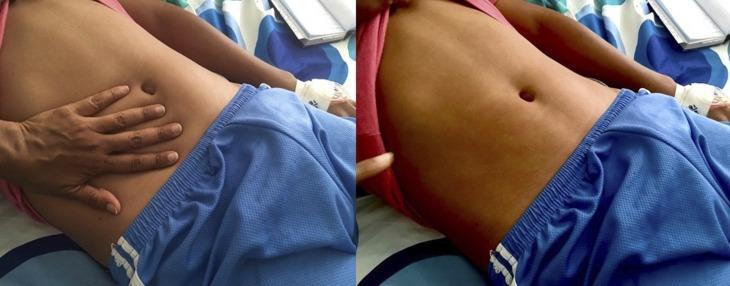
This clinical photograph displays a diffuse, erythematous maculopapular rash on the abdomen of a patient, characteristic of Dengue fever. The image is presented in a two-part sequence to demonstrate clinical blanching. On the left, a clinician's hand is shown applying direct pressure to the right side of the patient's abdomen. On the right, after the hand is removed, a clear, pale handprint-shaped outline remains where the pressure was applied, indicating that the redness fades (blanches) as blood is temporarily displaced from the superficial vessels. This finding confirms the rash is inflammatory rather than hemorrhagic (petechial/purpuric). The rash is widespread across the torso but typically spares the palms and soles in this clinical presentation. This image serves as a teaching tool for medical students and clinicians to distinguish between vascular inflammatory rashes and non-blanching hemorrhagic lesions in the context of viral hemorrhagic fevers and infectious diseases.
dengue fever clinical features symptoms
Dengue Fever - Symptoms & Clinical Features
Three Phases of Dengue
1. Febrile Phase (Days 1-7)
- High fever (often 39-40°C) lasting 2-7 days
- Severe myalgia and bone/joint pain - the hallmark "breakbone fever"
- Headache, especially retro-orbital (behind the eyes) pain
- Facial flushing and erythema
- Injected oropharynx (red throat)
- Nausea and vomiting
- Leukopenia (low white blood cell count)
- Thrombocytopenia - platelet count <100,000 in ~50% of patients
- Elevated liver enzymes (LFTs ~3x normal on average)
- Appears in 90% of rash cases between days 3-5, often as fever breaks
- Macular or morbilliform (measles-like), usually confluent
- Classic appearance: "islands of white in a sea of red" - small patches of normal skin surrounded by erythema
- Distribution: generalized (50%), extremities (30%), or trunk (20%)
- Mildly pruritic or asymptomatic
- Tourniquet test - inflating a BP cuff for 5 minutes, then counting petechiae; 10 or more per square inch is positive, a clue to dengue
2. Critical Phase (Days 3-7, around defervescence)
- Persistent vomiting
- Severe abdominal pain or tenderness
- Clinical fluid accumulation (ascites, pleural effusion)
- Mucosal bleeding (gums, nose)
- Rapid decline in platelet count + rising hematocrit (hemoconcentration)
- Lethargy or restlessness
- Liver enlargement >2 cm
- Difficulty breathing, early signs of shock
3. Convalescent Phase
- Gradual improvement and hemodynamic stabilization
- A second rash may appear ("convalescent rash" with "white islands")
- Complete recovery in 7-10 days in uncomplicated disease
WHO Classification of Dengue Severity
| Category | Criteria |
|---|---|
| Dengue without warning signs | Fever + 2 of: nausea/vomiting, rash, aches/pains, leukopenia, positive tourniquet test |
| Dengue with warning signs | Above + abdominal pain, persistent vomiting, fluid accumulation, mucosal bleeding, lethargy, liver enlargement |
| Severe dengue | Severe plasma leakage with shock, severe bleeding, severe organ involvement (AST/ALT ≥1000 IU/L, impaired consciousness, organ failure) |
Severe Dengue (Dengue Hemorrhagic Fever / Dengue Shock Syndrome)
- Widespread hemorrhages throughout the body
- Hepatitis and liver failure
- Massive plasma extravasation leading to hypovolemic shock
- Pleural effusions, ascites
- Respiratory distress (diffuse alveolar damage)
- Impaired consciousness
Less Common / Atypical Manifestations
- Myocarditis
- Pancreatitis
- Hemophagocytic lymphohistiocytosis (HLH) - a 2025 meta-analysis (PMID 40872761) documented this rare but severe complication
- Acute meningoencephalitis
- Post-dengue ADEM (acute disseminated encephalomyelitis)
Key Lab Findings
| Test | Finding |
|---|---|
| CBC | Leukopenia, thrombocytopenia |
| Hematocrit | Rising hematocrit signals plasma leakage |
| Liver enzymes | Elevated ALT/AST (~3x normal) |
| Tourniquet test | Positive (≥10 petechiae/sq inch) |