
Penis according to bdc
Reading File
Now let me search the BDC (BD Chaurasia) anatomy textbook for the penis section.
Finding Sources
Finding Sources
Let me search specifically for BD Chaurasia in the library:
Finding Sources
Finding Sources
BD Chaurasia's Human Anatomy is not in this library. The library has Gray's Anatomy for Students, Color Atlas of Human Anatomy (Thieme), and Netter's Atlas - but not BDC. Let me get the penis content from Gray's Anatomy for Students (the closest authoritative anatomy text available) and also check the Color Atlas for penis anatomy.
Reading File
Reading File
Let me also read the continuation of the Color Atlas content for blood supply, nerve supply, and lymphatics, plus check for any images:
Reading File
Finding Sources
Let me fetch the anatomical diagram image and also get the Gray's arterial supply detail:
Loading Image
Reading File
I now have comprehensive information. Note that BD Chaurasia's Human Anatomy is not present in this library. I'll provide a thorough answer based on standard anatomy textbooks available (which cover the same content as BDC), clearly noting this. Let me compile the full answer.
Note: BD Chaurasia's Human Anatomy (BDC) is not available in this textbook library. The anatomy content below is sourced from Gray's Anatomy for Students and the Color Atlas of Human Anatomy (Thieme, Vol. 2) - both standard references that cover the same material as BDC on this topic.
Penis - Anatomy
Overview
The penis is the male copulatory and urinary organ. It is composed of three erectile bodies:
- Two corpora cavernosa (paired, dorsal)
- One corpus spongiosum (single, ventral) - contains the urethra
Parts of the Penis
1. Root (Attached Part)
The root consists of three structures:
- Two crura - proximal extensions of the corpora cavernosa, attached to the inferior pubic rami; each is surrounded by the ischiocavernosus muscle
- Bulb of the penis - the proximal thickened part of the corpus spongiosum; firmly connected to the perineal membrane (urogenital diaphragm) and covered by the bulbospongiosus muscle
2. Body (Free Part)
- Made up of the two fused corpora cavernosa (forming the bulk) and the corpus spongiosum lying in the ventral groove
- Covered by thin skin that contains no fat and is freely movable
- Supported by two ligaments:
- Suspensory ligament of the penis - attached superiorly to the pubic symphysis
- Fundiform ligament of the penis - attached above to the linea alba; splits below into two bands passing on either side of the penis
3. Glans Penis
- Formed by the expanded distal end of the corpus spongiosum projecting beyond the corpora cavernosa
- The rounded margin at its base is the corona of the glans
- The tip bears the external urethral orifice (slit-like opening)
- The skin over the body folds forward over the glans as the prepuce (foreskin); its inner layer forms the frenulum of the prepuce inferiorly
Internal Structure
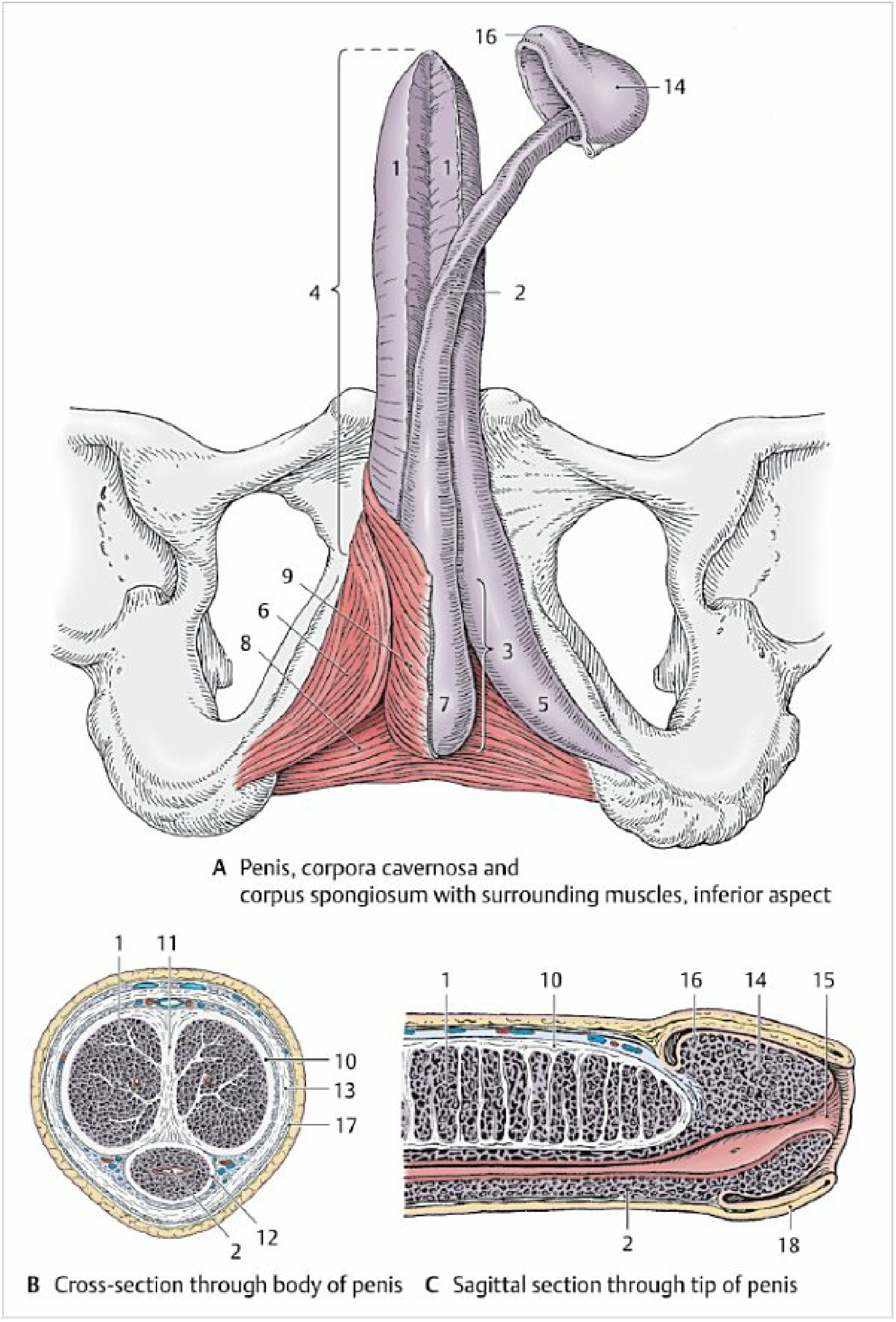
Corpora Cavernosa
- Each enclosed in a thick fibrous sheath - tunica albuginea of the corpora cavernosa
- A median septum penis partially separates the two chambers
- Internally: vascular (cavernous) spaces lined by endothelium, embedded in a trabecular framework of collagen, elastic fibers, and smooth muscle
- Fed by helicine arteries (from the deep artery of the penis); these act as resistance vessels
- Blood drains to subfascial and epifascial veins
Corpus Spongiosum
- Enclosed in a thinner tunica albuginea of the corpus spongiosum
- Vascular spaces are viewed as continuations of the venous system
- Contains less prominent trabeculae than the corpora cavernosa
- Filling leads only to "soft" swelling - keeps the urethra open during ejaculation
Fascial Layers
- Fascia of the penis (Buck's fascia) - tough deep fascia surrounding all three corpora collectively
- Subcutaneous tissue of the penis - thin layer beneath the skin, contains no fat
Blood Supply
All three erectile bodies are supplied by branches of the internal pudendal artery (itself a branch of the anterior division of the internal iliac artery):
| Branch | Supplies |
|---|---|
| Dorsal artery of the penis | Passes deep to fascia on dorsum; supplies glans, foreskin, and skin |
| Deep artery of the penis | Runs in the center of each corpus cavernosum; gives off helicine arteries |
| Artery of the bulb of the penis | Supplies corpus spongiosum and urethra |
| Urethral artery | Supplies urethra |
Venous drainage: Mostly via the unpaired superficial and deep dorsal veins of the penis (both with numerous valves) draining into the prostatic venous plexus and vesical venous plexus.
Nerve Supply
- Sensory: Dorsal nerve of the penis (branch of the pudendal nerve, S2-S4) - general somatic sensory
- Parasympathetic (erection): Pelvic splanchnic nerves (S2-S4) via the inferior hypogastric plexus - cause vasodilation and erection
- Sympathetic (emission/ejaculation): Lumbar sympathetic fibers (L1-L3) via inferior hypogastric plexus
Lymphatic Drainage
Lymph from the penis drains to the superficial inguinal lymph nodes.
Erection
Erection is a vascular event:
- Parasympathetic stimulation (S2-S4) causes relaxation of helicine arteries
- Blood fills the cavernous spaces of the corpora cavernosa
- Venous outflow is simultaneously reduced (veno-occlusive mechanism)
- The ejaculation reflex center (L2/L3) initiates emission and ejaculation when stimulation reaches threshold
Sources: Color Atlas of Human Anatomy Vol. 2 (Thieme), pp. 430-433; Gray's Anatomy for Students, pp. 496-517
This is a shared conversation. Sign in to Orris to start your own chat.