
Blood supply of heart
Reading File
This is a broad anatomy topic. I'll search the medical library using headings and paragraphs FTS in parallel.
Finding Sources
Finding Sources
Good - found Gray's Anatomy for Students and Fuster/Hurst's The Heart. Let me read the anatomy content and also check for a dedicated coronary circulation section.
Reading File
Finding Sources
Good content found. Let me continue reading the coronary artery details from Gray's and also check Fuster & Hurst for more detailed coronary anatomy.
Reading File
Finding Sources
Excellent! Let me read the detailed coronary artery sections from Fuster & Hurst's The Heart and Bailey & Love simultaneously, plus the Gray's Anatomy coronary section.
Reading File
Reading File
Excellent content. Now let me read the Gray's Anatomy dedicated coronary artery section and the cardiac veins section.
Finding Sources
Reading File
Now let me look for Gray's Anatomy dedicated coronary artery and venous drainage sections:
Finding Sources
Finding Sources
Now let me read Gray's Anatomy coronary arteries section and Fischer's cardiac veins section:
Reading File
Reading File
Excellent - I now have comprehensive content. Let me also read the remaining Gray's section on coronary artery variations and fetch the key diagram images:
Reading File
Loading Image
Now let me fetch the coronary arteries diagram from Gray's:
Loading Image
Loading Image
I now have comprehensive, well-sourced content with excellent diagrams. Let me compile the full answer.
Blood Supply of the Heart
The heart receives its arterial supply from the right and left coronary arteries, both arising from the ascending aorta. Venous drainage is primarily via the coronary sinus into the right atrium.
Arterial Supply
Origin
Both coronary arteries arise from the aortic sinuses (sinuses of Valsalva) just above the aortic valve, immediately below the sinotubular ridge. They run in the subepicardial fat within the sulci of the heart.
Right Coronary Artery (RCA)
The RCA originates from the right aortic sinus of the ascending aorta. It passes anteriorly between the right atrial appendage and the pulmonary trunk, then descends in the coronary sulcus between the right atrium and right ventricle. On reaching the inferior (acute) margin of the heart, it turns posteriorly onto the diaphragmatic surface, traveling toward the crux of the heart.
Branches of the RCA:
| Branch | Supply |
|---|---|
| Sino-atrial (SA) nodal artery | SA node (arises early via atrial branch) |
| Right marginal branch | Runs along the acute margin toward the apex |
| AV nodal branch | Atrioventricular node (just before the crux) |
| Posterior interventricular artery (PDA) | Lies in the posterior interventricular sulcus |
Territory supplied: Right atrium, right ventricle, SA and AV nodes, interatrial septum, a portion of the left atrium, the posteroinferior one-third of the interventricular septum, and a portion of the posterior left ventricle.
Left Coronary Artery (LCA)
The LCA originates from the left aortic sinus. It passes between the pulmonary trunk and the left auricle before entering the coronary sulcus. The left main stem is short and quickly divides into two main branches:
1. Anterior Interventricular Artery (LAD - Left Anterior Descending)
- Descends in the anterior interventricular sulcus toward the apex
- Gives off diagonal branches that cross the anterior surface of the left ventricle
- Supplies the anterior wall of the left ventricle, anterior two-thirds of the interventricular septum, and the bundle branches (AV bundle + its branches)
2. Circumflex Branch
- Courses left in the coronary sulcus and onto the base/diaphragmatic surface
- Gives off the left marginal artery (runs across the rounded obtuse margin)
- Usually ends before reaching the posterior interventricular sulcus
- In left dominant hearts (~10-15%), gives rise to the posterior descending artery
Territory supplied: Most of the left atrium and left ventricle, and most of the interventricular septum including the bundle of His and its branches.
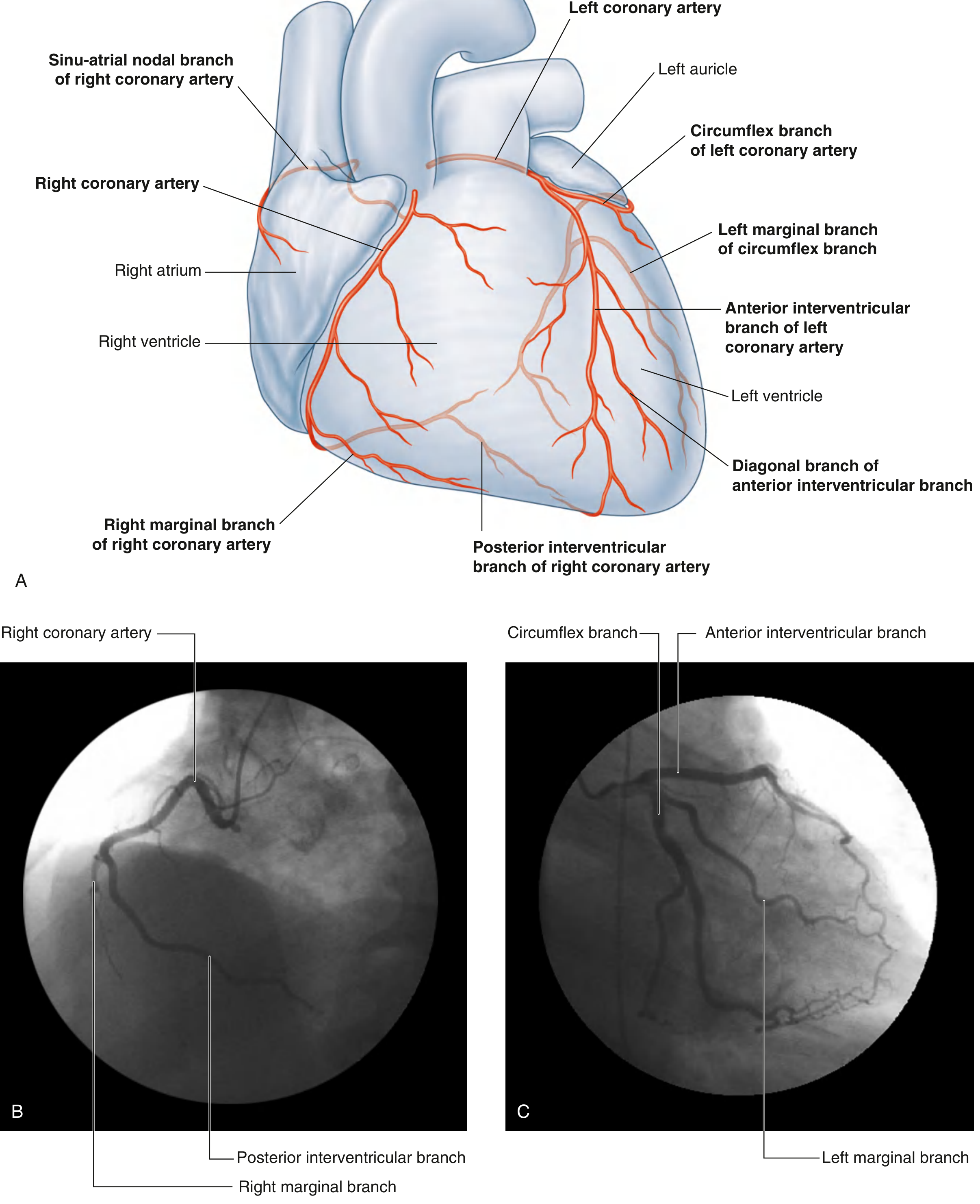
Fig. 3.80 - Coronary artery distribution (anterior view and angiographic views). Note the RCA, LAD, circumflex, and their branches. - Gray's Anatomy for Students
Coronary Dominance
Dominance is defined by which artery supplies the posterior descending artery (PDA), which determines the blood supply to the inferior wall of the left ventricle and the posterior interventricular septum.
| Pattern | Artery Supplying PDA | Frequency |
|---|---|---|
| Right dominant | RCA | ~85-90% |
| Left dominant | Circumflex branch of LCA | ~10-15% |
| Co-dominant | Both RCA and circumflex | ~5% |
In right-dominant hearts, the RCA also supplies the AV node. In left-dominant hearts, the enlarged circumflex supplies the posterior wall of the left ventricle and often the AV node.
Venous Drainage
The heart drains primarily via veins that empty into the coronary sinus, which runs left-to-right in the posterior part of the coronary sulcus and opens into the right atrium.
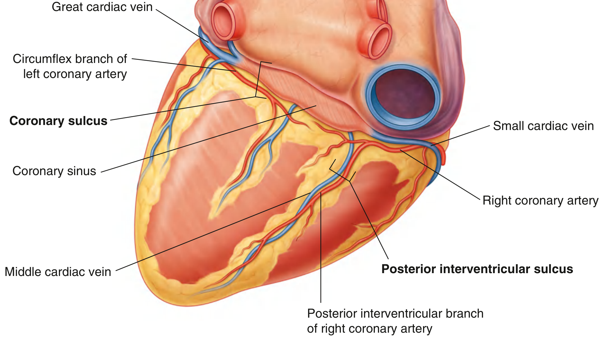
Posterior view showing the coronary sinus, middle cardiac vein, circumflex branch, and small cardiac vein - Gray's Anatomy for Students
Major Cardiac Veins (draining into the coronary sinus):
| Vein | Course | Companion Artery |
|---|---|---|
| Great cardiac vein | Starts at apex, ascends in anterior interventricular sulcus, then turns into the left coronary sulcus | LAD |
| Middle cardiac vein | Starts at apex, runs in posterior interventricular sulcus to the right end of the coronary sinus | Posterior interventricular artery (PDA) |
| Small cardiac vein | Begins as marginal vein along the right margin, turns posteriorly in coronary sulcus to right end of coronary sinus | Right marginal branch |
| Posterior vein of left ventricle | Runs on the diaphragmatic surface of the left ventricle | - |
| Oblique vein of left atrium (of Marshall) | Runs on the left atrium; remnant of the embryonic left superior vena cava | - |
Veins NOT draining into the coronary sinus:
- Anterior cardiac veins (3-4 small veins): Arise from the anterior wall of the right ventricle, cross the RCA, and drain directly into the right atrium
- Venae cordis minimae (Thebesian veins): Tiny veins that start in the myocardial capillary bed and open directly into the cardiac chambers (mainly the atria)
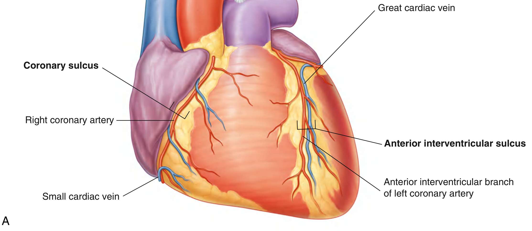
Anterior surface of the heart showing coronary arteries (red) and cardiac veins (blue) - Gray's Anatomy for Students
Clinical Relevance
- The LAD is the most frequently diseased coronary artery and most often bypassed during CABG surgery - Bailey and Love's Short Practice of Surgery, 28th Ed.
- Left main stem disease (stenosis of the short left main coronary artery) carries the worst prognosis without surgical intervention
- Occlusion of the RCA typically causes inferior MI and may damage the SA and AV nodes, producing bradyarrhythmias
- Occlusion of the LAD ("widow maker") causes anterior MI and can disrupt the bundle branches
- In right-dominant hearts, the RCA supplies both the SA and AV nodes; their ischemia can cause sick sinus syndrome or complete heart block
- Myocardial bridges: The LAD occasionally runs an intramyocardial course; the overlying muscle may compress it during systole, potentially mimicking stenosis on angiography
Sources:
- Gray's Anatomy for Students, pp. 228-242
- Fischer's Mastery of Surgery, 8th Ed., Ch. 273
- Bailey and Love's Short Practice of Surgery, 28th Ed.
- Fuster and Hurst's The Heart, 15th Ed., Ch. 3
This is a shared conversation. Sign in to Orris to start your own chat.