
Babinski sign
The Babinski sign is a plantar reflex test where stroking the lateral sole causes upward (extensor) movement of the big toe and fanning of other toes, indicating upper motor neuron (corticospinal tract) damage. It's normal in infants under ~2 years but pathological in adults.
Searching sources...
Reading File
Finding Sources
Finding Sources
Reading File
Reading File
Reading File
Reading File
Reading File
Loading Image
Loading Image
Loading Image
Here is a complete overview of the Babinski Sign, drawn from Adams and Victor's Principles of Neurology (12th Ed.), Neuroanatomy through Clinical Cases (3rd Ed.), and Neuroscience: Exploring the Brain (5th Ed.).
Babinski Sign
Background
Named after the French neurologist Joseph Babinski, who first described the great toe sign in 1896 and the toe abduction (fan sign) in 1903. These two components are now commonly conflated and referred to simply as "the Babinski sign." - Adams and Victor's Principles of Neurology, 12th Ed., p. 74
How to Elicit It
Technique: Apply a firm but not necessarily painful stimulus along the lateral plantar surface of the foot - starting from the lateral heel, sweeping upward toward the small toe, and then arcing medially toward the ball of the foot.
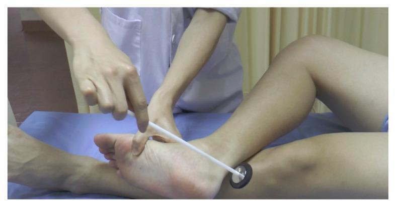
Responses
| Response | Interpretation |
|---|---|
| Toes curl downward (plantarflexion) | Normal in anyone older than ~2 years |
| Big toe extends upward (dorsiflexion) + fanning of other toes | Positive Babinski sign - pathological in adults |
| Toes are "silent" (neither up nor down) | If unilateral, the silent side is considered abnormal |
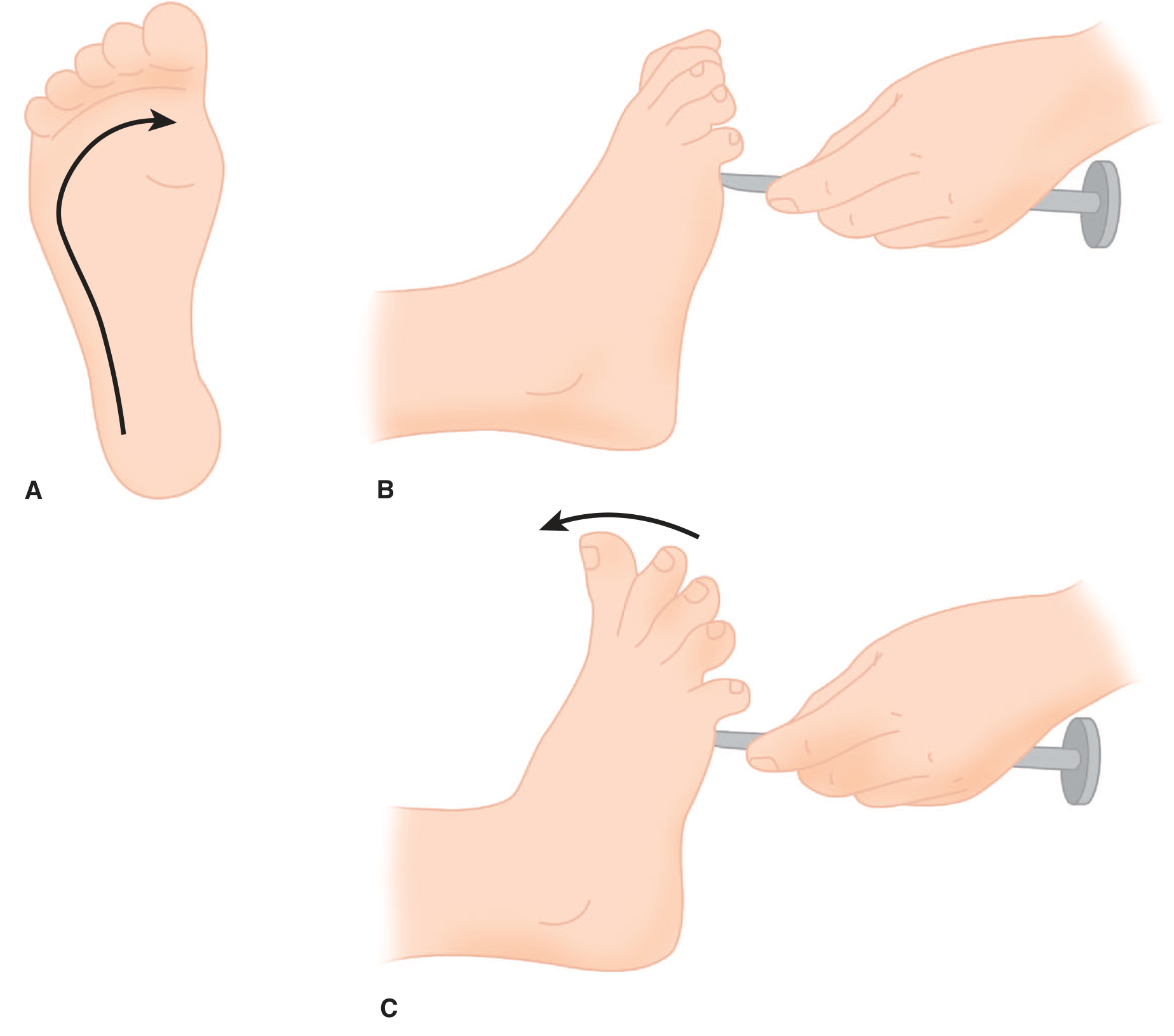
Figure: A. The plantar response is elicited by stroking from the heel to the ball of the foot. B. Normal plantar response - flexion of the toes. C. Positive Babinski sign - extension of the great toe with fanning of the remaining digits. - Adams and Victor's, p. 74
Mechanism / Pathophysiology
The extension of the great toe is physiologically a component of the spinal flexion (nocifensive/protective) reflex - a "triple flexion response" involving hip, thigh, and ankle dorsiflexion. This reflex is normally suppressed by descending corticospinal inhibition. When the corticospinal tract is damaged, that inhibition is removed ("released"), and the flexor withdrawal program of spinal origin becomes disinhibited.
Key pathway:
-
Corticospinal tract damage removes inhibitory control
-
The dorsal reticulospinal tract (normally inhibitory to stretch reflexes) is also usually affected in spinal cord lesions
-
The result is release of the spinal flexion reflex, manifesting as the Babinski sign
-
Adams and Victor's, p. 73-74
Clinical Significance
-
The Babinski sign is the most reliable indicator of an upper motor neuron (UMN) / corticospinal tract lesion
-
It is pathological in adults and children beyond ~18 months
-
It may be present with lesions anywhere along the corticospinal tract - from the motor cortex down to the spinal cord
-
It may not be elicitable if there is severe weakness of the toe extensors
-
A unilateral Babinski sign localizes the lesion to the contralateral corticospinal system
-
Neuroanatomy through Clinical Cases, 3rd Ed.
Normal in Infants
The Babinski sign is normal in infants up to approximately 1-2 years of age because the descending corticospinal motor tracts have not yet fully myelinated. As myelination completes, the plantar response switches to the normal downgoing (flexor) pattern. Persistence beyond this age is pathological. - Neuroscience: Exploring the Brain, 5th Ed., p. 1327
Associated UMN Findings (Context)
When Babinski sign is present, look for other corticospinal tract signs:
- Spasticity with clasp-knife phenomenon
- Hyperreflexia (exaggerated deep tendon reflexes) and possibly clonus
- Weakness in an antigravity pattern (arm flexors and leg extensors)
- Loss of superficial abdominal and cremasteric reflexes
- "Triple flexion response" (hip, knee, ankle dorsiflexion to noxious stimuli)
Surrogate Signs (Same Significance)
Several eponymous variants use alternative stimulation sites but carry the same pathological meaning:
| Sign | Stimulus |
|---|---|
| Chaddock | Stroke the lateral dorsum of the foot |
| Oppenheim | Downward scraping along the shin |
| Gordon | Squeezing the calf |
| Bing | Pricking the dorsum of the foot |
| Gonda/Stransky | Flicking or pressing the 4th toe |
These are useful when foot hypersensitivity (ticklishness or allodynia) prevents standard elicitation. - Adams and Victor's, p. 74
Key Teaching Points
- Positive = UMN lesion - anywhere from motor cortex to spinal cord
- Normal in infants (<~18 months); pathological at any age beyond that
- The sign reflects release of a primitive spinal flexion reflex normally suppressed by corticospinal influence
- Always interpret in context - a withdrawal response (quick, non-pathological) must not be confused with the true, slower Babinski response
- If toes are silent on one side when clearly downgoing on the other, the silent side is abnormal
This is a shared conversation. Sign in to Orris to start your own chat.