
Treatment for phimosis
phimosis foreskin
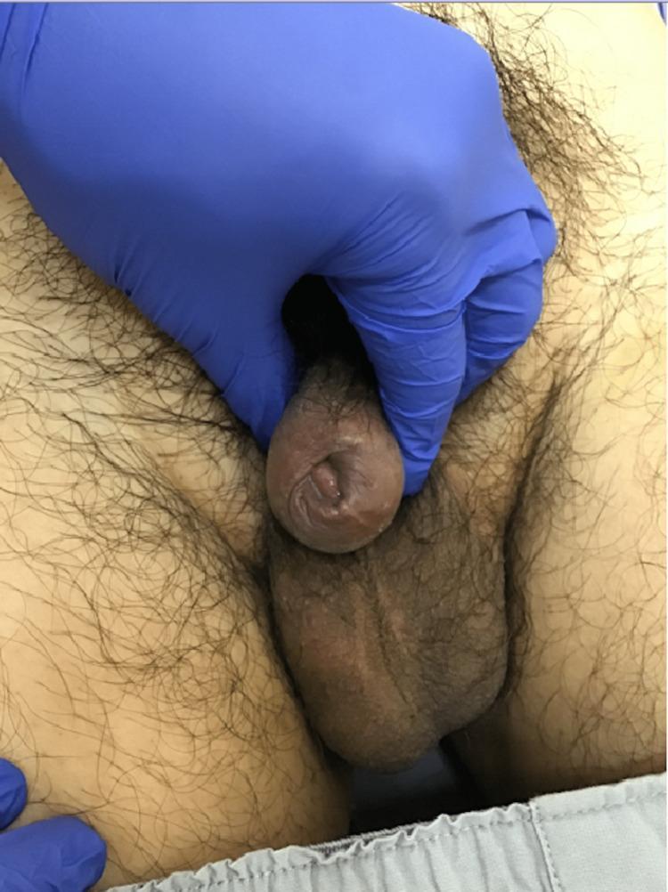
Clinical photograph of the male genitalia showing a case of severe phimosis. The penile foreskin is prominently thickened and constricted, with a significantly narrowed preputial opening. There is a visible firm, localized induration of the foreskin tissue approximately 5 mm from the external urethral orifice. The foreskin is tightly adherent to the underlying glans penis, preventing manual retraction and obscuring the visualization of the glans and coronal sulcus. The skin of the prepuce appears hyperpigmented and fibrotic in texture. No active ulcerations, purulent discharge, or overt erythema are visible at the distal aspect. A gloved hand is shown for examination, illustrating the lack of preputial mobility. This image is used for educational purposes to demonstrate the clinical presentation of preputial induration and secondary phimosis, which may be associated with inflammatory, infectious (such as syphilis), or neoplastic conditions like penile carcinoma.
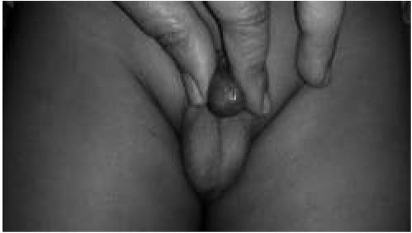
This grayscale clinical photograph depicts a physical examination of the pediatric male genitalia, demonstrating a non-retractile prepuce consistent with Grade 4 phimosis on the Kikiros scale. The image shows a clinician's fingers attempting to retract the foreskin. There is minimal retraction, with the preputial orifice remaining narrow and constricted. Only a small portion of the glans penis is visible through the stenotic opening of the foreskin. The surrounding inguinal and scrotal skin appears normal without evidence of inflammation or secondary lesions. This visual represents a diagnostic evaluation of pathological or physiological phimosis in a pediatric patient, emphasizing the clinical assessment of preputial mobility and the degree of urethral meatus exposure.
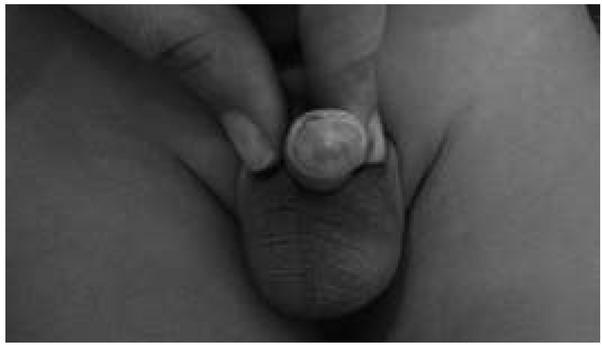
A grayscale clinical photograph of a pediatric male patient's genitals demonstrating a non-retractile prepuce, classified as Kikiros Grade 1. The image shows manual retraction of the foreskin using a finger to expose the glans penis. Only a small, central portion of the glans is visible through a narrowed preputial orifice. The surrounding foreskin appears smooth but tight, indicating significant phimosis where the preputial opening is insufficient for full retraction over the glans corona. This visual serves as an educational example for the Kikiros and Woodward classification system used in pediatric urology to grade the severity of phimosis. The educational focus is on identifying the degree of prepuce extensibility and the visible proportion of the glans in a clinical examination context.
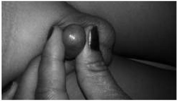
A greyscale clinical photograph depicting a close-up view of a pediatric penis with severe phimosis, classified as Kikiros Grade 5. The image shows the prepuce being manually retracted by an examiner's fingers. The foreskin is tight, non-retractile, and forms a rounded, spherical shape at the distal end, with no visibility of the glans penis or the urethral meatus. The preputial opening is extremely constricted, appearing as a tiny, pinpoint orifice. The skin of the prepuce appears smooth and tense due to the resistance against retraction. This visual serves as a diagnostic reference for severe physiological or pathological phimosis in pediatric urology, illustrating the highest degree of preputial non-retractability where the foreskin is completely tight and the glans is entirely obscured.
phimosis treatment
phimosis treatment guidelines topical steroid circumcision
Phimosis: Treatment
- Physiologic - normal in uncircumcised infants/young boys due to natural adhesions between prepuce and glans; not a disease
- Pathologic - due to scarring/fibrosis (often from balanitis xerotica obliterans, recurrent balanoposthitis, or forceful retraction attempts), causing a true stenotic ring
1. Physiologic phimosis (children)
2. Symptomatic or pathologic phimosis - first-line: topical corticosteroids
- Regimen: A potent topical corticosteroid (e.g., betamethasone 0.025-0.1%, clobetasol 0.05%, or triamcinolone) applied to the tight distal foreskin ring once or twice daily for 4-8 weeks (some protocols extend to ~3 months), combined with gentle stretching exercises after application.
- Efficacy: Reported success (resolution of phimosis) ranges widely, roughly 33-95% depending on the study and severity, with pooled rates often cited around 65-90%. Combining steroid cream with stretching improves outcomes.
- Once the ring loosens, the patient is advised to wean the frequency/potency and continue gentle retraction to prevent recurrence.
- Topical steroids are now considered first-line in European and Canadian pediatric urology guidelines, largely replacing early circumcision for uncomplicated cases.
3. Surgical treatment
- Failure of adequate medical therapy
- True pathologic phimosis with significant scarring (e.g., balanitis xerotica obliterans / lichen sclerosus)
- Recurrent paraphimosis, recurrent balanoposthitis, or urinary obstruction
- Patient/family preference, or religious/cultural indications
- Circumcision - definitive treatment; removes the foreskin entirely (Bailey and Love's Short Practice of Surgery: "Symptomatic phimosis is treated by circumcision," while "physiological phimosis does not need circumcision")
- Preputioplasty (dorsal slit or ventral slit with plasty) - a foreskin-preserving alternative that widens the preputial ring without full circumcision, useful when preservation of the foreskin is desired (Hinman's Atlas of Urologic Surgery)
- Dorsal slit - can also be used as an emergency temporizing procedure if there is acute urinary retention from phimosis
4. Emergency considerations
Clinical image
Summary
| Scenario | First-line management |
|---|---|
| Physiologic phimosis, asymptomatic | Reassurance, routine hygiene, no forced retraction |
| Symptomatic phimosis (mild-moderate) | Topical corticosteroid (4-8 weeks) + gentle stretching |
| Steroid failure / severe scarring (e.g., BXO) | Circumcision or preputioplasty |
| Acute urinary retention from phimosis | Emergency catheterization/decompression, dorsal slit if needed |
| Paraphimosis | Emergency manual reduction; surgery if reduction fails |
Proper Dosage
betamethasone 0.05% phimosis dosing regimen application frequency duration
Proper Dosage: Topical Corticosteroids for Phimosis
Standard regimen
- Drug: Betamethasone dipropionate/valerate 0.05% (alternatives: clobetasol propionate 0.05%, triamcinolone 0.01-0.5%, fluticasone propionate 0.05%)
- Application: A thin layer applied directly to the tight/stenotic distal ring of the foreskin (not the whole penis) - not just the outer skin, but massaged into the phimotic band itself
- Frequency: Twice daily (BID) is the most common regimen
- Duration: 4-8 weeks (most protocols use 4-6 weeks; some extend up to a maximum of 3-4 months if response is partial)
- Combine with stretching: Gentle manual retraction/stretching exercises 1-2 times daily (often started after ~5-7 days of cream use, once the skin has thinned) significantly improves success rates - cream alone is less effective than cream + stretching
Evidence-based variations (equally effective)
| Regimen | Duration | Source |
|---|---|---|
| BID for 30 days (60 doses) | 1 month | Palmer & Palmer comparison study |
| TID for 21 days (63 doses) | 3 weeks | Palmer & Palmer comparison study |
| BID for 15 days, then once daily for 15 more days | 1 month | Larger case series (247 boys), ~77% resolution after 1 cycle, +20% after a repeat cycle |
| BID | Minimum 30 days, up to 4 months if needed | Brazilian cohort study |
- Half-strength betamethasone (0.025%) has also been shown effective in young children if minimizing steroid exposure is a concern.
- Success rates: Daily/BID application for at least 4 weeks resolves phimosis completely in roughly 65-95% of boys in various series, especially when combined with stretching.
- If no meaningful improvement after one full course, a second course can be tried before considering surgical referral (circumcision or preputioplasty).
Practical/safety notes
- Use the lowest effective potency for the shortest effective duration - prolonged use of potent steroids on genital skin (which absorbs more readily) carries a theoretical risk of local skin atrophy, so courses are typically capped around 6-12 weeks and not routinely repeated indefinitely.
- Adults with symptomatic phimosis may need a more potent steroid (e.g., clobetasol 0.05%, "Dermovate") for 2 months or more, per UK primary care guidance, with weaning of frequency/potency once retraction is achieved.
- Warn patients/parents about paraphimosis if the foreskin is retracted during treatment and not returned to its normal forward position.
- If there's no response after an adequate steroid trial, or if pathologic scarring (e.g., balanitis xerotica obliterans/lichen sclerosus) is suspected, refer for circumcision or preputioplasty rather than prolonging medical therapy.