


Explain this case and why is haematemesis caused in this case
esophageal varices portal hypertension upper GI bleeding
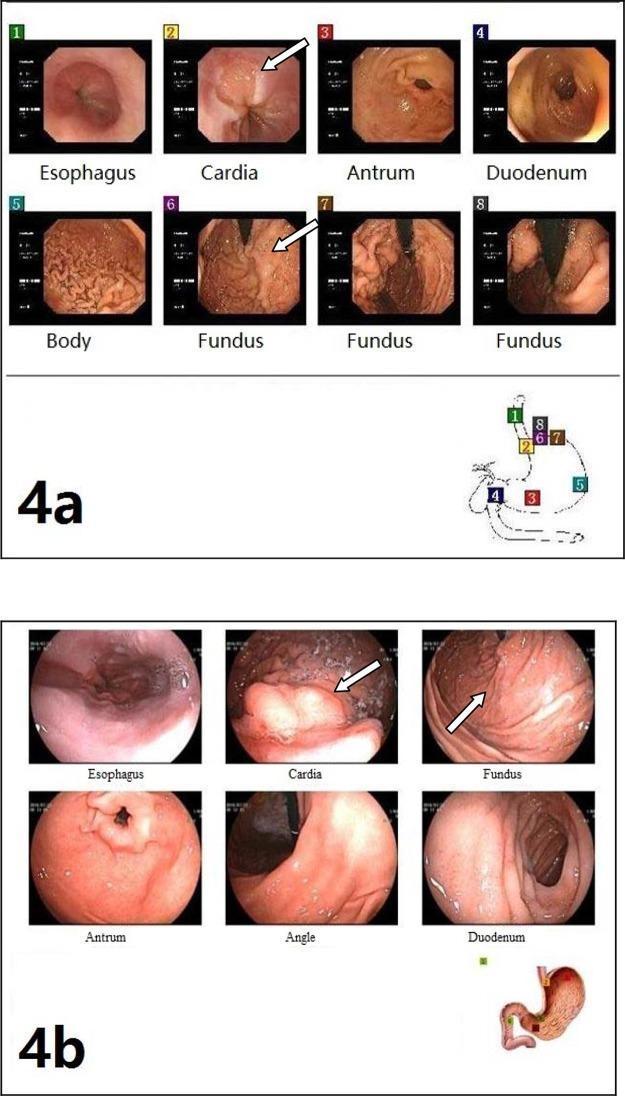
This comparative clinical imaging figure presents two sets of upper gastrointestinal endoscopy panels (4a and 4b) demonstrating the treatment progression of gastroesophageal varices. Panel 4a (Preoperative): Shows multiple views of the upper GI tract, including the esophagus, cardia, fundus, body, antrum, and duodenum. The cardia and fundus images specifically highlight prominent, tortuous, and elevated gastric varices (marked with arrows) and irregular mucosal texture characteristic of portal hypertension. The gastric body displays typical rugal folds, while the antrum and duodenum provide anatomical reference. Panel 4b (Postoperative): Displays a follow-up endoscopy of the same regions after shunt surgery and foam sclerotherapy. There is a marked reduction in the size and prominence of gastric varices in the cardia and fundus (indicated by arrows), showing a smoother mucosal surface. However, residual esophageal varices remain visible. Small anatomical maps accompany each panel to indicate the endoscopic orientation within the stomach. This figure serves as an educational tool for assessing the efficacy of surgical and sclerotherapeutic interventions in managing portal hypertension-related variceal bleeding.
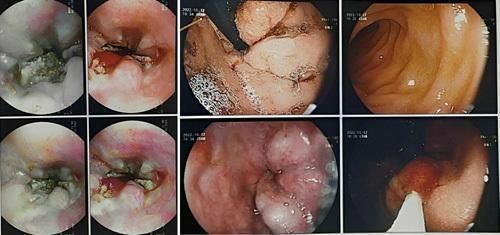
A series of endoscopic images from an upper gastrointestinal endoscopy (esophagogastroduodenoscopy) demonstrating severe esophageal pathology. The images reveal multiple deep esophageal ulcers with irregular borders and fibrinopurulent exudates. Significant esophageal varices (Grade IV) are visible as tortuous, dilated blueish-pink protrusions within the esophageal lumen, indicating portal hypertension. Some frames show congestive gastropathy with erythematous, mottled mucosa typical of portal hypertensive changes. Signs of recent or active hemorrhage and luminal narrowing due to mucosal edema and variceal bulk are also observed. These findings are clinically significant in the context of extrahepatic portal vein obstruction, highlighting the risk of upper GI bleeding and the compensatory collateral circulation development secondary to portal venous system thrombosis.
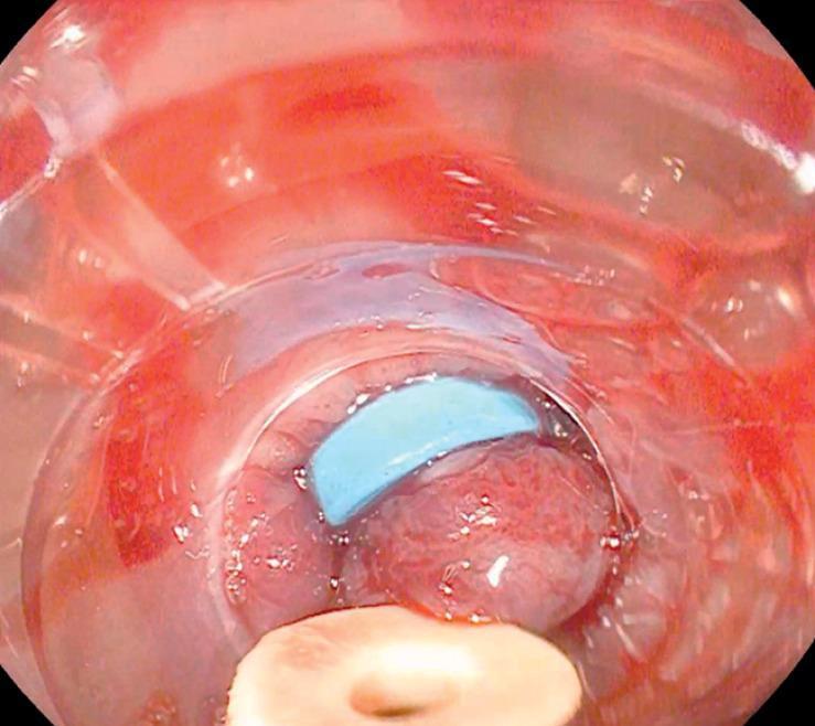
This clinical photograph shows an endoscopic view of an upper gastrointestinal procedure focused on the management of esophageal varices. The image depicts the esophageal lumen through a transparent plastic banding cap attached to the end of the endoscope. Within the center of the field, a large, reddish-pink, distended varix is visible. A bright blue elastic ligation band is seen partially encircling the base of the variceal tissue, demonstrating a key step in Endoscopic Variceal Ligation (EVL). The tissue appears congested and irregular, characteristic of portal hypertension. The banding cap is positioned to provide suction and visualization, facilitating the mechanical ligation process. This procedural image illustrates the technique of band-on-band ligation, where a secondary band is applied to ensure hemostasis and secure tissue prolapse when initial placement is suboptimal. The medical domain is Gastroenterology, and the target audience is intermediate-to-advanced medical students and surgical residents specializing in endoscopic interventions for GI bleeding.
Case Explanation: Ravi - Haematemesis in a 38-Year-Old Chronic Alcoholic
Patient Summary
| Detail | Info |
|---|---|
| Name | Ravi |
| Age/Sex | 38 years, Male |
| DOA | 8-6-26 |
| Social History | Alcoholic since 5 years |
| Chief Complaints | Vomiting large amounts of blood (haematemesis) + blood in stools (malaena) |
Vitals
| Parameter | Day 1 (8/6) | Day 2 (9/6) |
|---|---|---|
| BP | 145/70 mmHg | 130/80 mmHg |
| PR | 85 BPM | 78 BPM |
| SpO2 | 98% | 99% |
Lab Investigations - Analysis
- Hb: 13.3 g/dL - within normal range (not significantly anaemic yet, suggesting acute rather than chronic bleeding)
- WBC (TC): 6,600 cells/cumm - normal
- Platelets: 2.7 lakh/cumm - normal
- Creatinine: 0.5 mg/dL - normal
- Blood Urea: 2.6 mg/dL - normal
- Na+: 138 mmol/L - normal
- K+: 3.9 mmol/L - normal
- Total Bilirubin: 0.11 mg/dL - normal
- SGOT: 10 U/L - normal
- SGPT: 21 U/L - normal
- Total Protein: 5.4 U/L - low-normal (albumin 4.2 is borderline)
- ALP: 81 mg/dL - normal
Important clinical note: The relatively preserved liver function tests are a bit atypical for advanced alcoholic liver disease - this patient may be in an early-to-moderate stage of alcoholic liver disease or the tests were drawn before decompensation became fully evident. The diagnosis of haematemesis is clinical, not purely lab-based.
Diagnosis: Haematemesis
Why Is Haematemesis Caused in This Case?
Step-by-Step Pathophysiology
- Chronic alcohol causes hepatocyte injury through acetaldehyde toxicity and oxidative stress
- This leads to alcoholic hepatitis → hepatic fibrosis → cirrhosis
- Fibrosis and regenerative nodules distort the liver architecture and compress the hepatic sinusoids and portal venules
- This increases resistance to portal blood flow - the blood backs up into the portal system
- Portal venous pressure rises (normal portal pressure: 5-10 mmHg; portal hypertension: >10 mmHg)
- Varices develop when the hepatic venous pressure gradient (HVPG) exceeds 10 mmHg; they bleed when HVPG exceeds 12 mmHg
- When portal pressure rises, blood seeks alternate routes back to the heart via portosystemic collateral vessels
- The most clinically significant collateral is at the gastro-oesophageal junction, where the coronary (left gastric) vein connects the portal system to the oesophageal submucosal venous plexus
- These thin-walled veins become engorged, tortuous, and dilated - forming oesophageal varices
- In the distal 5 cm of the oesophagus (the palisade zone), these veins are especially vulnerable because they have no perforating branches to decompress them into peri-oesophageal veins
- The fundus of the stomach drains via short gastric veins into the splenic vein - these may also form gastric varices
- As varices enlarge under continued portal hypertension, the wall tension increases (LaPlace's law: wall tension = pressure × radius)
- Physical triggers (retching, increased intra-abdominal pressure) or spontaneous rupture leads to sudden, massive upper GI bleeding
- Blood vomited = haematemesis (vomiting large amounts of blood, as described in this case)
- Blood passing through the GI tract = malaena / blood in stools (as also reported here)
Visual Reference
Treatment Given (Explained)
| Drug | Class | Rationale |
|---|---|---|
| Inj. Pantoprazole (PPI) | Proton pump inhibitor | Inhibits H+/K+-ATPase in parietal cells; reduces gastric acid that worsens mucosal damage and impairs clot stability at bleeding sites |
| Inj. Ondansetron | 5-HT3 antagonist | Blocks serotonin-mediated activation of the vomiting reflex; prevents further retching which can re-rupture fragile varices |
| Inj. Thiamine (100 mg BD) | B1 vitamin | Alcoholics are chronically thiamine-deficient; supplementation prevents Wernicke's encephalopathy |
| Inj. Tranexamic acid | Antifibrinolytic | Inhibits plasminogen activation, prevents clot breakdown at bleeding sites, reduces blood loss |
| Inj. Multivitamin | Nutritional support | Replenishes multiple vitamins depleted by chronic alcoholism |
Note: Ideally, a vasoconstrictor such as terlipressin or octreotide (somatostatin analogue) would also be used in variceal haematemesis to reduce splanchnic blood flow and portal pressure. Endoscopic variceal ligation (EVL) or sclerotherapy would be the definitive management.
Summary Flowchart
Alcohol (5 years)
      ↓
Hepatocyte damage → Fibrosis → Cirrhosis
      ↓
Increased hepatic resistance → Portal Hypertension (HVPG >12 mmHg)
      ↓
Collateral vessel formation via coronary vein → Oesophageal Varices
      ↓
Variceal wall tension increases → Rupture
      ↓
HAEMATEMESIS (vomiting blood) + Blood in stools (malaena)
- Sleisenger and Fordtran's Gastrointestinal and Liver Disease - Collateral Circulation and Varices section
- Mulholland and Greenfield's Surgery - Varices (portal hypertension chapter)