
25 mark answer on controversies for management of ameloblastoma
ameloblastoma management treatment controversy
ameloblastoma resection recurrence treatment
ameloblastoma jaw mandible radiology X-ray panoramic
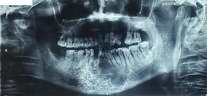
This orthopantomogram (panoramic X-ray) of the human mandible reveals a significant diagnostic finding in the right mandibular body extending toward the symphysis. The image displays a large, well-defined multilocular radiolucent lesion characterized by two distinct internal architectural patterns: a 'honeycomb' appearance, consisting of numerous small, uniform radiolucent compartments separated by thin radiopaque septa, and a 'soap bubble' pattern featuring larger, more irregular circular radiolucencies with thicker internal septations. The lesion demonstrates cortical expansion and involves a substantial portion of the vertical mandibular height, commonly associated with odontogenic tumors such as ameloblastoma or odontogenic keratocysts. The radiographic presentation is indicative of a slow-growing but locally aggressive intraosseous pathology. Educational focus includes the recognition of multilocular radiolucencies in oral and maxillofacial radiology and the differential diagnosis of mandibular cystic and neoplastic lesions based on internal trabeculation patterns.
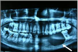
This diagnostic image is an Orthopantomogram (OPG), a panoramic dental X-ray, demonstrating a large pathological lesion in the mandible. The primary finding is a well-defined yet irregular radiolucent lesion located within the left mandibular body, indicated by a white arrow. The lesion exhibits significant osteolytic activity, resulting in the destruction of the surrounding medullary and cortical bone. The adjacent dentition shows clinical complications, including missing teeth in the left mandibular quadrant and evidence of dental caries in the remaining teeth bordering the mass. The radiographic appearance of the radiolucent area, combined with the irregular margins and localized bone destruction, is suggestive of an odontogenic tumor or carcinoma, such as clear cell odontogenic carcinoma or ameloblastoma. This image is used in dental and maxillofacial education to illustrate the radiographic presentation of aggressive mandibular neoplasms and the importance of panoramic imaging in identifying bone-destroying lesions of the jaw.
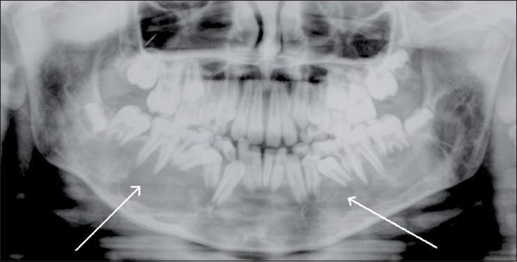
This orthopantomogram (panoramic dental X-ray) of a pediatric patient displays a large, bilateral expansile osteolytic lesion within the body of the mandible. The pathology is characterized by a significant area of radiolucency extending across the midline, involving both the right and left mandibular bodies. The lesion features well-defined, sclerotic margins, indicating a slow-growing process. Its expansile nature is evidenced by the thinning and outward bowing of the mandibular cortex. Notably, the lesion causes displacement of developing permanent teeth and molar roots, which is a common finding in intraosseous vascular malformations (AVMs) or giant cell lesions. Two white arrows highlight the lateral extents of the radiolucent area. This diagnostic image is used to illustrate primary dental/maxillofacial radiology findings for osteolytic jaw lesions and is essential for the differential diagnosis of mandibular bone expansion in young patients.
Controversies in the Management of Ameloblastoma
Introduction
Controversy 1: Conservative vs. Radical Surgery for Intraosseous (Conventional) Ameloblastoma
The Case for Radical/Resective Surgery
- Resection (en bloc removal) with a 1 cm margin beyond the radiographic extent of the tumor is favored by most North American and European authorities.
- After resection, the specimen allows intraoperative radiographic assessment and postoperative histological confirmation of margins - something impossible with enucleation.
- Recurrence rates after enucleation/curettage range from 55% to 90% in some series, compared to 10-15% after resection with adequate margins.
- Cummings Otolaryngology states: "Simple enucleation of ameloblastomas is not considered standard of care in the United States, and even combined with curettage ameloblastomas are known to recur."
- Even after en bloc resection reportedly well past radiographic tumor margins, recurrence rates of 10-15% have been reported, highlighting the tumor's aggressive biology.
The Case for Conservative Surgery
- Functional preservation and avoidance of the morbidity of major jaw resection (particularly in younger patients).
- Some modern reports have shown success with enucleation combined with aggressive curettage or peripheral osteotomy with a rotary bur.
- The theory is that aggressive enough curettage achieves efficient medullary bone removal, approximating the outcome of formal resection.
- Critics of this approach note that ensuring uniform bone removal is technically difficult and margins cannot be assessed grossly or histologically.
The Unresolved Question of Margin Width
Controversy 2: Conservative vs. Resective Surgery for Unicystic Ameloblastoma
Definitional Controversy
- Unilocular radiographically
- Unicystic histologically
- Displaying no connective tissue (mural) invasion
- Multilocular lesions classified as "cystic" or "cystogenic" - Cummings states this is incorrect; multilocularity increases recurrence risk and such lesions should be classified and treated as conventional ameloblastoma.
- Lesions with connective tissue wall (mural) invasion classified as "Type 3 unicystic" (Ackerman and Shear) - again these should be managed like standard ameloblastoma.
Management by Subtype
| Subtype | Features | Treatment |
|---|---|---|
| Luminal | Ameloblastomatous epithelium lines the cyst lumen only | Enucleation with clear margins (conservative acceptable) |
| Intraluminal | Nodule projects into cyst lumen, no wall invasion | Enucleation with clear margins |
| Mural (Type 3) | Tumor invades connective tissue wall | Must be treated like conventional ameloblastoma - resection with 1 cm margins |
Role of Lesion Size
Controversy 3: Role of Marsupialisation and Decompression
- Paediatric patients where jaw growth must be preserved
- Very large lesions where immediate resection would cause extreme morbidity
- Reduces tumour size prior to definitive surgery, potentially allowing a smaller resection.
- May allow conservative resection in a lesion that would otherwise require a very radical procedure.
- It changes the histopathological character of the specimen (inflammation disrupts the diagnostic features).
- It does not eradicate the tumour and must always be followed by definitive surgery.
- No randomised controlled trial has established the benefit of neoadjuvant marsupialisation vs. primary resection.
- Cummings notes that "inflammation may disrupt the diagnostic features of unicystic ameloblastoma," potentially leading to under-diagnosis of mural invasion.
Controversy 4: Mandibular vs. Maxillary Ameloblastoma - Differing Approaches
- The dense compact bone of the inferior mandibular border and outer periosteum act as natural barriers.
- The periosteum usually contains the tumour and serves as a backup barrier.
- 1 cm medullary bone margins are the standard recommendation (proximal and distal).
- The lingual and buccal cortex are sacrificed, but this does not imply 1 cm of soft tissue outside the periosteum.
- Only a thin cortical plate exists between contiguous bones, making it a poor barrier.
- The posterior maxilla can extend into the orbit, pterygoid space, infratemporal fossa, and skull base - areas difficult to clear.
- Some authors recommend 1-2 cm margins in the maxilla, though the larger margins are not practical near the orbit.
- The general rule of thumb is "excision to extend two biologic barriers past the tumour."
- Frozen section margin assessment is essential for soft tissue margins.
- Follow-up of maxillary lesions is especially important; radiotherapy may occasionally play a role in unresectable tumours.
Controversy 5: Management of Recurrent Ameloblastoma
- Re-resection vs. conservative salvage: Most authorities agree that recurrent disease after conservative treatment should be managed with resection. However, recurrence after prior resection is even more challenging.
- BRAF-targeted therapy: Approximately 90% of conventional ameloblastomas carry mutations in the MAPK pathway (most commonly BRAF V600E). In recurrent ameloblastoma with confirmed BRAF V600E mutation, treatment with BRAF inhibitors (e.g. vemurafenib, dabrafenib) has been reported in case series. This represents an emerging controversy - whether molecular targeted therapy can serve as definitive treatment or only as palliative/bridging therapy. A 2022 narrative review in Cureus highlighted this as an area of active investigation.
- Radiotherapy: Not traditionally recommended given the relative radio-resistance of ameloblastoma, but may have a role in unresectable or recurrent disease, particularly maxillary lesions.
Controversy 6: Timing and Method of Reconstruction
Immediate vs. Delayed Reconstruction
- Immediate reconstruction (at the time of resection) with a fibula free flap or other osseous graft is now standard in most high-volume centres. Benefits include single-stage surgery, psychological benefit, and maintenance of facial contour.
- Delayed reconstruction was historically preferred to ensure clear margins and allow radiological monitoring for recurrence before placing a large osseous reconstruction.
- The controversy is unresolved in resource-limited settings where the infrastructure for free flap surgery is unavailable, and where patients may travel great distances for follow-up.
Type of Reconstruction
- Vascularised free flaps (fibula most common) vs. non-vascularised bone grafts
- Non-vascularised grafts are cheaper but have higher failure rates, particularly in the posterior mandible and maxilla
Controversy 7: Follow-Up Duration and Protocol
- The overall recurrence rate is approximately 22%, with nearly half of all recurrences occurring within 5 years.
- However, recurrences can appear many years, even decades, after treatment.
- Malignant transformation (to ameloblastic carcinoma) or distant metastasis (malignant ameloblastoma to lungs or lymph nodes) are rare but documented.
- SOX2 marker has been proposed as a potential marker for transformation to ameloblastic carcinoma.
- There is no consensus on the optimal follow-up interval or total duration. Most centres recommend lifelong clinical and radiographic surveillance, but the frequency and imaging modality (OPG vs. CT vs. MRI) vary widely.
Controversy 8: Peripheral Ameloblastoma Management
- Conservative local excision with histopathologically confirmed margins is the treatment of choice - Cummings states "treatment is ensured excision with histopathologically confirmed margins."
- The controversy lies in correct diagnosis: if there is any intraosseous component, the lesion is NOT a peripheral ameloblastoma and must be managed as an intraosseous ameloblastoma.
Summary Table: Key Controversies
| Controversy | Conservative View | Radical View | Current Consensus |
|---|---|---|---|
| Intraosseous conventional | Enucleation + curettage | Resection with 1 cm margins | Resection favoured (USA/Europe) |
| Unicystic luminal/intraluminal | Enucleation acceptable | Resection | Enucleation with clear margins |
| Unicystic mural | Conservative | Resection as for conventional | Treat as conventional |
| Margin width | Minimal margin | 1-2 cm | 1 cm past radiographic margin |
| Maxillary tumour near orbit | Function preservation | Radical clearance | Case-by-case, two biologic barriers |
| Reconstruction timing | Delayed | Immediate | Immediate in high-volume centres |
| Recurrent disease | BRAF inhibitors | Re-resection | Re-resection ± molecular therapy |
Conclusion
- Cummings Otolaryngology Head and Neck Surgery, Chapters 88 & 90
- K.J. Lee's Essential Otolaryngology, Chapter on Odontogenic Tumours
- Scott-Brown's Otorhinolaryngology Head & Neck Surgery, Vol. 4
- Ghai S. Ameloblastoma: An Updated Narrative Review of an Enigmatic Tumor. Cureus. 2022 [PMID: 36127985]
- Chen C et al. Surgical Resection and Reconstruction of Ameloblastoma: A 13-Year Retrospective Review. J Oral Maxillofac Surg. 2024 [PMID: 38636548]
25 mark answer on controversies for management of ameloblastoma with diagrams
ameloblastoma jaw mandible panoramic OPG X-ray multilocular soap bubble
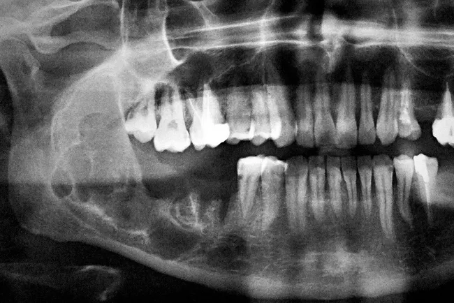
Imaging modality: dental panoramic radiography (Orthopantomogram, OPG) provides a single broad, two-dimensional view of the jaws and dentition. The lesion is located in the mandible, most likely in the posterior region, with a conspicuous radiolucent, multiloculated pattern described as a soap-bubble appearance. The borders are corticated but expanded, indicating benign but locally aggressive behavior. Teeth in the vicinity show displacement and possible root resorption; there are no radiopaque calcifications within the lesion. This radiographic phenotype is characteristic of ameloblastoma, an epithelial odontogenic tumor that comprises about 1% of jaw tumors and cysts. The differential diagnosis includes odontogenic keratocyst, central giant cell granuloma, odontogenic myxoma, and other multilocular jaw lesions. Clinically, ameloblastoma presents in young to middle-aged adults as slow-growing swelling with facial asymmetry, often without pain. Imaging assessment informs surgical planning, as complete en bloc resection or wide local excision is often required due to high recurrence rates if the lesion is incompletely removed. Correlate radiographic findings with histopathology for definitive diagnosis. The image is credited to Bulent Celasun, MD, Ankara, Turkey, with permission. This depiction reinforces radiographic hallmarks used in educational and diagnostic workflows. Biopsy confirmation remains essential to establish definitive histology and guide definitive management and long-term surveillance.
This orthopantomogram (panoramic X-ray) of the human mandible reveals a significant diagnostic finding in the right mandibular body extending toward the symphysis. The image displays a large, well-defined multilocular radiolucent lesion characterized by two distinct internal architectural patterns: a 'honeycomb' appearance, consisting of numerous small, uniform radiolucent compartments separated by thin radiopaque septa, and a 'soap bubble' pattern featuring larger, more irregular circular radiolucencies with thicker internal septations. The lesion demonstrates cortical expansion and involves a substantial portion of the vertical mandibular height, commonly associated with odontogenic tumors such as ameloblastoma or odontogenic keratocysts. The radiographic presentation is indicative of a slow-growing but locally aggressive intraosseous pathology. Educational focus includes the recognition of multilocular radiolucencies in oral and maxillofacial radiology and the differential diagnosis of mandibular cystic and neoplastic lesions based on internal trabeculation patterns.
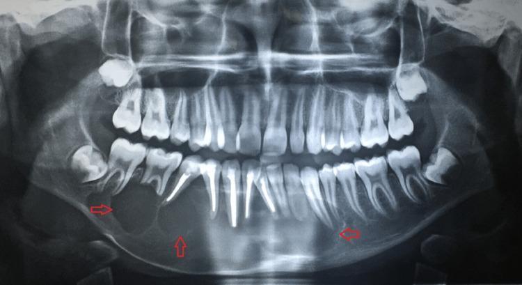
This diagnostic image is an orthopantomogram (OPG) or panoramic radiograph of the human mandible and maxilla. The central finding is a large, well-defined multilocular radiolucency with a characteristic 'soap bubble' appearance, prominently highlighted by red arrows. The lesion involves a significant portion of the mandibular body, extending bilaterally across the symphysis from approximately the lower left first premolar (tooth 34) to the lower right second molar (tooth 47). Notable secondary effects include the obliteration of root apices for teeth 31 through 34 and 41 through 46, and significant thinning of the inferior cortex of the mandible. Several anterior teeth exhibit endodontic filling material. The radiographic presentation is highly suggestive of an odontogenic tumor, such as an ameloblastoma. The image demonstrates key concepts in dental radiology, including multilocular lesion morphology, root resorption, and cortical thinning, serving as a critical educational example for oral and maxillofacial pathology.
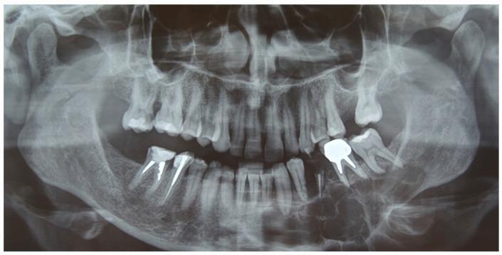
This diagnostic image is a preoperative orthopantomography (panoramic X-ray) of an adult human skull focusing on the dentomaxillofacial complex. The primary pathology is a large, well-defined multilocular radiolucency located in the left mandible. The lesion exhibits a 'soap bubble' or 'honeycomb' appearance characteristic of an ameloblastoma, extending from the left mandibular lateral incisor (tooth 3.2) posteriorly through the molar region (tooth 3.8) and into the ascending ramus. Significant cortical thinning and displacement of the surrounding dental roots are visible. Associated dental findings include several radiopaque endodontic treatments with intraradicular posts in the right mandibular premolars and a large radiopaque crown restoration in the left maxillary molar region. Anatomical landmarks such as the maxillary sinuses, nasal cavity, mandibular condyles, and temporomandibular joints are visible bilaterally. The image serves as a clinical example of odontogenic tumor presentation and is intended for intermediate to advanced oral and maxillofacial pathology education.
ameloblastoma histology follicular plexiform microscopy photomicrograph
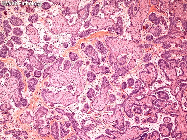
Histology: Light microscopy of a jaw lesion shows classic ameloblastoma morphology on hematoxylin and eosin stained tissue. The neoplasm is composed of multiple epithelial islands and cords embedded in a fibrous connective tissue stroma. Follicular pattern predominates, with nests of odontogenic epithelium displaying peripheral palisading of columnar cells and reversal of polarity, nuclei oriented away from the basement membrane. Central cells resemble stellate reticulum with loosely arranged, angular morphology. Some islands exhibit a loosely arranged, anastomosing arrangement compatible with plexiform architecture. Overall cellularity is moderate, with minimal pleomorphism and no overt mitotic activity in the field, consistent with benign but locally aggressive behavior. Basal layer cells are darkly basophilic with prominent nucleoli, while surrounding stroma is densely fibrous and occasionally myxoid, producing a subtle desmoplastic impression in portions of the section. The tumor borders appear infiltrative into adjacent connective tissue, reflecting its known propensity for local invasion despite benign cytology. There is an absence of keratinization, dysplasia, or metastatic features. The image highlights the diagnostic hallmarks: enamel organ–like islands, reverse polarity, stellate reticulum–like center, and a fibrous stroma. These histologic features underline confirmation of diagnosis and guide surgical planning in odontogenic tumors and jaw neoplasms. For educational and diagnostic purposes.
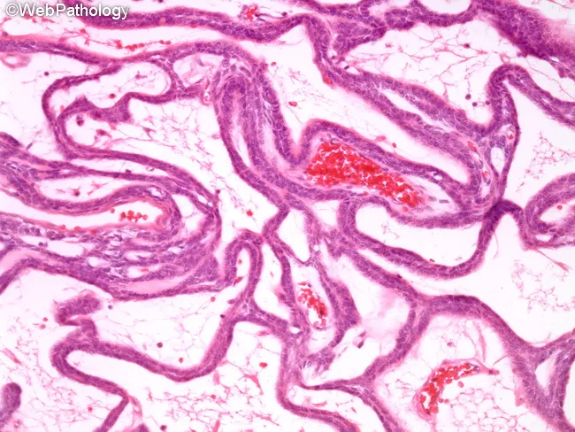
This histopathology slide, stained with Hematoxylin and Eosin and viewed under brightfield light microscopy at low to mid magnification, demonstrates classic plexiform ameloblastoma architecture within the jaw. Neoplastic odontogenic epithelium forms interconnected cords and anastomosing strands embedded in a loose, highly vascular stroma of fibrous connective tissue. The epithelial cords are composed of basaloid cells with peripheral cells often lacking the typical palisading and reverse nuclear polarity; within the cords, cells are more loosely arranged, resembling an inner stellate reticulum. Cystic change is uncommon in this plexiform subtype. The overall pattern shows benign cytology but a propensity for local invasion and bone destruction clinically. These features support the diagnosis of ameloblastoma, plexiform type, and help distinguish it from other odontogenic tumors and jaw cysts. Diagnostic significance includes confirming a locally aggressive, benign neoplasm that necessitates wide surgical excision with clear margins to minimize recurrence. Clinically, this pattern correlates with gradual cortical expansion and radiographic lytic lesions in the mandible or maxilla. In educational settings, the image is valuable for teaching odontogenic histology, subtyping ameloblastoma, and illustrating variations among follicular, plexiform, and desmoplastic patterns. Correlation with radiographs (multilocular radiolucencies) and clinical jaw swelling reinforces a multidisciplinary management approach.
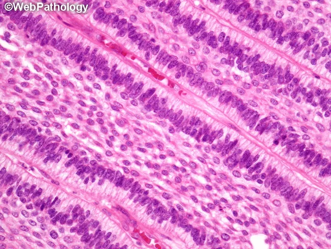
This histology image depicts ameloblastoma of the jaw, captured on a hematoxylin and eosin–stained biopsy section examined under light microscopy. The dominant pattern shown is follicular ameloblastoma, with oval to rounded islands of odontogenic epithelium embedded in a dense fibrous stroma. Peripheral columnar cells exhibit classic reverse polarity and peripheral palisading, while the central cells resemble the stellate reticulum of the developing tooth germ. In some regions, the epithelial nests are triangular or elongated and dispersed, consistent with alternate patterns that may coexist in the same tumor, including plexiform, acanthomatous, basaloid, granular cell, or desmoplastic variants. The nests are separated by a mature, hyalinized connective tissue stroma without prominent inflammatory infiltrate. The myxoid or hyalinized stroma and highly organized epithelial architecture help distinguish ameloblastoma from other odontogenic lesions. Clinically, these histologic features correlate with locally aggressive behavior despite benign cytology, underscoring the need for complete surgical excision with clean margins to minimize recurrence risk. The image emphasizes the diagnostically relevant hallmarks—pigmented? No, not pigmented; but rather cohesive islands with peripheral palisading and stellate reticulum core. This slide serves educational value for surgical planning, differential diagnosis, and radiologic-pathologic correlation in jaw lesions. It is suitable for teaching, research, and case documentation.
ameloblastoma surgical resection jaw reconstruction fibula free flap
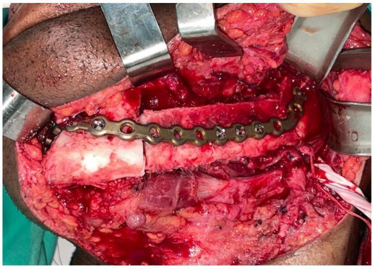
This intraoperative clinical photograph captures a surgical field during a complex maxillofacial reconstruction. The image shows a microvascularized fibula bone flap being used to reconstruct a mandibular defect following tumor resection for ameloblastoma. A contoured metallic reconstruction plate is secured across the osteotomized bone segments with multiple surgical screws, providing rigid internal fixation and maintaining the anatomical contour of the lower jaw. The surgical site demonstrates exposed skeletal structures, subcutaneous fat, and muscle tissue, with several metal retractors positioned to maintain exposure. A vascular pedicle is visible, essential for the survival of the free flap. This visual serves as an educational example of advanced reconstructive surgery, highlighting the application of rigid fixation plates in stabilizing bone grafts during mandibular continuity restoration.
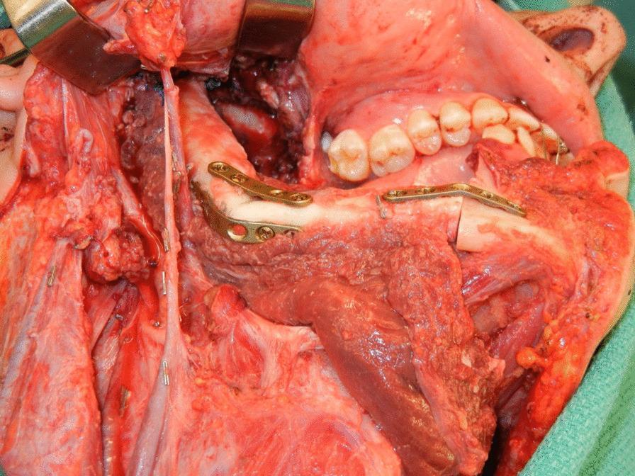
This clinical photograph captures an intraoperative view of a complex oral and maxillofacial reconstruction using a fibula free flap (FFF). The surgical site reveals a large mandibular defect being restored with a vascularized fibula bone graft. Key anatomical landmarks include the maxillary teeth and hard palate providing superior orientation. The transplanted fibula is contoured at its distal end to function as a neo-condyle, positioned within the native glenoid fossa to reconstruct the temporomandibular joint (TMJ). Rigorous internal fixation is achieved via metallic osteosynthesis plates and screws, securing the fibula segments to the remnant mandible. The image also demonstrates the soft tissue component of the flap and the associated vascular pedicle, essential for microvascular anastomosis. This procedure illustrates a standard approach for functional and aesthetic restoration following radical mandibular resection, highlighting the integration of the neo-condyle with the remnant joint capsule to maintain jaw mobility.
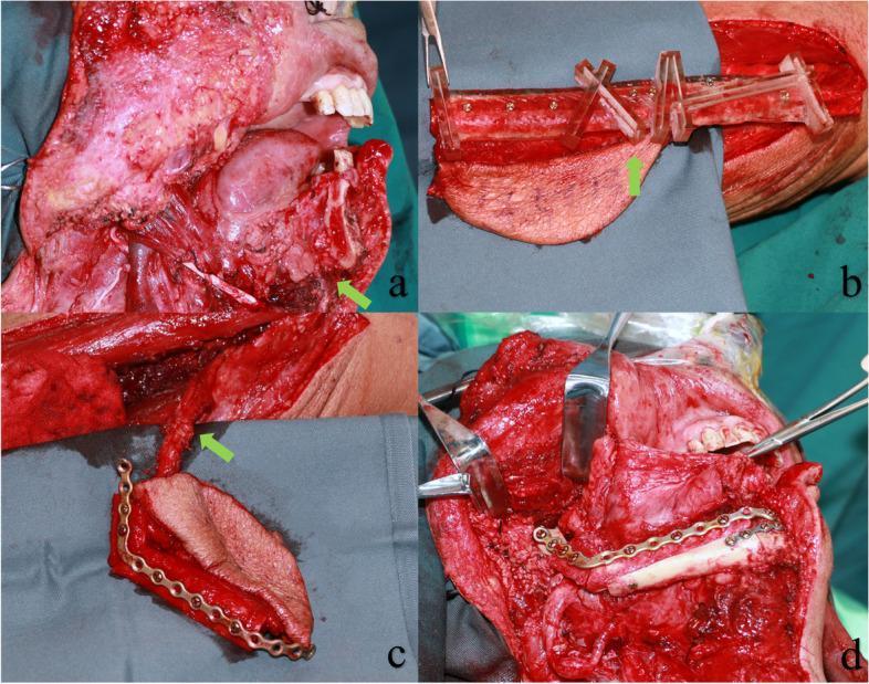
This composite of four clinical photographs illustrates a mandibular reconstruction using a free fibula flap guided by MOPITS (mandibular and osteotomy precision-improving template system). (a) Intraoperative view of a significant mandibular defect following tumor resection. (b) Preparation of the donor fibula segment; a clear resin surgical template is secured with screws to guide precise osteotomies and shaping of the bone. (c) The harvested fibular segment and associated skin paddle, featuring a pre-bent titanium reconstruction plate affixed to the bone to replicate the mandibular contour. The green arrow indicates the vascular pedicle containing the peroneal artery and veins for microsurgical anastomosis. (d) The final reconstruction stage, showing the vascularized fibular flap secured to the native mandibular remnants with the titanium plate and screws, restoring the anatomical continuity of the lower jaw. This surgical series demonstrates oral and maxillofacial reconstructive techniques, emphasizing the use of 3D-planned surgical guides for accuracy in free tissue transfer.
ameloblastoma resection recurrence treatment
unicystic ameloblastoma subtypes luminal intraluminal mural diagram classification
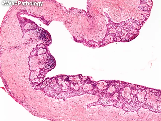
Histopathology slide of a jaw-based unicystic ameloblastoma, luminal subtype. Modality: light microscopy on hematoxylin and eosin-stained tissue, displaying a simple unilocular cyst with a fibrous wall. The cyst lining comprises ameloblastic epithelium with a tall columnar or cuboidal basal cell layer showing reverse nuclear polarity and peripheral palisading. The superficial cells toward the lumen resemble loosely cohesive stellate reticulum-like cells. The epithelium demonstrates luminal confinement, with no clear invasion into the cyst wall in this field. Intraluminal nodules or mural islands may be seen in other variants, but this specimen emphasizes luminal growth. Surrounding stroma is generally desmoplastic and collagenous without frank invasion by island in this view. Alternative terminology includes “odontogenic tumor” and “ameloblastoma variant” for educational clarity. Diagnostic significance: pattern supports a unicystic, luminal configuration with relatively favorable prognosis compared with multicystic forms, though recurrence risk persists if mural or intraluminal components are present. Relevant differential diagnoses include dentigerous cyst, inflammatory odontogenic cyst, and conventional solid/multicystic ameloblastoma. Clinically, histopathology guides surgical planning (enucleation vs resection) and informs prognosis; this image is valuable for oral pathology education, radiology-pathology correlation, and case-based learning in maxillofacial tumor pathology. This content supports learners in recognizing histologic patterns and correlating with radiographs globally.
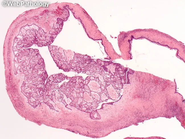
Histology: Light microscopy; Hematoxylin and Eosin (H&E) stained section of a unicystic ameloblastoma arising in the mandible. The specimen is a cystic lesion, typically associated with an unerupted third molar, showing a well circumscribed cavity lined by ameloblastomatous epithelium. The lining displays basal palisading of columnar cells with reversed nuclear polarity and an overlying zone resembling stellate reticulum. In many fields there are intraluminal or mural tumor nests within the cyst wall, representing the proliferative component. The surrounding stroma is usually fibrous and vascular, sometimes with mild inflammation. Clinically and radiographically, the lesion can mimic a dentigerous cyst, primordial cyst, radicular cyst, or residual cyst; histopathology confirms ameloblastic differentiation and classifies it as unicystic variant. Pathologic diagnosis has prognostic significance because involvement limited to the cyst lining (luminal or simple unicystic) has a favorable prognosis with conservative enucleation, while invasion of the cyst wall (mural unicystic) or solid nodules increases recurrence risk and may require more aggressive resection. Potential clinical uses include surgical planning, radiologic-pathologic correlation, and educational differentiation of odontogenic cystic lesions. This image exemplifies the diagnostic interface between histology and radiology in juvenile jaw lesions and odontogenic tumors.
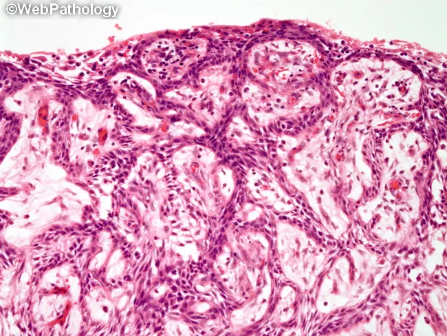
Modality: Brightfield light microscopy of hematoxylin and eosin stained histologic section from a jaw lesion. Specimen: cyst wall/tissue obtained from a presumed unicystic ameloblastoma of the mandible. Location: odontogenic tumor arising in the jaw; microscopic segment shows cystic architecture lined by ameloblastoma‑type epithelium with islands of tumor cells embedded in a fibrous stroma. The epithelial lining exhibits basal palisading of columnar cells with reverse polarity and a loosely formed stellate reticulum‑like center; in the mural variant, tumor islands infiltrate the fibrous cyst wall. The surrounding stroma is densely collagenous with scant inflammation; keratinization is minimal. The slide demonstrates classic features that overlap with odontogenic cysts but, in focal areas, demonstrates proliferating ameloblastomatous nests consistent with unicystic ameloblastoma. Clinically, unicystic ameloblastoma is often misdiagnosed as a dentigerous cyst; definitive diagnosis requires microscopic sampling and histopathologic subtyping into luminal, intraluminal, or mural variants. Prognosis and treatment depend on subtype; luminal and intraluminal lesions are typically managed by enucleation with close follow‑up, whereas mural invasion warrants more extensive resection due to higher recurrence (reported 10–20%, greatest risk with mural type). Long-term surveillance is essential given the potential for late recurrences and the need for radiographic monitoring.
Controversies in the Management of Ameloblastoma
Introduction
Classification (WHO 2022)
AMELOBLASTOMA
│
├── Conventional (Intraosseous) Ameloblastoma  ← most common, most controversial
│     Subtypes: Follicular, Plexiform, Acanthomatous,
│               Granular Cell, Desmoplastic, Basal Cell
│
├── Unicystic Ameloblastoma (~15%)
│     ├── Luminal
│     ├── Intraluminal
│     └── Mural (Type 3)  ← treated as conventional
│
├── Peripheral (Extraosseous) Ameloblastoma
│
└── Metastasising Ameloblastoma / Ameloblastic Carcinoma (rare)
Radiology
OPG: Classic "Soap Bubble" / Honeycomb Appearance
Histopathology
Follicular Pattern (Most Common)
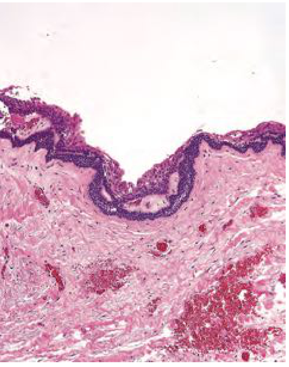
CONTROVERSY 1: Conservative vs. Radical Surgery for Conventional (Intraosseous) Ameloblastoma
Why the Tumour Demands Adequate Margins
- Infiltrates medullary bone trabeculae microscopically, well beyond radiographic margins
- Cannot infiltrate compact cortical bone well - the cortex and periosteum act as natural barriers
- When the periosteum is breached, access to vital structures (orbit, skull base, pterygoids) follows
Conservative Approach: Enucleation ± Curettage
- Avoids the major morbidity of jaw resection (functional loss, need for reconstruction)
- Preserves jaw continuity - especially important in children and young adults
- Some modern reports show acceptable outcomes with aggressive curettage + peripheral osteotomy (rotary bur)
- Widely used in sub-Saharan Africa, Southeast Asia, and resource-limited settings
- Recurrence rates of 55-90% with enucleation alone
- Uniform medullary bone removal cannot be assured - small tumour islands persist
- No intraoperative or postoperative margin assessment is possible
- Cummings states definitively: "Simple enucleation of ameloblastomas is not considered standard of care in the United States"
Radical Approach: En Bloc Resection
- Allows intraoperative radiographic assessment of margins on the specimen
- Allows postoperative histological margin confirmation (whole specimen demineralisation)
- Recurrence rates of 10-15% even after adequate resection
- Most predictable long-term outcome
- Major functional and cosmetic morbidity
- Requires complex reconstruction (free flaps, plates)
- Even "adequate" resection has a 10-15% recurrence rate, questioning if more margin helps
TREATMENT DECISION - CONVENTIONAL AMELOBLASTOMA

                    ┌─────────────────────────┐
                    │  Conventional Intraosseous │
                    │      Ameloblastoma        │
                    └────────────┬────────────┘
                                 │
           ┌─────────────────────┼─────────────────────┐
           │                     │                     │
    Mandibular              Mandibular            Maxillary
    Anterior/Body            Ramus/Posterior       (any)
           │                     │                     │
    En bloc resection     En bloc resection     En bloc resection
    1 cm medullary         1 cm medullary       1–2 cm / 2 biologic
    bone margins            bone margins        barriers past tumour
    ± immediate            ± immediate          Frozen section
    reconstruction        reconstruction        essential
CONTROVERSY 2: The Question of Margin Width
- No study has correlated presurgical and postsurgical radiographs with actual histological tumour extent in resected specimens
- Marx made initial attempts to accumulate such data but no well-documented study has been published
- Even after resection reportedly well past radiographic margins, recurrence rates of 10-15% are reported
- Some surgeons advocate larger margins; others argue the periosteum is the functional margin, not a measured centimetre of bone
CONTROVERSY 3: Unicystic Ameloblastoma - Definition and Management
The Definitional Problem
- Unilocular radiographically
- Unicystic histologically
- Ameloblastomatous lining of the lumen ONLY
- NO connective tissue (mural) wall invasion
- Including multilocular lesions as "cystic" or "cystogenic" (Cummings: "multilocularity increases the chance of recurrence and such lesions should be treated as standard ameloblastoma")
- Including mural invasion as "Type 3 unicystic" (Ackerman and Shear) - these must be treated as conventional ameloblastoma
Unicystic Subtype Classification and Treatment
UNICYSTIC AMELOBLASTOMA - SUBTYPE AND TREATMENT

  Luminal UA          Intraluminal UA         Mural UA (Type 3)
  ──────────────      ──────────────────      ──────────────────────
  Epithelium          Nodule projects          Tumour invades
  lines cyst          into lumen only          fibrous wall
  lumen only          (no wall invasion)
       │                    │                        │
       ▼                    ▼                        ▼
  Enucleation         Enucleation             Treat as CONVENTIONAL
  with clear          with clear              AMELOBLASTOMA
  margins             margins                 En bloc resection
  (conservative       (conservative           with 1 cm margins
  acceptable)         acceptable)
       │                    │                        │
  Recurrence ~10%     Recurrence ~10-20%      High recurrence
                                              if only enucleated
Lesion Size Controversy in UA
CONTROVERSY 4: Marsupialisation and Decompression as a Treatment Strategy
Proponents argue:
- Reduces tumour size in very large lesions, converting an extremely morbid resection into a smaller, more manageable operation
- Allows jaw growth to continue in paediatric patients
- Some centres use it as definitive treatment for luminal unicystic ameloblastoma
Critics argue:
- Does NOT eradicate the tumour - must always be followed by definitive surgery
- Marsupialisation causes inflammation, which Cummings notes "may disrupt the diagnostic features of unicystic ameloblastoma" - obscuring whether mural invasion is present
- The histopathological specimen after marsupialisation may be uninterpretable, risking under-treatment
- No RCT has established benefit of neoadjuvant marsupialisation vs. primary resection
CONTROVERSY 5: Mandibular vs. Maxillary Ameloblastoma
Mandibular Ameloblastoma
- Dense compact bone at inferior border and ramus acts as a natural first-line barrier
- Outer periosteum serves as a backup barrier - once breached, access to vital structures occurs
- Standard: 1 cm medullary bone margins (proximal and distal)
- The 1 cm margin refers to bone ONLY - it does NOT imply 1 cm of soft tissue outside the periosteum
- Overlying mucoperiostem, teeth, gingiva included in specimen; muscle attachment areas: periosteum is the margin
Maxillary Ameloblastoma
- Only a thin cortical plate separates adjacent compartments - a poor barrier compared to the mandible
- Posterior maxillary lesions can extend into orbit, pterygoid space, infratemporal fossa, and skull base
- Some authors recommend 1-2 cm margins; the rule of thumb is "two biologic barriers past the tumour"
- Frozen section assessment of soft tissue margins is essential
- Tumours near the orbit: complete oncological clearance may be impossible without sacrificing vision - this creates an irreducible tension between function preservation and tumour control
- Radiotherapy may play a role for unresectable maxillary lesions (not routine due to relative radio-resistance)
MANDIBLE vs MAXILLA: SURGICAL DIFFERENCES

        MANDIBULAR                    MAXILLARY
        ──────────────────────────────────────────────────
Barrier  Dense cortex + periosteum    Thin cortex only
Margin   1 cm medullary bone          1-2 cm / 2 biologic barriers
Assessment  Specimen radiograph       CT/MRI + frozen sections
Extension  Limited                    Orbit / skull base / pterygoids
Prognosis  Better                     Worse (harder to clear)
CONTROVERSY 6: Management of Recurrent Ameloblastoma
Surgical Re-resection
- Standard recommendation for recurrence after prior conservative treatment: escalate to resection
- Recurrence after prior resection: even more challenging - requires careful re-imaging (CT/MRI) to define extent
BRAF-Targeted Therapy
- BRAF inhibitors (vemurafenib, dabrafenib) have shown activity in BRAF V600E-mutant recurrent ameloblastoma in case series and small cohorts
- K.J. Lee recommends: "May consider recurrent ameloblastoma treatment with BRAF-targeted therapy with confirmed MAPK gene mutation in lesion"
- Core controversy: Can BRAF inhibitors serve as definitive treatment, or only as palliative/bridging therapy? No RCT data exist. Resistance is expected based on experience in melanoma.
- Molecular testing of the primary specimen is now increasingly recommended to guide future salvage options
BRAF/MAPK PATHWAY IN AMELOBLASTOMA

  BRAF V600E mutation (~90%) ──► ERK/MAPK activation
                                       │
                              Cell proliferation
                              Tumour growth
                                       │
                         BRAF inhibitors (vemurafenib)
                                       │
                              Tumour response
                         (recurrent/unresectable disease)
                                       │
                              Risk of resistance
                         (as seen in BRAF-mutant melanoma)
Radiotherapy
- Not routinely recommended - ameloblastoma has relative radio-resistance
- May be considered for unresectable recurrent disease, particularly maxillary lesions, as a palliative measure
CONTROVERSY 7: Timing and Method of Reconstruction
Immediate vs. Delayed Reconstruction
- Single-stage surgery
- Maintains facial contour and psychological benefit
- Fibula free flap: gold standard for mandibular reconstruction
- Ensures clear margins before committing to major reconstruction
- Allows monitoring for early recurrence before placing a large bony construct
- Still preferred in some centres due to resources or surgeon preference
CONTROVERSY 8: Peripheral Ameloblastoma - How Conservative is Safe?
- Treatment: Conservative local excision with histopathologically confirmed margins
- The controversy: If even the faintest intraosseous involvement is identified, the lesion is reclassified as intraosseous ameloblastoma and must be treated accordingly
CONTROVERSY 9: Malignant Ameloblastoma vs. Ameloblastic Carcinoma
| Feature | Malignant Ameloblastoma | Ameloblastic Carcinoma |
|---|---|---|
| Histology primary site | Benign-appearing | Cytologic atypia, mitoses, pleomorphism |
| Malignancy established by | Distant metastasis (lungs, nodes) | Cytopathology at primary site |
| Latency to metastasis | Years to decades after treatment | N/A |
| Treatment | Variable; excision preferred, RT if unresectable | Resection; data limited |
| Marker | - | SOX2 (potential carcinomatous transformation marker) |
CONTROVERSY 10: Follow-Up Duration and Protocol
- Recurrence rate ~22%; half within 5 years - but late recurrences (>10 years) are well documented
- Malignant transformation can occur (rare)
- No consensus on follow-up frequency, imaging modality (OPG vs. CT vs. MRI), or total duration
- Most authorities: lifelong follow-up with clinical and radiographic surveillance
- K.J. Lee: "Good with regular clinical and radiographic surveillance"
- Cummings: "Lifelong follow-up is recommended because, although it is rare, malignant transformation can occur"
Summary of All Controversies
| Controversy | Conservative View | Radical View | Current Position |
|---|---|---|---|
| Conventional ameloblastoma | Enucleation + curettage | Resection + 1 cm margin | Resection (USA/Europe standard) |
| Unicystic luminal/intraluminal | Enucleation acceptable | Resection | Enucleation with clear margins |
| Unicystic mural | Conservative possible | Treat as conventional | Treat as conventional (WHO 2022) |
| Margin width | Minimal | >1 cm | 1 cm past radiographic margin |
| Maxillary vs. mandibular | Same approach | 2 biologic barriers + frozen | Case-by-case; more aggressive in maxilla |
| Marsupialisation | Definitive option | Neoadjuvant only | Neoadjuvant or not at all |
| Reconstruction timing | Delayed | Immediate | Immediate (high-volume centres) |
| Recurrent disease | BRAF targeted therapy | Re-resection | Re-resection ± BRAF inhibitors if mutant |
| Follow-up | 5 years | Lifelong | Lifelong |
Conclusion
- Cummings Otolaryngology Head and Neck Surgery, Chapters 88 & 90
- K.J. Lee's Essential Otolaryngology, Chapter on Odontogenic Tumours
- Scott-Brown's Otorhinolaryngology Head & Neck Surgery
- Ghai S. Ameloblastoma: An Updated Narrative Review. Cureus. 2022 [PMID: 36127985]
- Chen C et al. Surgical Resection and Reconstruction of Ameloblastoma: A 13-Year Retrospective Review. J Oral Maxillofac Surg. 2024 [PMID: 38636548]
- Ouyang S et al. Risk Factors and Nomogram for Predicting Recurrence of Conventional Ameloblastoma. Oral Dis. 2025 [PMID: 40296385]