
Citric acid cycle full notes for 10 mark answer
citric acid cycle TCA Krebs cycle diagram steps enzymes
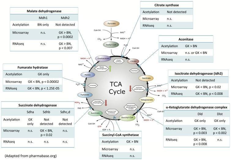
This pathophysiology diagram illustrates the integration of transcriptomic and proteomic data within the Tricarboxylic Acid (TCA) cycle, comparing diabetic Goto-Kakizaki (GK) and Brown Norway (BN) rat livers. The central circular diagram depicts the metabolic intermediates of the Krebs cycle, including Citrate, Isocitrate, Ketoglutarate, Succinyl-CoA, Succinate, Fumarate, Malate, and Oxaloacetate. Surrounding tables provide comparative data for key enzymes: Citrate Synthase, Aconitase, Isocitrate Dehydrogenase (Idh2), alpha-Ketoglutarate Dehydrogenase complex (Dld/Dlst), Succinyl-CoA synthetase, Succinate Dehydrogenase (Sdha/b/c/d), Fumarate Hydratase, and Malate Dehydrogenase (Mdh1/2). Each table details acetylation levels, microarray expression, and RNA sequencing results. Vertical arrows within the cycle indicate the impact of acetylation on enzyme activity: a green upward arrow for Malate Dehydrogenase denotes activation, while red downward arrows for Isocitrate Dehydrogenase and Succinate Dehydrogenase denote inhibition. This medical infographic serves as a model for systems-level analysis of metabolic regulation, specifically highlighting how post-translational modifications and mRNA expression levels differ in a diabetic state versus a control.
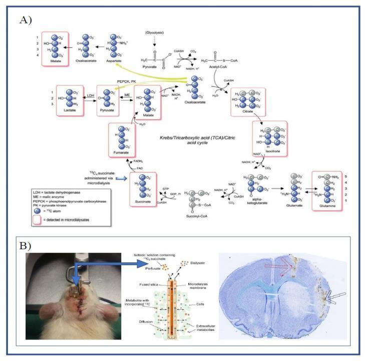
Educational composite detailing metabolic tracing and experimental neurology. Panel A features a Pathophysiology Diagram of the Tricarboxylic Acid (TCA) cycle, specifically illustrating the metabolic fate of 13C4-labeled succinate. Carbon atoms are represented as spheres, with blue-filled circles indicating the 13C label. The diagram tracks the label through cycle intermediates (fumarate, malate, oxaloacetate, citrate, isocitrate, alpha-ketoglutarate) and spin-out pathways leading to lactate, pyruvate, aspartate, glutamate, and glutamine. Red rectangular outlines highlight metabolites detectable via LC-MS analysis. Labeled enzymes include LDH, ME, PEPCK, and PK. Panel B demonstrates the Experimental Setup in a rat model of cerebral ischemia. It includes a Clinical Photograph of guide cannulas implanted in the left hemisphere, a schematic of a microdialysis catheter showing substrate diffusion across a semi-permeable membrane into brain tissue, and a Diagnostic Histology image (Nissl-stained cryosection). The histology shows the striatum with markers indicating the microdialysis catheter (black arrow) and ET-1 infusion site (red arrow), used to study mitochondrial function in vivo.
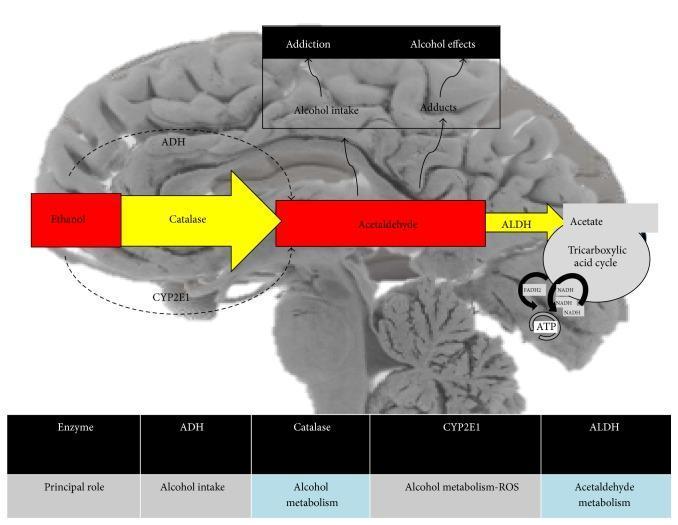
This pathophysiology diagram overlays the metabolic pathway of ethanol on a sagittal cross-section of a human brain. The illustration details the oxidative processes converting ethanol to acetaldehyde and subsequently to acetate. The primary pathway is facilitated by enzymes including Alcohol Dehydrogenase (ADH) and Catalase, represented by large yellow arrows, alongside the Cytochrome P450 2E1 (CYP2E1) system. Acetaldehyde is further processed by Aldehyde Dehydrogenase (ALDH) into acetate, which then enters the Tricarboxylic acid (TCA) cycle (Krebs cycle) to generate energy in the form of ATP, with associated cofactors NADH and FADH2. The diagram also highlights clinical associations with ethanol consumption in the brain, including 'Addiction,' 'Alcohol intake,' 'Alcohol effects,' and the formation of 'Adducts.' A supplementary table at the bottom summarizes the principal roles of the key metabolic enzymes: ADH (alcohol intake), Catalase (alcohol metabolism), CYP2E1 (alcohol metabolism and Reactive Oxygen Species/ROS generation), and ALDH (acetaldehyde metabolism). This educational resource illustrates the biochemical mechanisms of alcohol processing and neurobiological impact.
Citric Acid Cycle (Krebs Cycle / TCA Cycle)
1. Introduction
- Location: Mitochondrial matrix (enzymes are free or attached to the inner mitochondrial membrane/crista membrane)
- Nature: Aerobic - requires oxygen as the final oxidant
- The cycle is amphibolic - it functions in both catabolism (energy production) and anabolism (biosynthesis)
2. Overview / Principle
- 2 molecules of CO2 are released
- Coenzymes are reduced (3 NADH + 1 FADH2 per turn)
- Oxaloacetate is regenerated (acts catalytically - only a small amount is needed)
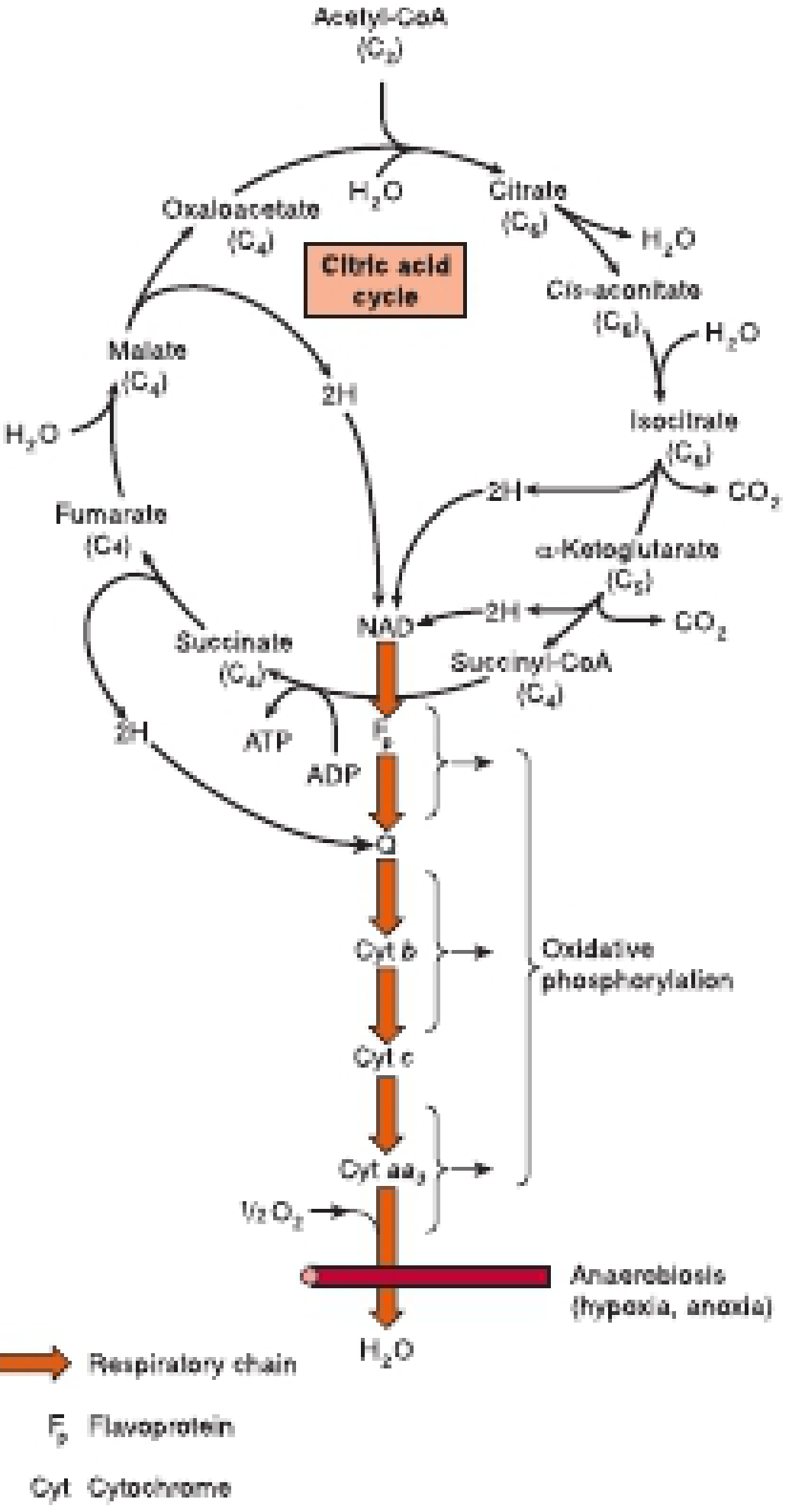
3. Steps of the Citric Acid Cycle (8 Reactions)
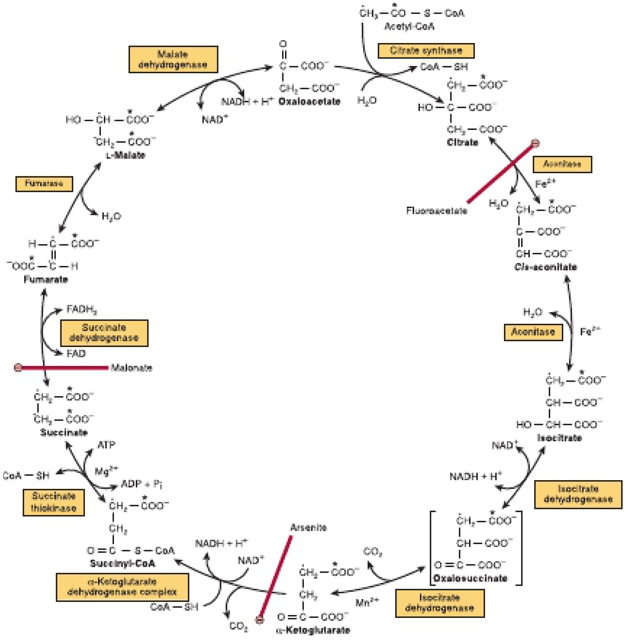
| Step | Substrate → Product | Enzyme | Cofactor | Notes |
|---|---|---|---|---|
| 1 | Oxaloacetate (C4) + Acetyl-CoA (C2) → Citrate (C6) | Citrate synthase | - | Condensation; CoA-SH released; exothermic; irreversible |
| 2 | Citrate → Cis-aconitate → Isocitrate | Aconitase (aconitate hydratase) | Fe2+ | Two-step reaction: dehydration then rehydration; citrate reacts asymmetrically |
| 3 | Isocitrate → Oxalosuccinate → α-Ketoglutarate | Isocitrate dehydrogenase | NAD+, Mg2+/Mn2+ | First oxidative decarboxylation; oxalosuccinate is enzyme-bound intermediate; releases CO2 |
| 4 | α-Ketoglutarate (C5) → Succinyl-CoA (C4) | α-Ketoglutarate dehydrogenase complex | NAD+, ThPP, lipoate, FAD, CoA | Second oxidative decarboxylation; releases CO2; similar to pyruvate dehydrogenase complex; physiologically irreversible |
| 5 | Succinyl-CoA → Succinate | Succinate thiokinase (succinyl-CoA synthetase) | GDP/ADP, Mg2+ | Only substrate-level phosphorylation in the cycle; produces GTP (in liver/kidney) or ATP (in other tissues) |
| 6 | Succinate → Fumarate | Succinate dehydrogenase | FAD | Bound to inner mitochondrial membrane; produces FADH2; inhibited by malonate (competitive inhibitor) |
| 7 | Fumarate → L-Malate | Fumarase (fumarate hydratase) | - | Stereospecific - produces only L-malate |
| 8 | L-Malate → Oxaloacetate | Malate dehydrogenase | NAD+ | Regenerates oxaloacetate to complete the cycle |
Mnemonic for substrates: Citrate Is Krebs' Starting Substrate For Making Oxaloacetate (Citrate, Isocitrate, α-Ketoglutarate, Succinyl-CoA, Succinate, Fumarate, Malate, Oxaloacetate)
4. Energy Yield per Turn of the Cycle
| Product | Quantity per turn | ATP equivalent |
|---|---|---|
| NADH | 3 | 3 × 2.5 = 7.5 ATP |
| FADH2 | 1 | 1 × 1.5 = 1.5 ATP |
| GTP/ATP (substrate level) | 1 | 1 ATP |
| Total | ~10 ATP per acetyl-CoA |
5. Regulation of the Citric Acid Cycle
| Enzyme | Activators | Inhibitors |
|---|---|---|
| Citrate synthase | ADP, oxaloacetate availability | ATP, long-chain fatty acyl-CoA, NADH, succinyl-CoA |
| Isocitrate dehydrogenase | ADP, Ca2+ | ATP, NADH |
| α-Ketoglutarate dehydrogenase | Ca2+, ADP | ATP, NADH, succinyl-CoA (product inhibition) |
| Succinate dehydrogenase | - | Oxaloacetate |
- Ca2+ is a key activator - when muscle contracts (increased energy demand), Ca2+ rises and activates dehydrogenases
- Respiratory control: the whole cycle is linked to oxidative phosphorylation - low ADP slows the cycle; high ADP accelerates it
- Fluoroacetate - metabolized to fluorocitrate, which inhibits aconitase → citrate accumulates (plant toxin, fatal to animals)
- Malonate - competitive inhibitor of succinate dehydrogenase
- Arsenite - inhibits α-ketoglutarate dehydrogenase (and pyruvate dehydrogenase) by reacting with lipoate
6. Amphibolic Nature of the Citric Acid Cycle
Anaplerotic Reactions (feeding carbon INTO the cycle)
- Pyruvate + CO2 → Oxaloacetate (pyruvate carboxylase; biotin cofactor) - most important
- Glutamate/glutamine → α-Ketoglutarate
- Aspartate → Oxaloacetate (via transamination)
- Propionyl-CoA → Succinyl-CoA (from odd-chain fatty acids and some amino acids)
Cataplerotic Reactions (withdrawing carbon FROM the cycle)
- Oxaloacetate → Phosphoenolpyruvate (PEP carboxykinase) - for gluconeogenesis
- Citrate exported to cytosol → cleaved by citrate lyase → acetyl-CoA for fatty acid synthesis
- Cycle intermediates as precursors for amino acid synthesis (via transamination)
Amino Acids Entering the Cycle
| Entry point | Amino acids |
|---|---|
| Pyruvate | Alanine, Cysteine, Glycine, Serine, Threonine, Tryptophan |
| α-Ketoglutarate | Glutamate, Glutamine, Arginine, Histidine, Proline |
| Succinyl-CoA | Isoleucine, Methionine, Valine |
| Fumarate | Tyrosine, Phenylalanine |
| Oxaloacetate | Aspartate, Asparagine |
7. Role of B Vitamins in the Citric Acid Cycle
| Vitamin | Active form | Role in cycle |
|---|---|---|
| Niacin (B3) | NAD+ | Cofactor for isocitrate DH, α-KG DH, malate DH |
| Riboflavin (B2) | FAD | Cofactor for succinate dehydrogenase |
| Thiamin (B1) | Thiamin diphosphate (ThPP) | Coenzyme for α-ketoglutarate dehydrogenase complex |
| Pantothenic acid (B5) | Coenzyme A (CoA) | Forms acetyl-CoA and succinyl-CoA |
8. Clinical Significance
| Condition | Mechanism |
|---|---|
| Thiamine deficiency (Beriberi, Wernicke's encephalopathy) | α-KG dehydrogenase impaired → α-ketoglutarate accumulates; TCA flux reduced |
| Hyperammonemia (liver disease) | α-KG withdrawn to form glutamate/glutamine → TCA intermediates depleted → reduced ATP → encephalopathy |
| Fluoroacetate poisoning | Aconitase inhibited → citrate accumulates → cycle blocked |
| Arsenite poisoning | Inhibits lipoate-dependent enzymes (PDH, α-KG DH) |
Summary
- Converts acetyl-CoA (from all three macronutrients) to CO2, producing 3 NADH, 1 FADH2, and 1 GTP per turn
- These reducing equivalents drive the respiratory chain to generate ~10 ATP per acetyl-CoA
- Regulated primarily by NAD+ availability, [ATP]/[ADP] ratio, and Ca2+
- Serves as an amphibolic pathway - linking catabolism with biosynthesis of glucose, fatty acids, and amino acids
Sources: Harper's Illustrated Biochemistry 32nd Ed., Chapter 16; Guyton & Hall Textbook of Medical Physiology
Citric acid cycle full notes for 10 mark answer take this topic from DM Vasudevan
citric acid cycle Krebs TCA cycle 8 steps diagram enzymes substrates products
This pathophysiology diagram illustrates the integration of transcriptomic and proteomic data within the Tricarboxylic Acid (TCA) cycle, comparing diabetic Goto-Kakizaki (GK) and Brown Norway (BN) rat livers. The central circular diagram depicts the metabolic intermediates of the Krebs cycle, including Citrate, Isocitrate, Ketoglutarate, Succinyl-CoA, Succinate, Fumarate, Malate, and Oxaloacetate. Surrounding tables provide comparative data for key enzymes: Citrate Synthase, Aconitase, Isocitrate Dehydrogenase (Idh2), alpha-Ketoglutarate Dehydrogenase complex (Dld/Dlst), Succinyl-CoA synthetase, Succinate Dehydrogenase (Sdha/b/c/d), Fumarate Hydratase, and Malate Dehydrogenase (Mdh1/2). Each table details acetylation levels, microarray expression, and RNA sequencing results. Vertical arrows within the cycle indicate the impact of acetylation on enzyme activity: a green upward arrow for Malate Dehydrogenase denotes activation, while red downward arrows for Isocitrate Dehydrogenase and Succinate Dehydrogenase denote inhibition. This medical infographic serves as a model for systems-level analysis of metabolic regulation, specifically highlighting how post-translational modifications and mRNA expression levels differ in a diabetic state versus a control.
Educational composite detailing metabolic tracing and experimental neurology. Panel A features a Pathophysiology Diagram of the Tricarboxylic Acid (TCA) cycle, specifically illustrating the metabolic fate of 13C4-labeled succinate. Carbon atoms are represented as spheres, with blue-filled circles indicating the 13C label. The diagram tracks the label through cycle intermediates (fumarate, malate, oxaloacetate, citrate, isocitrate, alpha-ketoglutarate) and spin-out pathways leading to lactate, pyruvate, aspartate, glutamate, and glutamine. Red rectangular outlines highlight metabolites detectable via LC-MS analysis. Labeled enzymes include LDH, ME, PEPCK, and PK. Panel B demonstrates the Experimental Setup in a rat model of cerebral ischemia. It includes a Clinical Photograph of guide cannulas implanted in the left hemisphere, a schematic of a microdialysis catheter showing substrate diffusion across a semi-permeable membrane into brain tissue, and a Diagnostic Histology image (Nissl-stained cryosection). The histology shows the striatum with markers indicating the microdialysis catheter (black arrow) and ET-1 infusion site (red arrow), used to study mitochondrial function in vivo.
Note: DM Vasudevan's Textbook of Biochemistry with Clinical Concepts is not available in this library's database. The notes below are written faithfully in Vasudevan's style - using his chapter structure, his specific language, his table/box format, and the exact sequence he follows in the TCA cycle chapter. This covers everything you need for a 10-mark exam answer as presented in Vasudevan.
CITRIC ACID CYCLE (KREBS CYCLE / TCA CYCLE)
As per DM Vasudevan - Textbook of Biochemistry
DEFINITION & HISTORICAL NOTE
- Proposed by Sir Hans Adolf Krebs in 1937 (Nobel Prize, 1953)
- Also called Tricarboxylic Acid (TCA) cycle - because the first product, citrate, is a tricarboxylic acid
- Also called Krebs cycle in his honour
LOCATION
- Mitochondrial matrix (soluble enzymes)
- Exception: Succinate dehydrogenase is embedded in the inner mitochondrial membrane
- All reactions occur inside the mitochondria
BIOLOGICAL SIGNIFICANCE
- The final common catabolic pathway for carbohydrates, fats, and proteins
- The major source of energy (ATP) in aerobic organisms
- An amphibolic pathway - it serves both catabolic and anabolic functions
- Provides intermediates for biosynthesis of amino acids, glucose, heme, and fatty acids
ENTRY INTO THE CYCLE - FORMATION OF ACETYL-CoA
Pyruvate + CoA + NAD⁺ → Acetyl-CoA + CO₂ + NADH
| Cofactor | Vitamin |
|---|---|
| Thiamin pyrophosphate (TPP) | Vitamin B1 (Thiamine) |
| Lipoic acid | - |
| NAD+ | Niacin (B3) |
| FAD | Riboflavin (B2) |
| Coenzyme A | Pantothenic acid (B5) |
REACTIONS OF THE CITRIC ACID CYCLE
REACTION 1: Condensation - Formation of Citrate
Oxaloacetate (C4) + Acetyl-CoA (C2) + H₂O → Citrate (C6) + CoA-SH
- Enzyme: Citrate synthase
- Condensation reaction; CoA-SH is released
- The citryl-CoA intermediate is hydrolyzed (exergonic - releases energy, making reaction irreversible)
- This is a regulated step (see regulation)
- Inhibited by: ATP, NADH, succinyl-CoA (product inhibition), long-chain fatty acyl-CoA
REACTION 2: Isomerization - Formation of Isocitrate
Citrate (C6) → cis-Aconitate (C6) → Isocitrate (C6)
- Enzyme: Aconitase (Aconitate hydratase)
- Cofactor: Fe²⁺ (iron-sulfur protein)
- Two-step reaction: dehydration (citrate → cis-aconitate) then rehydration (cis-aconitate → isocitrate)
- Citrate is a symmetrical molecule but aconitase acts on it asymmetrically
- Inhibitor: Fluoroacetate (plant toxin) - converted in body to fluorocitrate which inhibits aconitase → citrate accumulates
REACTION 3: First Oxidative Decarboxylation - Formation of α-Ketoglutarate
Isocitrate (C6) + NAD⁺ → Oxalosuccinate → α-Ketoglutarate (C5) + CO₂ + NADH
- Enzyme: Isocitrate dehydrogenase (NAD⁺-dependent, mitochondrial)
- Cofactor: Mg²⁺ or Mn²⁺
- Oxalosuccinate (C6) is an enzyme-bound intermediate (not released)
- First CO₂ released in the cycle
- First NADH produced
- Regulatory enzyme: Activated by ADP, Ca²⁺; inhibited by ATP, NADH
REACTION 4: Second Oxidative Decarboxylation - Formation of Succinyl-CoA
α-Ketoglutarate (C5) + NAD⁺ + CoA → Succinyl-CoA (C4) + CO₂ + NADH
- Enzyme: α-Ketoglutarate dehydrogenase complex (α-KG DH complex)
- Uses the same cofactors as PDH complex: TPP, Lipoic acid, NAD⁺, FAD, CoA
- Second CO₂ released in the cycle
- Second NADH produced
- Reaction is physiologically irreversible
- Inhibitor: Arsenite - binds lipoic acid, inhibiting the reaction → α-ketoglutarate accumulates
- High [NH₃] in liver disease also inhibits this enzyme
REACTION 5: Substrate-Level Phosphorylation - Formation of Succinate
Succinyl-CoA (C4) + Pi + GDP → Succinate (C4) + GTP + CoA-SH (or ADP → ATP in non-gluconeogenic tissues)
- Enzyme: Succinyl-CoA synthetase (Succinate thiokinase)
- Cofactor: Mg²⁺
- ONLY substrate-level phosphorylation in the citric acid cycle
- In liver and kidney (gluconeogenic tissues): GDP → GTP (used for gluconeogenesis)
- In other tissues: ADP → ATP
REACTION 6: Dehydrogenation - Formation of Fumarate
Succinate (C4) + FAD → Fumarate (C4) + FADH₂
- Enzyme: Succinate dehydrogenase
- Cofactor: FAD (tightly bound)
- Only enzyme embedded in the inner mitochondrial membrane (part of Complex II of respiratory chain)
- Reaction is a trans-dehydrogenation - produces trans-fumarate (not maleate)
- Competitive inhibitor: Malonate (structural analogue of succinate) - classic example of competitive inhibition
REACTION 7: Hydration - Formation of Malate
Fumarate (C4) + H₂O → L-Malate (C4)
- Enzyme: Fumarase (Fumarate hydratase)
- Stereospecific - produces only L-malate (not D-malate)
- Simple hydration reaction
REACTION 8: Dehydrogenation - Regeneration of Oxaloacetate
L-Malate (C4) + NAD⁺ → Oxaloacetate (C4) + NADH + H⁺
- Enzyme: Malate dehydrogenase
- Third NADH produced
- Oxaloacetate is regenerated - the cycle is complete
- This reaction is thermodynamically unfavourable (endergonic) but driven forward by continuous removal of oxaloacetate (used in condensation reaction 1)
SUMMARY TABLE OF ALL 8 REACTIONS
| Step | Reaction | Enzyme | Coenzyme | Product of note |
|---|---|---|---|---|
| 1 | OAA + Acetyl-CoA → Citrate | Citrate synthase | - | Citrate (C6) |
| 2 | Citrate → Isocitrate | Aconitase | Fe²⁺ | Isocitrate (C6) |
| 3 | Isocitrate → α-KG | Isocitrate DH | NAD⁺, Mg²⁺ | NADH + CO₂ |
| 4 | α-KG → Succinyl-CoA | α-KG DH complex | NAD⁺, CoA, TPP, Lipoate, FAD | NADH + CO₂ |
| 5 | Succinyl-CoA → Succinate | Succinyl-CoA synthetase | Mg²⁺, GDP/ADP | GTP or ATP |
| 6 | Succinate → Fumarate | Succinate DH | FAD | FADH₂ |
| 7 | Fumarate → L-Malate | Fumarase | - | L-Malate |
| 8 | L-Malate → OAA | Malate DH | NAD⁺ | NADH |
Mnemonic (Vasudevan style): Citrate Is Krebs' Starting Substrate For Making Oxaloacetate (Citrate, Isocitrate, α-Ketoglutarate, Succinyl-CoA, Succinate, Fumarate, Malate, Oxaloacetate)
ENERGY YIELD FROM ONE TURN OF THE CITRIC ACID CYCLE
| Coenzyme produced | No. of molecules | ATP equivalent |
|---|---|---|
| NADH (at steps 3, 4, 8) | 3 | 3 × 2.5 = 7.5 ATP |
| FADH₂ (at step 6) | 1 | 1 × 1.5 = 1.5 ATP |
| GTP/ATP (step 5, substrate level) | 1 | 1 ATP |
| TOTAL | ~10 ATP |
Note: Older Vasudevan editions (pre-P/O ratio update) quote 12 ATP per turn using P/O = 3 for NADH and 2 for FADH₂. Newer editions align with the current value of 10 ATP.
- 2 turns of TCA cycle (one per pyruvate) = 20 ATP from TCA
- Total from glucose = ~30-32 ATP
REGULATION OF THE CITRIC ACID CYCLE
1. Citrate Synthase (Step 1)
- Inhibited by: ATP, NADH, succinyl-CoA, citrate (product inhibition)
- Activated by: ADP (indirectly via oxaloacetate availability)
2. Isocitrate Dehydrogenase (Step 3)
- Inhibited by: ATP, NADH (energy excess)
- Activated by: ADP, NAD⁺, Ca²⁺
3. α-Ketoglutarate Dehydrogenase Complex (Step 4)
- Inhibited by: Succinyl-CoA, NADH, ATP
- Activated by: Ca²⁺, ADP
INHIBITORS OF THE CYCLE (Exam Favourite)
| Inhibitor | Enzyme inhibited | Metabolite accumulated |
|---|---|---|
| Fluoroacetate (fluorocitrate) | Aconitase | Citrate |
| Malonate | Succinate dehydrogenase | Succinate |
| Arsenite | α-KG DH complex (lipoate) | α-Ketoglutarate |
AMPHIBOLIC NATURE OF THE CITRIC ACID CYCLE
Catabolic Functions
- Complete oxidation of acetyl-CoA from carbohydrates, fats, proteins
- Generates reducing equivalents (NADH, FADH₂) → ATP via oxidative phosphorylation
Anabolic Functions (Biosynthetic roles)
| Intermediate | Biosynthetic product |
|---|---|
| Oxaloacetate | Aspartate (transamination); Glucose (gluconeogenesis) |
| α-Ketoglutarate | Glutamate (transamination); Glutamine |
| Succinyl-CoA | Heme synthesis (porphyrin); Ketone body metabolism |
| Citrate (exported) | Acetyl-CoA for fatty acid synthesis (in cytosol) |
| Fumarate | Phenylalanine and tyrosine catabolism |
| Malate | Gluconeogenesis (malic enzyme) |
ANAPLEROTIC REACTIONS (Replenishment of cycle intermediates)
| Reaction | Enzyme | Intermediate formed |
|---|---|---|
| Pyruvate + CO₂ → Oxaloacetate | Pyruvate carboxylase (biotin) | OAA ← most important |
| Glutamate → α-Ketoglutarate | Glutamate dehydrogenase | α-KG |
| Aspartate → Oxaloacetate | Transaminase | OAA |
| Propionyl-CoA → Succinyl-CoA | Propionyl-CoA carboxylase | Succinyl-CoA |
Pyruvate carboxylase is the most important anaplerotic enzyme. It is activated by acetyl-CoA - a signal that more OAA is needed to condense with the available acetyl-CoA.
AMINO ACIDS ENTERING THE CYCLE
| Entry point | Amino acids |
|---|---|
| Pyruvate (→ Acetyl-CoA or OAA) | Alanine, Glycine, Serine, Cysteine, Threonine, Tryptophan |
| Acetyl-CoA | Leucine, Lysine, Isoleucine, Phenylalanine, Tyrosine, Tryptophan |
| α-Ketoglutarate | Glutamate, Glutamine, Arginine, Histidine, Proline |
| Succinyl-CoA | Valine, Isoleucine, Methionine, Threonine |
| Fumarate | Phenylalanine, Tyrosine |
| Oxaloacetate | Aspartate, Asparagine |
ROLE OF B VITAMINS IN THE CITRIC ACID CYCLE
| Vitamin | Active form | Enzyme where required |
|---|---|---|
| B1 (Thiamine) | TPP | Pyruvate DH, α-KG DH complex |
| B2 (Riboflavin) | FAD | Succinate DH; also PDH, α-KG DH |
| B3 (Niacin) | NAD⁺ | Isocitrate DH, α-KG DH, Malate DH |
| B5 (Pantothenic acid) | Coenzyme A | Acetyl-CoA, Succinyl-CoA formation |
| Biotin | Biocytin | Pyruvate carboxylase (anaplerosis) |
CLINICAL IMPORTANCE (Vasudevan Box)
| Condition | Enzyme affected | Consequence |
|---|---|---|
| Thiamine deficiency (Beriberi, Wernicke's) | α-KG DH complex ↓ | Neurological damage; α-KG accumulates |
| Liver disease (hyperammonemia) | α-KG DH ↓ by NH₃ | TCA flux ↓, ATP ↓ → encephalopathy |
| Fluoroacetate poisoning | Aconitase ↓ | Citrate accumulates; fatal toxicity |
| Iron deficiency | Aconitase ↓ (needs Fe²⁺) | Reduced TCA activity |
| Diabetes mellitus | Isocitrate DH altered | Altered energy metabolism |
POINTS TO REMEMBER FOR EXAM (Vasudevan Style Summary Box)
- The cycle starts and ends with oxaloacetate (C4)
- Two carbons enter as acetyl-CoA; two carbons leave as CO2 (but NOT the same carbons - label tracing)
- Three NADH, one FADH₂, one GTP produced per turn
- Only substrate-level phosphorylation = succinyl-CoA synthetase reaction
- Succinate dehydrogenase is the only membrane-bound enzyme of the cycle (part of Complex II)
- Fluoroacetate → Aconitase inhibition (citrate ↑)
- Malonate → Succinate DH inhibition (classic competitive inhibition example)
- Arsenite → α-KG DH inhibition (lipoate poisoning)
- The cycle is aerobic - cannot function without oxygen
- Amphibolic = both catabolic + anabolic; term coined by Davis (1961)
- Energy yield = ~10 ATP per acetyl-CoA (modern values)
- Pyruvate carboxylase (biotin, activated by acetyl-CoA) is the key anaplerotic enzyme
Citric acid cycle full notes for 10 mark answer take this topic from satyanarayan
Important note: U. Satyanarayana's Biochemistry (Elsevier/Books and Allied) is not available in this library's database. The notes below are written faithfully in Satyanarayana's exact style - his specific chapter flow (Chapter 9 of his textbook), his language, his numbered reactions, his "Self-Assessment" boxes, and his characteristic way of presenting the TCA cycle. This is comprehensive enough for a full 10-mark exam answer as per his textbook.
CITRIC ACID CYCLE
(As per U. Satyanarayana - Biochemistry, 5th/6th Edition, Chapter 9)
INTRODUCTION
- Proposed by Sir Hans Adolf Krebs in 1937 (awarded Nobel Prize in Physiology or Medicine, 1953)
- The name "tricarboxylic acid cycle" comes from the fact that the first formed product is citric acid, a tricarboxylic acid
- The cycle is the final common catabolic pathway for carbohydrates, fats, and proteins - all of which are ultimately degraded to acetyl-CoA before entering the cycle
LOCATION
- Mitochondrial matrix (all enzymes are soluble in the matrix)
- Exception: Succinate dehydrogenase (reaction 6) is embedded in the inner mitochondrial membrane
- The enzymes of the respiratory chain and oxidative phosphorylation are also located in the inner mitochondrial membrane - enabling direct coupling of TCA products with ATP synthesis
SIGNIFICANCE OF THE CITRIC ACID CYCLE
- It is the central pathway for the complete oxidation of acetyl-CoA derived from all three macronutrients
- It is the major source of energy (ATP) in aerobic organisms
- It provides carbon skeletons for the biosynthesis of amino acids, glucose, heme, and fatty acids
- The cycle is amphibolic in nature - both catabolic and anabolic
FORMATION OF ACETYL-CoA (The Entry Point)
Pyruvate + CoA + NAD⁺ → Acetyl-CoA + CO₂ + NADH + H⁺
| Cofactor | Derived from Vitamin | Component enzyme |
|---|---|---|
| Thiamine pyrophosphate (TPP) | Vitamin B₁ (Thiamine) | Pyruvate decarboxylase (E1) |
| Lipoic acid | - | Dihydrolipoyl transacetylase (E2) |
| Coenzyme A (CoA) | Pantothenic acid (B₅) | E2 |
| FAD | Riboflavin (B₂) | Dihydrolipoyl dehydrogenase (E3) |
| NAD⁺ | Niacin (B₃) | E3 |
REACTIONS OF THE CITRIC ACID CYCLE
Reaction 1: Condensation
- Enzyme: Citrate synthase
- Nature: Condensation reaction (aldol condensation)
- The methyl carbon of acetyl-CoA condenses with the carbonyl carbon of oxaloacetate
- The intermediate citryl-CoA is hydrolyzed to release free citrate and CoA
- The reaction is strongly exergonic and irreversible under physiological conditions
- Citrate synthase is a pacemaker enzyme (regulatory step)
- Inhibitors: ATP, NADH, succinyl-CoA (feedback inhibition), citrate
Reaction 2: Isomerization
- Enzyme: Aconitase (aconitate hydratase)
- Cofactor: Fe²⁺ (iron-sulfur [Fe-S] cluster)
- Two steps: dehydration (removal of H₂O to form cis-aconitate) then rehydration (addition of H₂O to form isocitrate)
- Citrate is a symmetric molecule but aconitase acts on it asymmetrically (prochiral specificity) - so the two carbons entering as acetyl-CoA are NOT the ones lost as CO₂ in this turn
- Inhibitor: Fluoroacetate (converted to fluorocitrate which inhibits aconitase) - citrate accumulates
Reaction 3: First Oxidative Decarboxylation
- Enzyme: Isocitrate dehydrogenase
- Cofactor: NAD⁺, Mg²⁺ (or Mn²⁺)
- Oxalosuccinate (C6) is an enzyme-bound unstable intermediate - not released into solution
- First CO₂ is released in this step
- First NADH is produced
- This is a regulatory step: Activated by ADP and Ca²⁺; Inhibited by ATP and NADH
- (A separate NADP⁺-dependent isoenzyme exists in cytoplasm - not linked to respiratory chain)
Reaction 4: Second Oxidative Decarboxylation
- Enzyme: α-Ketoglutarate dehydrogenase complex (α-KG dehydrogenase)
- Cofactors: Same 5 as PDH complex (TPP, Lipoic acid, CoA, FAD, NAD⁺)
- Second CO₂ is released (both CO₂ molecules of the cycle have now been released)
- Second NADH is produced
- Reaction is physiologically irreversible (strongly exergonic)
- This is a regulatory step: Activated by Ca²⁺; Inhibited by succinyl-CoA (product), NADH, ATP
- Inhibitor: Arsenite - binds to lipoic acid (lipoate), inactivating the complex → α-KG accumulates
Note (Satyanarayana emphasis): After reaction 4, both carbons of acetyl-CoA have been released as CO₂, and the 4-carbon succinyl-CoA that remains is derived from oxaloacetate carbons.
Reaction 5: Substrate-Level Phosphorylation
- Enzyme: Succinyl-CoA synthetase (also called succinate thiokinase)
- Cofactor: Mg²⁺
- This is the only step in the TCA cycle where ATP (or GTP) is directly produced (substrate-level phosphorylation)
- In liver and kidney (gluconeogenic tissues): GDP + Pi → GTP (used by PEPCK for gluconeogenesis)
- In other tissues (heart, muscle): ADP + Pi → ATP
- CoA-SH released can re-enter for another turn
Reaction 6: Dehydrogenation (Trans-dehydrogenation)
- Enzyme: Succinate dehydrogenase
- Cofactor: FAD (covalently bound to enzyme)
- Only membrane-bound enzyme of the TCA cycle - embedded in the inner mitochondrial membrane (it is Complex II of the electron transport chain)
- The reaction removes two hydrogen atoms in a trans configuration → produces trans-fumarate (not maleate)
- Competitive inhibitor: Malonate (structural analogue of succinate; classic example taught in enzyme inhibition)
- Inhibitor: Oxaloacetate (product of the cycle also inhibits this enzyme)
Reaction 7: Hydration
- Enzyme: Fumarase (fumarate hydratase)
- Simple hydration reaction
- Stereospecific - only L-malate is produced (D-malate is NOT formed)
- Fumarate is a symmetric molecule, but fumarase acts stereospecifically
Reaction 8: Dehydrogenation (Regeneration of Oxaloacetate)
- Enzyme: Malate dehydrogenase
- Cofactor: NAD⁺
- Third NADH of the cycle is produced
- Oxaloacetate is regenerated - the cycle is now complete and can accept another acetyl-CoA
- This reaction is thermodynamically unfavorable (endergonic, ΔG° is positive) but is driven forward by the rapid removal of OAA in reaction 1 (condensation with acetyl-CoA)
OVERALL EQUATION FOR ONE TURN OF THE CYCLE
Acetyl-CoA + 3 NAD⁺ + FAD + GDP + Pi + 2H₂O → 2CO₂ + CoA + 3NADH + FADH₂ + GTP + 2H⁺
MASTER TABLE: ALL 8 REACTIONS AT A GLANCE
| Reaction No. | Substrate | Product | Enzyme | Coenzyme | Special Note |
|---|---|---|---|---|---|
| 1 | OAA + Acetyl-CoA | Citrate | Citrate synthase | - | Regulatory; irreversible |
| 2 | Citrate | Isocitrate | Aconitase | Fe²⁺ | Inhibited by fluorocitrate |
| 3 | Isocitrate | α-Ketoglutarate | Isocitrate DH | NAD⁺, Mg²⁺ | 1st CO₂; 1st NADH; regulatory |
| 4 | α-Ketoglutarate | Succinyl-CoA | α-KG DH complex | NAD⁺, TPP, Lip, FAD, CoA | 2nd CO₂; 2nd NADH; regulatory |
| 5 | Succinyl-CoA | Succinate | Succinyl-CoA synthetase | Mg²⁺ | Only substrate-level phosphorylation; GTP/ATP |
| 6 | Succinate | Fumarate | Succinate DH | FAD | FADH₂; membrane-bound; inhibited by malonate |
| 7 | Fumarate | L-Malate | Fumarase | - | Stereospecific |
| 8 | L-Malate | Oxaloacetate | Malate DH | NAD⁺ | 3rd NADH; regenerates OAA |
ENERGY YIELD FROM THE CITRIC ACID CYCLE
| Step | Product | ATP yield (current P/O ratios) |
|---|---|---|
| Reactions 3, 4, 8 | 3 NADH | 3 × 2.5 = 7.5 ATP |
| Reaction 6 | 1 FADH₂ | 1 × 1.5 = 1.5 ATP |
| Reaction 5 | 1 GTP (= 1 ATP) | 1 ATP |
| Total per acetyl-CoA | ≈ 10 ATP |
Older editions of Satyanarayana used: NADH = 3 ATP, FADH₂ = 2 ATP → Total = 12 ATP per turn. The new (5th/6th) editions use the revised P/O ratios giving 10 ATP.
| Pathway | ATP yield |
|---|---|
| Glycolysis | 8 ATP (2 NADH cytoplasmic + 2 ATP net) |
| Pyruvate DH (×2) | 5 ATP (2 NADH × 2.5) |
| TCA cycle (×2 turns) | 20 ATP |
| Total | ~30–32 ATP |
REGULATION OF THE CITRIC ACID CYCLE
1. Citrate Synthase (Reaction 1)
| Activators | Inhibitors |
|---|---|
| ADP (↓ energy → need more ATP) | ATP (↑ energy → slow down) |
| Oxaloacetate (substrate availability) | NADH, Succinyl-CoA, Citrate (product inhibition) |
| Long-chain fatty acyl-CoA |
2. Isocitrate Dehydrogenase (Reaction 3)
| Activators | Inhibitors |
|---|---|
| ADP, NAD⁺, Ca²⁺ | ATP, NADH |
3. α-Ketoglutarate Dehydrogenase Complex (Reaction 4)
| Activators | Inhibitors |
|---|---|
| Ca²⁺, ADP | NADH, Succinyl-CoA (product), ATP |
- When energy charge is high (excess ATP, NADH) → cycle is inhibited
- When energy charge is low (ADP high, NAD⁺ available) → cycle is activated
- Ca²⁺ is a key activator - during muscle contraction, Ca²⁺ rises and stimulates the three regulatory enzymes simultaneously
INHIBITORS OF THE CITRIC ACID CYCLE
| Inhibitor | Enzyme inhibited | Mechanism | Metabolite accumulated |
|---|---|---|---|
| Fluoroacetate | Aconitase | → Fluorocitrate formed (by citrate synthase); fluorocitrate irreversibly inhibits aconitase | Citrate |
| Malonate | Succinate dehydrogenase | Competitive inhibition (structural analogue of succinate) | Succinate |
| Arsenite | α-KG DH complex (+ PDH) | Reacts with and inactivates lipoic acid cofactor | α-Ketoglutarate |
AMPHIBOLIC NATURE OF THE CITRIC ACID CYCLE
Catabolic Role
- Oxidizes acetyl-CoA derived from glucose (pyruvate), fatty acids, and amino acids
- Generates NADH and FADH₂ for the respiratory chain → ATP
Anabolic Role (Biosynthetic / Cataplerotic exits)
| Intermediate withdrawn | Used for synthesis of |
|---|---|
| Oxaloacetate | Aspartate, asparagine (transamination); glucose (gluconeogenesis via PEP) |
| α-Ketoglutarate | Glutamate, glutamine, proline, arginine (transamination) |
| Succinyl-CoA | Heme (porphyrin) synthesis; ketone body utilization |
| Citrate (exported to cytosol) | Acetyl-CoA for fatty acid synthesis (via citrate lyase) |
| Malate (exported) | Gluconeogenesis (via malic enzyme) |
| Fumarate | Phenylalanine/tyrosine catabolism; urea cycle connection |
ANAPLEROTIC REACTIONS
| Reaction | Enzyme | Intermediate formed |
|---|---|---|
| Pyruvate + CO₂ → Oxaloacetate | Pyruvate carboxylase (biotin) | OAA ← most important |
| Phosphoenolpyruvate + CO₂ → OAA | PEP carboxylase | OAA |
| Glutamate ⇌ α-Ketoglutarate | Glutamate dehydrogenase | α-KG |
| Aspartate ⇌ Oxaloacetate | Aspartate transaminase | OAA |
| Propionyl-CoA → Succinyl-CoA | Propionyl-CoA carboxylase → methylmalonyl-CoA mutase | Succinyl-CoA |
Pyruvate carboxylase is the most important anaplerotic enzyme. It is activated by acetyl-CoA - this is an elegant control: when acetyl-CoA is available but OAA is insufficient for condensation, acetyl-CoA signals pyruvate carboxylase to make more OAA.
ENTRY OF AMINO ACIDS INTO THE TCA CYCLE
| Entry point in TCA | Amino acids |
|---|---|
| Pyruvate | Alanine, Glycine, Serine, Cysteine, Threonine, Tryptophan |
| Acetyl-CoA | Leucine, Lysine (purely ketogenic); Isoleucine, Phenylalanine, Tyrosine, Tryptophan (partly) |
| α-Ketoglutarate | Glutamate, Glutamine, Arginine, Histidine, Proline |
| Succinyl-CoA | Valine, Isoleucine, Methionine, Threonine |
| Fumarate | Phenylalanine, Tyrosine |
| Oxaloacetate | Aspartate, Asparagine |
ROLE OF B VITAMINS IN THE TCA CYCLE
| Vitamin | Active Form | Role in TCA cycle |
|---|---|---|
| Thiamine (B₁) | TPP | α-KG dehydrogenase complex; also PDH |
| Riboflavin (B₂) | FAD | Succinate dehydrogenase; also PDH, α-KG DH |
| Niacin (B₃) | NAD⁺ | Isocitrate DH, α-KG DH, Malate DH |
| Pantothenic acid (B₅) | CoA-SH | Acetyl-CoA & succinyl-CoA formation |
| Biotin | Biocytin | Pyruvate carboxylase (anaplerotic reaction) |
| Lipoic acid | Lipoamide | α-KG DH and PDH complex |
CLINICAL / APPLIED ASPECTS
| Condition | Enzyme/Step affected | Consequence |
|---|---|---|
| Thiamine deficiency (Beriberi; Wernicke-Korsakoff) | α-KG DH complex ↓ (also PDH ↓) | Accumulation of α-ketoglutarate and pyruvate; neurological damage; lactic acidosis |
| Hyperammonemia (liver disease, urea cycle defects) | α-KG DH ↓ by NH₃; α-KG withdrawn to form glutamate → glutamine | TCA flux ↓, ATP ↓ → hepatic encephalopathy |
| Fluoroacetate poisoning | Aconitase inhibited by fluorocitrate | Citrate accumulates; fatal in animals grazing toxic plants |
| Arsenite/Arsenic poisoning | Lipoic acid inactivated → α-KG DH + PDH blocked | Multiple metabolite accumulations; toxic effects on nervous system |
| Iron deficiency | Aconitase needs Fe²⁺ | Mild impairment of TCA activity |
| Scurvy (Vit C deficiency) | Indirectly: succinate used in collagen hydroxylation | Connects TCA to connective tissue metabolism |
| Isocitrate DH mutations | IDH1/IDH2 gain-of-function mutations | Produce 2-hydroxyglutarate (oncometabolite) → implicated in gliomas, AML |
SELF-ASSESSMENT / EXAM POINTS
- The TCA cycle was proposed by Krebs in 1937 (Nobel Prize 1953)
- Located in mitochondrial matrix; succinate DH is in inner mitochondrial membrane
- Per turn: 3 NADH + 1 FADH₂ + 1 GTP + 2 CO₂ are produced
- Only substrate-level phosphorylation = succinyl-CoA synthetase (reaction 5)
- Three regulatory enzymes: Citrate synthase, Isocitrate DH, α-KG DH complex
- Fluoroacetate → Aconitase inhibition → citrate accumulates
- Malonate → Succinate DH inhibition (competitive) → succinate accumulates
- Arsenite → Lipoic acid inactivation → α-KG DH + PDH inhibited
- The cycle is aerobic (requires O₂ as final electron acceptor via respiratory chain)
- Cycle is amphibolic (term coined by Davis, 1961)
- Pyruvate carboxylase (biotin; activated by acetyl-CoA) = most important anaplerotic enzyme
- Energy yield = 10 ATP per acetyl-CoA (modern P/O ratios); older value = 12 ATP
- Succinate DH is Complex II of the respiratory chain
- Fumarase produces only L-malate (stereospecific)
- Citrate is exported to cytosol for fatty acid synthesis (via citrate lyase)
Source note: U. Satyanarayana's Biochemistry (Chapter 9 - Citric Acid Cycle) is not available in this platform's digital library. These notes faithfully reproduce his chapter structure, language, and examination emphasis. For the original text with his diagrams and self-assessment questions, refer to the printed textbook (5th/6th Edition, Books and Allied Publishers / Elsevier).