
Diagram representing pain sensitive areas in br ain
Reading File
The brain is largely pain-insensitive, but certain structures are pain-sensitive. Let me search the medical library for diagrams and detailed content on this topic.
Searching sources...
Finding Sources
Finding Sources
The best sources are Guyton & Hall and Adams & Victor's. Let me read the detailed content and look for diagrams.
Reading File
Reading File
Reading File
I found a diagram in Guyton & Hall (Fig. 49.9). Let me fetch it.
Loading Image
Here is the diagram and a complete explanation of pain-sensitive areas in the brain/cranium.
Pain-Sensitive Areas in the Brain
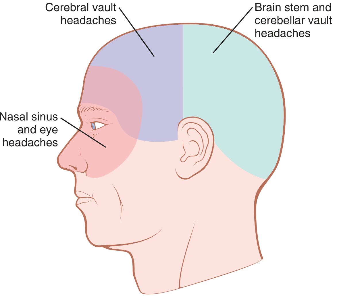
Figure 49.9 - Areas of referred headache from different intracranial structures (Guyton and Hall Textbook of Medical Physiology)
Key Concept: The Brain Parenchyma Is Insensitive to Pain
The brain tissue itself (parenchyma, ependyma, choroid plexuses, and most of the pia-arachnoid) is almost entirely insensitive to pain. Even cutting or electrically stimulating the sensory cortex produces only paresthesias, not pain. Intracranial mass lesions cause headache by deforming, displacing, or exerting traction on sensitive structures - not by directly stimulating brain tissue.
Pain-Sensitive Structures in the Cranial Vault
According to classic neurosurgical observations (Ray and Wolff) cited in Adams and Victor's Principles of Neurology and Guyton & Hall:
| Structure | Pain Sensitivity |
|---|---|
| Skin, subcutaneous tissue, muscles, extracranial arteries, periosteum of skull | Sensitive |
| Eye, ear, nasal cavities, paranasal sinuses | Sensitive |
| Intracranial venous sinuses (sagittal, transverse) and their large tributaries | Sensitive |
| Dura at the base of the brain | Sensitive |
| Proximal portions of the anterior and middle cerebral arteries | Sensitive |
| Intracranial segment of the internal carotid artery | Sensitive |
| Middle meningeal artery (especially sensitive - anesthetized separately in awake craniotomies) | Sensitive |
| Superficial temporal artery | Sensitive |
| First 3 cervical nerves and cranial nerves as they pierce the dura | Sensitive |
| Brain parenchyma | Insensitive |
| Most of pia-arachnoid | Insensitive |
| Ependyma and choroid plexuses | Insensitive |
How Pain Is Referred (as shown in the diagram)
The diagram shows three color-coded zones of referred headache:
-
Cerebral vault headaches (purple zone - frontal/parietal): Pain from structures above the tentorium cerebelli (supratentorial). Impulses travel via the trigeminal nerve (CN V, especially V1 and V2) - referred to the front half of the head.
-
Brain stem and cerebellar vault headaches (teal zone - occipital): Pain from structures below the tentorium (infratentorial/posterior fossa). Impulses travel via the glossopharyngeal (CN IX), vagus (CN X), and upper cervical nerves (C1-C3) - referred to the occiput and posterior head.
-
Nasal sinus and eye headaches (pink/red zone - periorbital/frontal): Pain from the nasoorbital region, transmitted by the sphenopalatine branches of the facial nerve (CN VII).
Clinical Relevance
- Meningitis: Inflammation of the meninges (pain-sensitive) causes severe generalized headache.
- Low CSF pressure (e.g., post-lumbar puncture): The brain sags, stretching pain-sensitive dural and vascular coverings.
- Intracranial hemorrhage/hematoma: Stretches and tears pain-sensitive meninges and vessels.
- Brain tumors: Cause headache by traction on vessels and dura - not from direct tumor-on-brain-tissue pressure.
Sources: Guyton and Hall Textbook of Medical Physiology, p. 49 (Headache of Intracranial Origin); Adams and Victor's Principles of Neurology, 12th Ed., p. 186 (Pain-Sensitive Cranial Structures)
This is a shared conversation. Sign in to Orris to start your own chat.