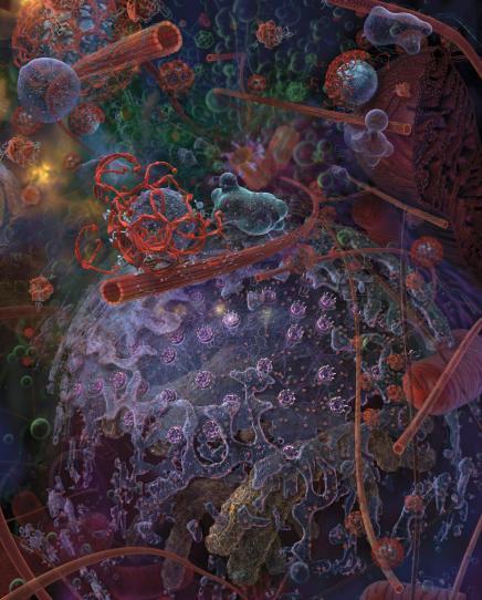
cell structure and differentiation with images
cell structure organelles diagram
stem cell differentiation lineage hematopoiesis
cell membrane lipid bilayer proteins structure
Cell Structure and Differentiation
1. The Cell: An Overview
2. The Cell Membrane (Plasma Membrane)
- 55% proteins
- 25% phospholipids
- 13% cholesterol
- 4% other lipids
- 3% carbohydrates
Lipid Bilayer
| Lipid | Role |
|---|---|
| Phospholipids | Most abundant; hydrophilic phosphate head faces outward, hydrophobic fatty acid tails face inward |
| Sphingolipids | Protection from harmful factors, signal transmission, adhesion |
| Cholesterol | Controls fluidity and permeability of the bilayer |
Membrane Proteins
3. Membranous Organelles
Endoplasmic Reticulum (ER)
- Rough (Granular) ER: Studded with ribosomes on its outer surface. Ribosomes (composed of RNA and proteins) synthesize new protein molecules. The rough ER is directly continuous with the nuclear membrane, allowing efficient information exchange.
- Smooth (Agranular) ER: Lacks attached ribosomes. Functions in synthesis of lipid substances, detoxification of drugs/poisons, and other intrareticular enzyme processes.
Golgi Apparatus
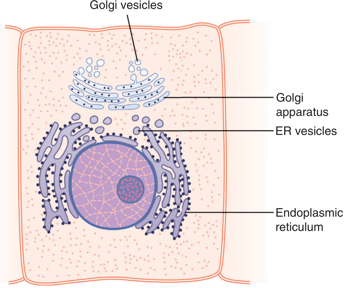
- Processes, packages, and routes proteins for export outside the cell
- Forms lysosomes and secretory vesicles
Lysosomes
- Damaged cellular structures
- Ingested food particles
- Unwanted matter (bacteria, viruses)
Mitochondria
Peroxisomes
4. The Nucleus
- Chromatin: DNA wound around histone proteins (nucleosomes). Condensed chromatin (heterochromatin) is transcriptionally silent; open chromatin (euchromatin) is active.
- Nucleolus: Site of ribosomal RNA synthesis and ribosome assembly.
- Nuclear envelope: Double membrane perforated by nuclear pores that regulate transport of RNA and proteins.
5. The Cytoskeleton
| Component | Diameter | Functions |
|---|---|---|
| Microtubules | ~25 nm | Cell shape, intracellular transport, mitotic spindle |
| Microfilaments (Actin) | ~6 nm | Cell motility, cytokinesis, muscle contraction |
| Intermediate Filaments | ~10 nm | Mechanical strength, nuclear lamina |
6. Transport Across the Cell Membrane
- Diffusion (passive): Fat-soluble substances (O₂, CO₂, alcohol) pass freely through the bilayer. Water-soluble ions and molecules require channels.
- Active transport: Protein pumps carry substances against concentration gradients, requiring ATP.
- Endocytosis:
- Pinocytosis: Ingestion of minute fluid particles and macromolecules via vesicle formation (coated pits with clathrin); requires ATP and Ca²⁺.
- Phagocytosis: Ingestion of large particles (bacteria, dead cells) by macrophages and white blood cells; opsonization by antibodies facilitates this process.
- Exocytosis: Secretory vesicles fuse with the cell membrane to release contents.
7. Cell Differentiation
Definition
"Most cells of the fetus undergo a specialization process called differentiation in which they predominantly express sets of genes that mediate specific cytoplasmic activities, becoming efficiently organized in tissues with specialized functions and usually changing their shape accordingly." — Junqueira's Basic Histology, 17e
Mechanism: Selective Gene Repression
Embryological Induction
- The primordial chordamesoderm (primary organizer) forms a mesodermal axis with segmentally arranged somites that induces formation of essentially all organs.
- Developing eye vesicles contact head ectoderm → ectoderm thickens into a lens plate → folds inward to form the lens of the eye.
Specialized Cell Types by Function
| General Cellular Activity | Specialized Cell(s) |
|---|---|
| Movement/contraction | Muscle cells |
| Form tight junctions | Epithelial cells |
| Synthesize extracellular matrix | Fibroblasts, osteoblasts, chondrocytes |
| Convert stimuli to action potentials | Sensory neurons |
| Phagocytosis | Macrophages, neutrophils |
| Hormone secretion | Endocrine cells |
8. Hematopoietic Cell Differentiation (Classic Example)
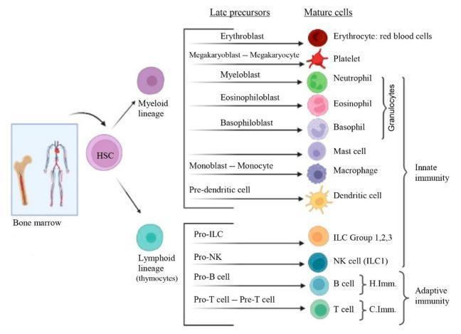
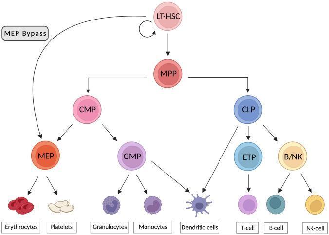
- MPP (Multipotent Progenitor) →
- CMP (Common Myeloid Progenitor) → MEP → Erythrocytes, Platelets; GMP → Granulocytes, Monocytes, Dendritic cells
- CLP (Common Lymphoid Progenitor) → T cells, B cells, NK cells
9. Apoptosis: Programmed Cell Death
- Is initiated by activation of caspases (proteases stored as inactive procaspases)
- Causes cell shrinkage, cytoskeletal disassembly, and membrane alteration
- Results in phagocytosis of the cell before leakage of contents
- Does not cause inflammation (unlike necrosis)
- Necrosis = acute injury → cells swell and burst → membrane integrity lost → contents spill → inflammation → neighboring cell injury.
- Apoptosis = controlled → no inflammation → neighboring cells remain healthy.
Summary Table: Major Cell Organelles
| Organelle | Membrane? | Key Function |
|---|---|---|
| Plasma membrane | Yes | Selective barrier, transport, signaling |
| Nucleus | Yes (double) | DNA storage, transcription, replication |
| Rough ER | Yes | Protein synthesis (ribosomes) |
| Smooth ER | Yes | Lipid synthesis, detoxification |
| Golgi apparatus | Yes | Protein processing, packaging, secretion |
| Mitochondria | Yes (double) | ATP production (oxidative phosphorylation) |
| Lysosomes | Yes | Intracellular digestion |
| Peroxisomes | Yes | Fatty acid oxidation, H₂O₂ detoxification |
| Ribosomes | No | Protein synthesis |
| Cytoskeleton | No | Structure, motility, transport |
create a well designed powerpoint with good animation and font with images on each organelles and other beautiful pictures with charts on different aspects with a video at last slide showing cell differentiation works pit it in pdf so as i can download it
mitochondria electron microscopy structure
nucleus nuclear envelope chromatin histology
An error occurred (ValidationException) when calling the ConverseStream operation: The model returned the following errors: prompt is too long: 201198 tokens > 200000 maximum
We encountered an error while processing your request. Please try again or reach out to us for help.
create a well designed powerpoint with good animation and font with images on each organelles and other beautiful pictures with charts on different aspects with a video at last slide showing cell differentiation works pit it in pdf so as i can download it just 20 slides
An error occurred (ValidationException) when calling the ConverseStream operation: The model returned the following errors: prompt is too long: 201198 tokens > 200000 maximum
We encountered an error while processing your request. Please try again or reach out to us for help.
create a well designed powerpoint with good animation and font with images on each organelles aspects with a video at last slide showing cell differentiation works put it in pdf so as i can download it
An error occurred (ValidationException) when calling the ConverseStream operation: The model returned the following errors: prompt is too long: 201198 tokens > 200000 maximum
We encountered an error while processing your request. Please try again or reach out to us for help.
biolojia
An error occurred (ValidationException) when calling the ConverseStream operation: The model returned the following errors: prompt is too long: 201198 tokens > 200000 maximum
We encountered an error while processing your request. Please try again or reach out to us for help.