
Draw schematically indirect elisa direct elusa sandwich elusa and competitive elisa
ELISA types direct indirect sandwich competitive diagram
ELISA enzyme immunoassay antibody antigen well plate
A clean scientific schematic diagram comparing four types of ELISA (Enzyme-Linked Immunosorbent Assay) in a 2x2 grid layout on microtiter plate wells: 1) Direct ELISA - showing antigen coated on well bottom, with an enzyme-labeled primary antibody binding directly to it, then substrate turning into colored product. 2) Indirect ELISA - showing antigen coated on well, a primary unlabeled antibody binding to antigen, then an enzyme-labeled secondary antibody binding to the primary antibody, then color change. 3) Sandwich ELISA - showing a capture antibody coated on well bottom, antigen binding to capture antibody, then an enzyme-labeled detection antibody binding to a different epitope on top of the antigen, then color change. 4) Competitive ELISA - showing antigen coated on well competing with free antigen in sample for a labeled antibody, showing reduced color signal when more sample antigen is present. Each panel labeled clearly with title, layers labeled (antigen, primary antibody, secondary antibody, enzyme, substrate, capture antibody, detection antibody), using simple Y-shaped antibody icons, colored circles for antigens, and enzyme substrate color change shown as a color gradient in the well. Textbook style, labeled diagram, white background.
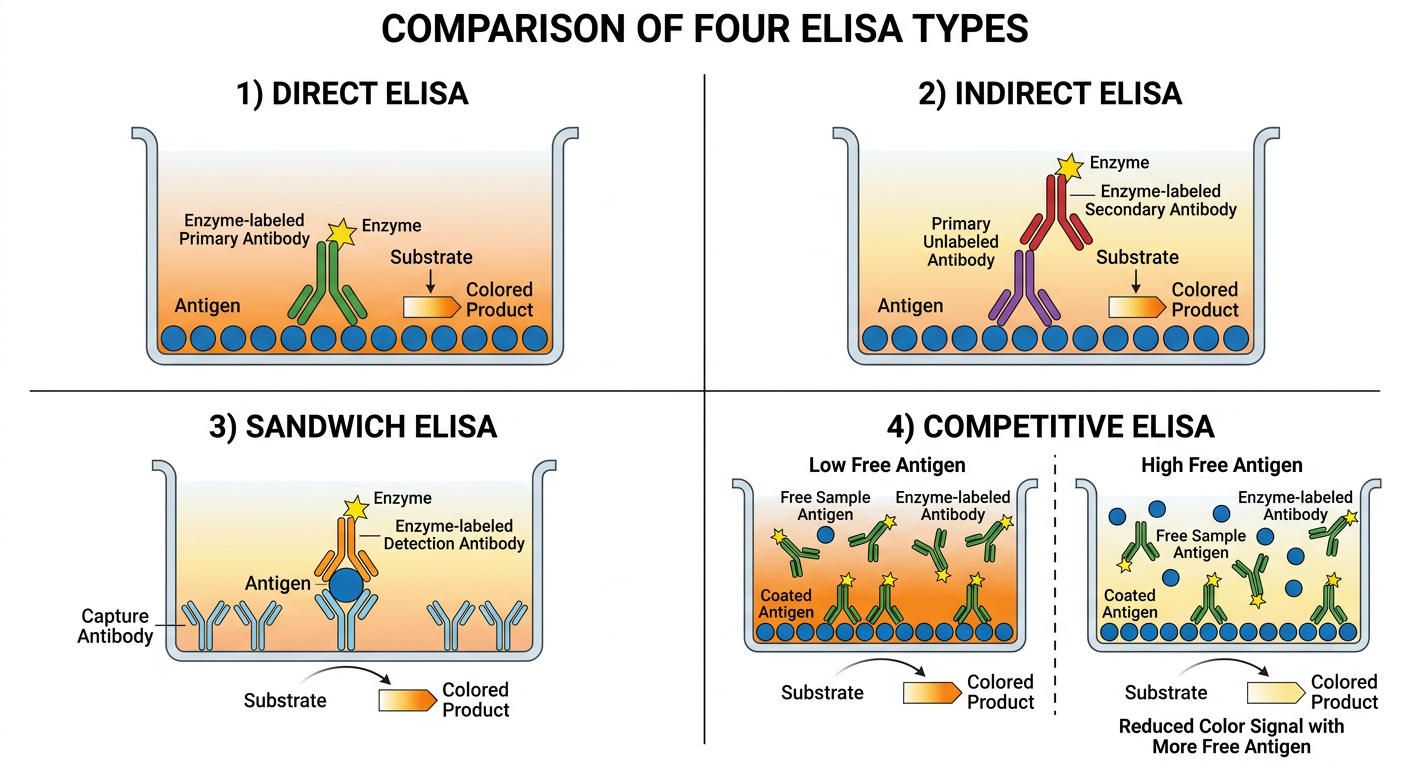
1. Direct ELISA
- The antigen from the sample is adsorbed directly onto the plastic well surface, and residual sticky sites are blocked with irrelevant protein.
- An enzyme-conjugated antibody specific for the antigen is added and binds directly to it.
- Unbound antibody is washed away, substrate is added, and the enzyme converts it into a colored product whose intensity is read spectrophotometrically.
- This is the simplest, fastest format but has lower sensitivity because there is no signal amplification step - Janeway's Immunobiology, 10e, Fig. A.5, p. 821-822.
2. Indirect ELISA
- Antigen is coated on the well as before.
- An unlabeled primary antibody binds the antigen.
- A labeled secondary antibody (anti-Ig) then binds the primary antibody. Because two or more secondary antibody molecules can bind each primary antibody molecule, the signal is amplified compared with direct ELISA.
- This format is also used to detect antibody titers in patient serum (e.g., autoantibody testing), where the antigen is fixed on the plate and the "unknown" is the patient's antibody - Janeway's Immunobiology, 10e, p. 822.
3. Sandwich (Capture) ELISA
- Instead of coating antigen directly, an antigen-specific "capture" antibody is bound to the plate first.
- The sample is added, and the capture antibody concentrates the antigen on the surface with high affinity, even at low antigen concentrations.
- A second, enzyme-labeled "detection" antibody that recognizes a different epitope on the same antigen is added, forming an antibody-antigen-antibody "sandwich."
- This format is widely used to measure secreted products such as cytokines, because it is highly sensitive and specific - Janeway's Immunobiology, 10e, p. 822.
4. Competitive ELISA
- A known amount of antigen is coated on the plate (or antibody, depending on the variant).
- The patient sample (containing unknown antigen) is pre-mixed with a fixed amount of labeled antibody before being added to the well.
- Labeled antibody in the mixture competes for binding: free antigen in the sample binds the labeled antibody in solution, preventing it from binding the plate-bound antigen.
- After washing, less color signal indicates more antigen was present in the original sample (inverse relationship between signal and analyte concentration).
- This principle is related to radioimmunoassay-style competitive inhibition assays - Janeway's Immunobiology, 10e, Fig. A-4/A.5 area, p. 821-823.
Key distinguishing logic
| Format | What's fixed to plate | Detection step | Signal relationship |
|---|---|---|---|
| Direct | Antigen | Labeled 1st Ab binds antigen directly | Direct |
| Indirect | Antigen | Unlabeled 1st Ab, then labeled 2nd Ab | Direct (amplified) |
| Sandwich | Capture Ab | Antigen captured, then labeled detection Ab (different epitope) | Direct |
| Competitive | Antigen or Ab | Sample antigen competes with labeled reagent | Inverse |


Answers
tuberculin test cattle single intradermal double intradermal Stormont test comparative cervical test ophthalmic test principle
E-rosette assay principle T lymphocytes sheep red blood cells applications
leucocyte migration inhibition test principle applications cell mediated immunity
https://tbhub.co.uk/tb-testing-cattle/skin-testing/tuberculi…
Answer
counting Questions: 1. Define rosette 2. Write the principle and applications of E-rosette assay 3. List out the methods which are commonly used to assess the cell mediated immunity. Explain with principles. 4. Enumerate the methods to separate B and T lymphocytes,Questions: Enumerate the tests, which are commonly used to diagnose the hypersensitivity reactions 1. 2. 3. in animals. Enumerate different types of tuberculin tests. Name the suitable sites and tests for demonstration of tuberculin reaction in various species of animals. c) OT 4. 5. Define the following b) Tuberculin a) PPD Define and write the advantages and disadvantages of the following test a) Single intra dermal test b) Double intra dermal test c) Stormont test d) Opthalmic test e) Mantoux test 6. What are the other diseases diagnosed by a similar intradermal skin test? Give details of the test.Questions 1. Write the principle and applications of leucocyte migration inhibiton test Answers