
A person suffering from fever and body pain she is feeling weak and has swelling in left calf suggest medicine
fever body pain weakness calf swelling DVT warning signs treatment 2025
deep vein thrombosis calf swelling DVT diagram
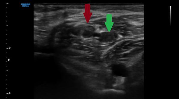
This diagnostic image is a B-mode grayscale ultrasound in the transverse plane demonstrating deep vein thrombosis (DVT) in the gastrocnemius veins of the lower extremity. The image captures a compression maneuver, a key diagnostic technique where external pressure is applied with the transducer. Two rounded venous structures are highlighted: the red arrow points to a vein containing visible echogenic material (thrombus), and the green arrow indicates a neighboring vein that remains patent but fails to collapse under pressure. The surrounding muscular and subcutaneous tissue exhibits a heterogeneous echotexture with hyperechoic fascial planes. The failure of venous coaptation (non-compressibility) despite active pressure is a definitive sonographic sign of acute or subacute venous thrombosis. This clinical finding is essential for medical education regarding vascular emergencies and the sonographic evaluation of calf pain or swelling.
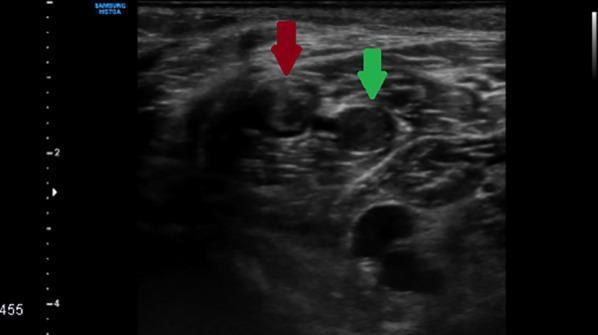
This diagnostic image is a grayscale B-mode ultrasound of the calf in a transverse plane, demonstrating gastrocnemius vein thrombosis. The image highlights two adjacent gastrocnemius veins, indicated by red and green arrows. Both vessels exhibit characteristic signs of Deep Vein Thrombosis (DVT), primarily the presence of internal echogenic material filling the lumens, which should normally appear anechoic (black). The vein on the left (red arrow) shows more distinct, brighter echogenic content compared to the vein on the right (green arrow), which appears slightly more heterogeneous. The surrounding musculature of the calf is visible, providing anatomical context. This visual is a classic educational example used to teach diagnostic criteria for venous thrombosis, emphasizing the identification of intraluminal echoes and increased vessel diameter as indicators of an occlusive or semi-occlusive thrombus.
⚠️ URGENT: This Is a Medical Emergency - Please Read First
Why This Is Serious
| Symptom | DVT Connection |
|---|---|
| Calf swelling (left leg) | Classic site of DVT |
| Fever | Inflammatory response to clot |
| Body pain / weakness | Systemic response |
| Weakness in the leg | Impaired venous drainage |
What To Do RIGHT NOW
- Order a Doppler Ultrasound of the leg (the gold standard test for DVT)
- Check D-dimer blood test (elevated in DVT)
- Assess for pulmonary embolism (shortness of breath, chest pain are danger signs)
Medical Treatment (Requires a Doctor's Prescription)
- First-line (preferred): Rivaroxaban (Xarelto) or Apixaban (Eliquis) - oral blood thinners
- Alternative: Low Molecular Weight Heparin (LMWH) like Enoxaparin (Clexane) injections
- Older option: Warfarin with Heparin bridge
- For fever/pain: Paracetamol (Acetaminophen) 500mg-1000mg every 6-8 hours is safe to use WHILE going to the hospital. Do NOT take NSAIDs like Ibuprofen/Aspirin without doctor advice as they affect blood clotting.
What NOT To Do
- Do NOT massage or rub the calf - this can dislodge the clot
- Do NOT apply heat to the swelling
- Do NOT take any anticoagulants without prescription
- Do NOT delay - hours matter with DVT
Emergency Warning Signs - Call Ambulance Immediately If:
- Sudden shortness of breath
- Chest pain
- Coughing up blood
- Rapid heartbeat or dizziness
- The leg turns blue or goes numb
Disclaimer: This information is for educational purposes only and does not replace professional medical advice. Please seek immediate in-person medical evaluation. Only a qualified doctor can diagnose and safely prescribe treatment for this condition.
she is also suffering from chest and backpain
🚨 CALL AN AMBULANCE RIGHT NOW — THIS IS A MEDICAL EMERGENCY 🚨
Why This Is Critical
| New Symptom | What It Means |
|---|---|
| Chest pain | Clot blocking blood flow to lungs / lung tissue dying |
| Back pain | Referred pain from lung infarction (tissue death) |
| Calf swelling | The original source clot (DVT) |
| Fever + weakness | Systemic inflammatory response |
Do These Things RIGHT NOW
1. Call Emergency Services Immediately
- India: 108 (ambulance) or 112
- Tell them: "Patient has chest pain, back pain, leg swelling and fever - possible blood clot in lungs"
2. While Waiting for Ambulance
- Have her sit upright (not lie flat) - this helps breathing
- Do NOT give her food or water - she may need immediate surgery/procedures
- Do NOT leave her alone
- Keep her calm and still - physical exertion can worsen clot travel
- If she loses consciousness - begin CPR
3. Do NOT Do These Things
- Do NOT massage her leg
- Do NOT give Aspirin or Ibuprofen
- Do NOT give any medicine without doctor direction at this point
What Doctors Will Do at the Hospital
- CT Pulmonary Angiography (CTPA) - confirms PE
- ECG - checks heart strain from PE
- D-dimer + troponin blood tests
- Oxygen therapy immediately
- Anticoagulation - IV Heparin or clot-busting drugs (thrombolytics like tPA) if massive PE
- ICU admission likely
Danger Signs of Imminent Collapse - If Any of These Appear, Start CPR
- She stops breathing or loses consciousness
- Lips or fingertips turn blue
- She cannot speak or respond
- Pulse becomes very faint or disappears
There is NO home medicine or over-the-counter drug that can treat a pulmonary embolism. Every minute without treatment increases the risk of death. Please call an ambulance immediately.