
Describe briefly about paediatric dengue with all the recent updates, treatment guidelines taking references from standard textbooks, ncbm, iap, pubmed
dengue fever children pediatric management
IAP NCBI dengue paediatric guidelines 2024 2025 treatment India
https://www.ncbi.nlm.nih.gov/books/NBK430732
dengue pediatric children fluid management platelet transfusion
dengue vaccine Dengvaxia children safety seroprevalence
dengue hemorrhagic fever shock syndrome children treatment outcomes
IAP dengue guidelines 2023 2024 paediatric India management classification
https://iapindia.org/pdf/Ch-138-Dengue-in-Children.pdf
PMID: 40830031
PMID: 40209729
dengue fever rash petechiae child dengue shock syndrome clinical signs
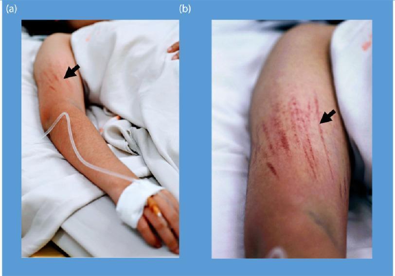
Clinical photograph of a patient's arm demonstrating bleeding manifestations characteristic of Dengue Shock Syndrome (DSS). The image is divided into two panels: (a) shows the full arm in a clinical setting with an intravenous (IV) line secured to the hand, and (b) provides a close-up view of the upper arm. Both panels highlight linear petechiae (indicated by black arrows), which are small, red to purplish hemorrhagic spots arranged in distinct linear patterns. These lesions represent microvascular fragility and are shown following the inflation of a blood pressure cuff (Hess test or capillary fragility test), a common diagnostic maneuver for assessing vascular permeability in dengue fever. The presence of an IV line and hospital bedding contextualizes the severity of the illness, illustrating the systemic plasma leakage and hemostatic dysfunction associated with severe viral hemorrhagic fevers. Key educational concepts include endothelial dysfunction, capillary fragility, and clinical signs of thrombocytopenia or vasculopathy.
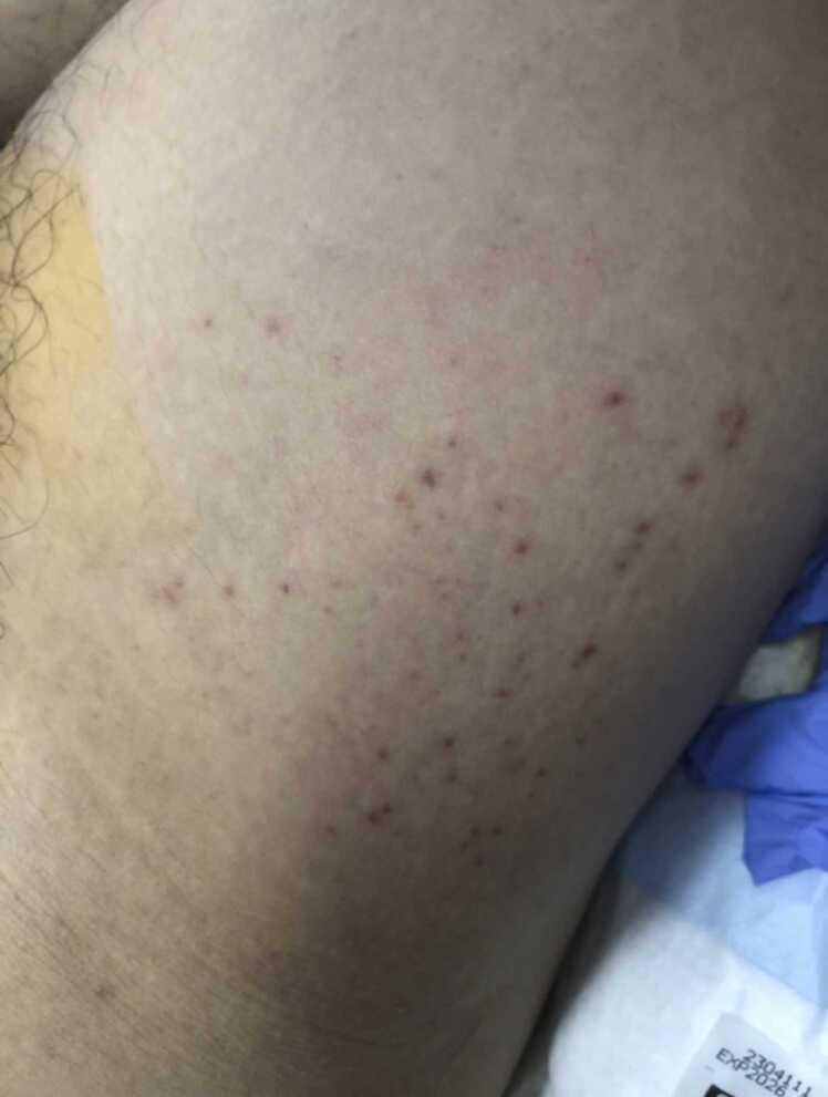
A clinical photograph displaying a close-up view of the right groin and anterior thigh of a patient. The primary finding consists of numerous, discrete, non-blanching macules identified as petechiae. These lesions are reddish-purple or dark red in color, measuring approximately 1-2 mm in diameter, and exhibit a punctate, irregular morphology. They are distributed in a scattered, non-confluent pattern across the skin surface. The surrounding skin appears light-toned with natural texture and focal areas of hair growth on the left. Clinical context suggests these findings are associated with a systemic viral infection, specifically Dengue Hemorrhagic Fever/Dengue Shock Syndrome. Peripheral elements in the frame include a strip of yellowish adhesive medical dressing, a blue medical glove, white hospital bedding, and a medical supply label with an expiration date, indicating a clinical setting.
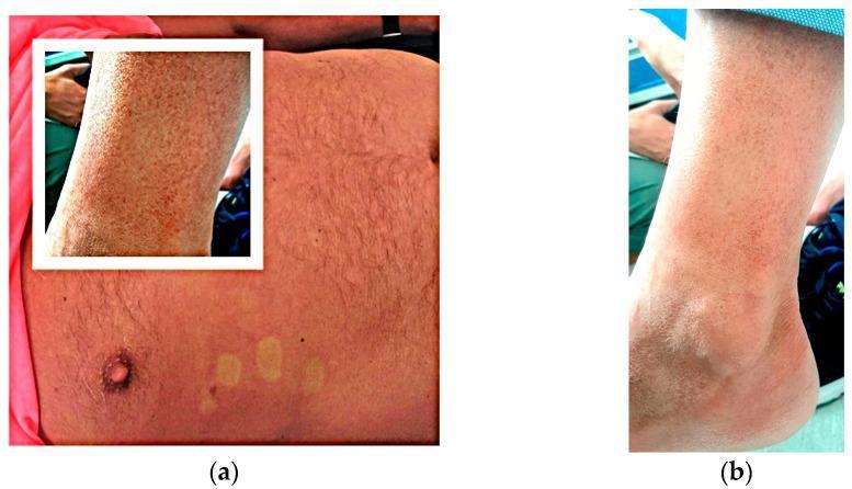
Clinical photographs of a 62-year-old male demonstrating dermatologic manifestations of an infectious disease, likely Dengue fever. Panel (a) shows a generalized nonpruritic macular rash on the thorax and abdomen characterized by small, erythematous spots. Notably, there are visible 'islands of skin sparing,' which are pale, circular areas of normal skin within the erythematous rash, a classic diagnostic sign for Dengue. An inset shows a dense distribution of this macular exanthema on the arm. Panel (b) illustrates purpuric lesions on the lower extremity (ankle and leg). These lesions appear as non-blanching, reddish-purple petechiae and ecchymoses, indicating capillary fragility or thrombocytopenia. The combination of generalized macular exanthema and distal purpura in the context of recent travel to a tropical region is highly suggestive of an arboviral infection. The clinical context involves differential diagnosis of febrile viral exanthems in patients with recent travel history to Southeast Asia.
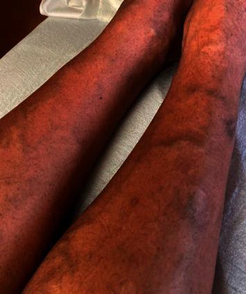
This clinical photograph shows the bilateral lower extremities of a 47-year-old male presenting with a diffuse dermatologic manifestation of thrombocytopenia. The skin demonstrates widespread, non-blanching petechiae, appearing as numerous pinpoint-sized (1-2 mm), reddish-purple macules. These lesions are densely scattered across the anterior and lateral aspects of the shins and calves. In addition to the petechial rash, several larger, coalescent purpuric lesions and subtle ecchymoses are visible, indicating small-vessel bleeding into the dermis. The background skin exhibits generalized erythema. This visual finding is characteristic of severe thrombocytopenia, in this clinical context associated with a viral hemorrhagic fever such as dengue. The image serves as an educational example of the cutaneous signs of low platelet counts and the systemic progression of infectious diseases like dengue fever in a hospital setting.
Paediatric Dengue - A Comprehensive Review
1. Introduction & Epidemiology
2. Pathophysiology
- Plasma leakage - sudden, massive extravasation of intravascular fluid due to increased vascular permeability, triggered by the immune response (complement activation, cytokines, NS1 glycoprotein-mediated endothelial injury)
- Thrombocytopenia - bone marrow suppression + platelet destruction by immune complexes
- ADE - pre-existing sub-neutralising antibodies from a prior dengue infection enhance viral uptake into macrophages via Fc receptors, amplifying viral replication and cytokine release
- Hyperinflammatory phenotype - in severe cases, a cytokine storm resembling secondary HLH, with extreme hyperferritinaemia and multi-organ failure
3. Clinical Phases
| Phase | Timing | Features |
|---|---|---|
| Febrile | Days 1-3 (up to day 7) | Abrupt high fever, headache, retro-orbital pain, severe myalgia, arthralgia, facial flush, maculopapular rash, leukopenia, petechiae |
| Critical (Plasma Leakage) | Days 3-7 (24-48 h around defervescence) | Sudden improvement in fever PLUS plasma leakage - pleural effusion, ascites, haemoconcentration; rapid platelet fall; risk of shock |
| Recovery (Convalescent) | Days 7-10+ | Reabsorption of extravasated fluids, bradycardia, confluent rash with "islands of white skin sparing," diuresis |
4. WHO/IAP Classification (2009, still current standard)
Category A - Dengue without Warning Signs
Category B - Dengue with Warning Signs
- Abdominal pain or tenderness
- Persistent vomiting
- Clinical fluid accumulation (ascites, pleural effusion)
- Mucosal bleeding
- Lethargy or restlessness
- Liver enlargement >2 cm
- Laboratory: rising haematocrit + rapid fall in platelet count
Category C - Severe Dengue
- Severe plasma leakage → DSS (shock) or fluid accumulation with respiratory distress
- Severe bleeding (clinician-assessed)
- Severe organ involvement: AST/ALT ≥1,000 IU/L; impaired consciousness; cardiac, renal, or other organ failure
5. Clinical Features Specific to Children
- Infants may present atypically with only irritability and poor feeding
- Hepatosplenomegaly is more common in children than adults
- Febrile convulsions can occur during the febrile phase
- Neonatal dengue (vertical transmission ~20%) presents as acute febrile illness with thrombocytopenia; risk is highest when maternal dengue occurs close to delivery
- The tourniquet test (Hess test) - inflate BP cuff to midpoint between SBP and DBP for 5 minutes; ≥10 petechiae per 1 inch² = positive - remains a useful bedside tool in resource-limited settings

6. Diagnosis
Timing-based approach (critical to get right):
| Test | Best Window | Sensitivity (2025 Meta-analysis) |
|---|---|---|
| RT-PCR | Days 0-4 (up to day 7) | 95% (95% CrI 77-99%) |
| NS1 ELISA/RDT | Days 0-4 (up to day 7) | 90% (95% CrI 68-98%) |
| IgM ELISA | Days 3-5 onwards; 99% by day 10 | 71% early (days 1-7) |
| IgG ELISA | After day 7; remains elevated lifelong | Confirms secondary infection |
7. Management - IAP & WHO Guidelines
Management Groups (IAP STG Ch.138)
Group A - Outpatient Management
- Paracetamol 10-15 mg/kg/dose q4-6h (max 60 mg/kg/day, 4g/day) - first-line antipyretic
- Avoid NSAIDs, aspirin, ibuprofen - risk of bleeding
- Oral rehydration - encourage fluids (ORS, coconut water, fruit juices, clear soups)
- CBC when clinically indicated
- Daily monitoring, return immediately if any warning sign develops
- Explain danger signs to parents: severe abdominal pain, persistent vomiting, bleeding, rapid breathing, lethargy, refusal to feed
Group B - Inpatient Management (Warning Signs Present)
- Baseline haematocrit (HCT)
- Platelet count, FBC
- Blood glucose, LFTs, renal function
- Chest X-ray, abdominal ultrasound
- If oral intake tolerated: oral fluids; if not tolerated:
- 0.9% NS or Ringer's Lactate (isotonic crystalloid only)
- Start at 5-7 mL/kg/h for 1-2 h
- Reduce to 3-5 mL/kg/h for 2-4 h
- Reduce to 2-3 mL/kg/h until oral intake adequate
- Serial HCT monitoring every 2-4 hours guides up- or down-titration
- Avoid hypotonic fluids - risk of hyponatraemia and worsening plasma leakage
- Cautious fluid resuscitation is mandatory to avoid "fluid creep" and worsening respiratory status
- Lung ultrasound for B-lines and intra-abdominal pressure monitoring where available
- Fluid balance monitoring is as important as fluid administration
Group C - PICU Management (Dengue Shock)
- Severe plasma leakage with hypoperfusion/hypotension (DSS)
- Fluid accumulation with respiratory distress
- Severe bleeding
- Severe organ impairment: myocardial dysfunction, AKI, CNS dysfunction (altered GCS, seizures), hepatic dysfunction (ALT/AST >1,000 IU/L), HLH
- Isotonic crystalloid (Ringer's Lactate preferred over NS to avoid hyperchloraemic acidosis)
- Initial: 10-20 mL/kg over 15-30 minutes as a bolus; reassess
- After 20 mL/kg crystalloid/colloid: perform echocardiography to assess myocardial function (current PICU standard per IAP)
- Where advanced monitoring unavailable: continuous clinical monitoring (HR, BP, CRT, urine output, mental status)
- If HCT rises despite adequate resuscitation: consider colloid (albumin 5%)
- Reduce and stop fluids promptly as plasma-leakage phase resolves to prevent overload
- Platelet transfusion is NOT recommended prophylactically, regardless of platelet count alone
- Platelet transfusion only for active significant bleeding OR significant coagulopathy
- Evidence does not support prophylactic transfusion; it may cause volume overload
- Packed RBC: for confirmed significant blood loss with haemodynamic compromise
- FFP/Cryoprecipitate: for documented coagulopathy with bleeding
8. Management of Complications
Dengue with Hepatic Dysfunction
- Manage as for acute liver failure - avoid hepatotoxic drugs
- Judicious fluids, correct coagulopathy only if bleeding
- Monitor blood glucose closely (hypoglycaemia risk)
Dengue with AKI
- Judicious fluid management to target urine output >0.5 mL/kg/h
- Early renal replacement therapy (RRT) when indicated
- Continuous Veno-Venous Haemofiltration (CVVH) preferred modality in PICU
Dengue with Respiratory Complications
- Avoid pleural tap unless massive effusion causes ventilatory failure
- Children with refractory shock may require intubation + mechanical ventilation
- Lung-protective ventilation for ARDS
Dengue with Neurological Involvement (Dengue Encephalopathy/ADEM)
- Cerebral oedema management: avoid hypotonic fluids, consider mannitol
- Post-dengue ADEM: immunotherapy (steroids, IVIG) reported in case series
- Monitor electrolytes - hyponatraemia is a common cause of seizures
Dengue-associated HLH / Cytokine Storm - 2025 Update
- Median age: 48 months; median ferritin: 16,433 ng/mL
- Overall survival: 73%; when excluding those dying within 24h of admission: 80% survival
- Anakinra led to improvement in fluid requirements, ferritin, and transaminases
- Mortality was 100% in children with ferritin >100,000 ng/mL
- Conclusion: Anakinra may be a valuable adjunct in dengue with hyperinflammation; prospective trials needed
9. Dengue Vaccine - Current Status
- FDA approved for ages 9-16 years in endemic regions with prior laboratory-confirmed dengue infection
- 3-dose schedule at 0, 6, and 12 months
- Critical safety caveat: seronegative (dengue-naive) recipients who are vaccinated have an increased hazard ratio for severe dengue upon subsequent natural infection (ADE mechanism). For this reason, pre-vaccination seroprevalence screening is mandatory
- Not recommended for seronegative individuals, travellers from non-endemic regions, or those with unknown prior infection status
- Available in ~17 countries as of 2019 - Red Book 2021, p.522
- JNJ-1802 (mosnodenvir) - oral NS3-NS4B inhibitor; showed prophylactic efficacy in controlled human infection models; Phase 1 safety established; development was de-prioritised by sponsor in 2024 (NCBI StatPearls 2025)
- Dengue therapeutics consortium 2025 is evaluating dexamethasone, baricitinib, and N-acetylcysteine in a platform trial (Lancet Infect Dis 2026, PMID 41072450)
- No antiviral drug is currently approved for clinical use in dengue
10. Prevention & Vector Control
- Aedes aegypti bites mainly during daytime - bed nets alone are insufficient; insect repellent (DEET or picaridin-based) and full-coverage clothing are important for children
- Eliminate standing water - containers, tyres, plant pots; dengue vector can breed in as little as a few millimetres of water
- India's National Vector Borne Disease Control Programme (NVBDCP) coordinates case management, surveillance, and vector control nationally
- Dengue has been a nationally notifiable disease in the USA since 2010; India mandates reporting under IDSP
11. Differential Diagnosis
- Chikungunya (arthritis more prominent, less plasma leakage)
- Zika (milder illness, microcephaly risk in neonates if maternal)
- Malaria - must always be excluded in endemic areas (blood smear/RDT)
- Leptospirosis (jaundice, renal involvement, conjunctival suffusion)
- Rickettsial infections (eschar, prominent rash)
- Scrub typhus (eschar, lymphadenopathy)
- Measles (Koplik's spots, prodrome, immunisation history)
- ITP (isolated thrombocytopenia, no fever phase, no haemoconcentration)
- Ebola/Marburg/yellow fever (relevant for travellers from Africa)
12. Key Points - Summary Table
| Parameter | Key Fact |
|---|---|
| Serotypes | DENV 1-4; second infection with different serotype = highest risk |
| High-risk children | Infants, secondary infection, comorbidities, immunosuppressed |
| Critical phase window | 24-48 hours around defervescence (days 3-7) |
| Best diagnostic test (early) | NS1 antigen (sensitivity 90%) ≈ RT-PCR (95%) |
| Fluid of choice (shock) | Ringer's Lactate / 0.9% NS (isotonic crystalloid) |
| Avoid | NSAIDs, aspirin, prophylactic platelet transfusion, hypotonic fluids |
| Platelet transfusion threshold | Only for active significant bleeding, not prophylactically |
| Antipyretic | Paracetamol only |
| New 2025 therapeutic | Anakinra for dengue-HLH/cytokine storm (retrospective data, India) |
| Vaccine | Dengvaxia (9-16y, seropositive only); TAK-003 (Qdenga, 4y+, newer) |
References
- Red Book 2021: AAP Committee on Infectious Diseases. Report of the Committee on Infectious Diseases, 32nd Ed. pp.520-525.
- Harrison's Principles of Internal Medicine 22E (2025), McGraw Hill Medical.
- Rosen's Emergency Medicine: Concepts and Clinical Practice, 9th Ed. pp.2630.
- NCBI StatPearls - Dengue Fever (2025 update): NCBI Bookshelf NBK430732
- IAP Standard Treatment Guidelines, Ch.138 - Dengue in Children: iapindia.org STG
- WHO Guidelines for Dengue Diagnosis, Treatment, Prevention and Control (2009) - remains the international classification standard
- Pillay K et al. (2025) - NS1 ELISA vs RT-PCR meta-analysis. Lancet Microbe. PMID 40209729
- Bhat C et al. (2025) - Anakinra in children with severe dengue. Arch Dis Child. PMID 40830031
- Ong LT & Balasubramaniam R (2024) - HLH in dengue meta-analysis. Trans R Soc Trop Med Hyg. PMID 38721675
- Yacoub S et al. (2026) - DEN-CORE outcome set for dengue trials. Lancet Infect Dis. PMID 41072450
- Indian Pediatrics (Sept 2024, Vol 61) - IAP President's page: "Tackling Dengue: A Comprehensive Approach to Prevention and Management"