
What is MI
Myocardial infarction (MI), commonly called a "heart attack," occurs when blood flow to part of the heart muscle is blocked long enough to cause cell death. This is most often due to rupture of an atherosclerotic plaque in a coronary artery, leading to thrombus formation and vessel occlusion.
Searching sources...
Reading File
Finding Sources
Searching the Web
myocardial infarction definition types pathophysiology 2024
Reading File
Reading File
Reading File
Reading File
Reading File
Loading Image
Reading File
Reading File
Here is a thorough overview of Myocardial Infarction (MI):
Myocardial Infarction (MI)
Definition
MI, commonly called a "heart attack," is necrosis (death) of heart muscle resulting from prolonged ischemia. The 2018 joint task force of U.S. and European cardiology groups defines it as "the presence of acute myocardial injury detected by abnormal cardiac biomarkers in the setting of evidence of acute myocardial ischemia."
Roughly 800,000 people in the United States experience an MI each year (nearly one every 40 seconds), causing almost 400,000 deaths annually.
- Robbins, Cotran & Kumar Pathologic Basis of Disease
Causes and Risk Factors
- The major underlying cause is atherosclerosis (coronary artery disease)
- ~10% of MIs occur before age 40; ~45% before age 65
- Men are at greater risk than women, though the gap narrows after menopause (decline in estrogen leads to worsening coronary artery disease)
- In ~10% of cases, MI occurs without classic atherothrombosis, due to:
- Coronary vasospasm (e.g., cocaine, ephedrine use)
- Embolism (from left atrial thrombus in atrial fibrillation, endocarditis vegetations, prosthetic valves)
- Microvessel pathology (vasculitis, amyloid, sickle cell disease)
Pathogenesis - How MI Develops
The typical sequence:
- An atheromatous plaque is disrupted by endothelial injury, intraplaque hemorrhage, or mechanical forces, exposing subendothelial collagen
- Platelets adhere, aggregate, and activate - releasing thromboxane A2, ADP, and serotonin, causing further aggregation and vasospasm
- Coagulation is activated via tissue factor exposure, enlarging the thrombus
- Within minutes, the thrombus completely occludes the coronary artery lumen
Angiography within 4 hours of MI onset demonstrates coronary thrombosis in ~90% of cases. Early thrombolysis and/or angioplasty can be highly effective in limiting the extent of necrosis.
Types of MI (by Location/Extent)
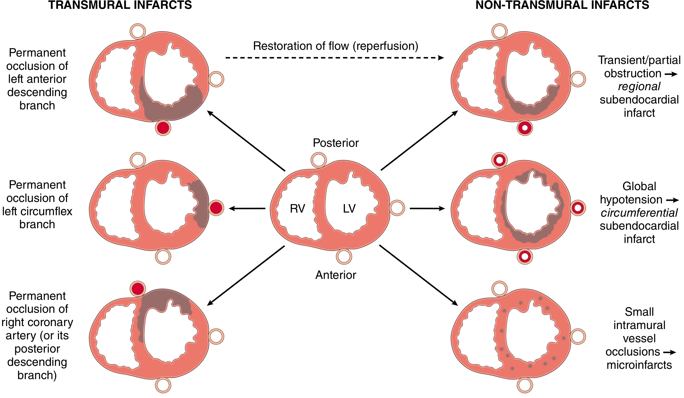
| Type | Cause | Pattern |
|---|---|---|
| Transmural (STEMI) | Complete occlusion of epicardial vessel | Full-thickness wall necrosis in distribution of one coronary artery |
| Subendocardial (NSTEMI) | Partial/transient occlusion, or global hypotension | Inner layer necrosis; may be regional or circumferential |
| Microinfarction | Small intramural vessel occlusion (embolism, vasculitis) | Multifocal scattered infarcts |
Coronary artery territory:
- Left Anterior Descending (LAD): anterior wall + anterior interventricular septum (most common)
- Right Coronary Artery (RCA): posterior wall, posterior septum, inferior-posterior left ventricle
- Left Circumflex (LCX): lateral wall of left ventricle
ECG Changes
Three major electrical changes occur (per Ganong's Review of Medical Physiology):
| Defect in Infarcted Cells | Current Flow | ECG Change |
|---|---|---|
| Rapid repolarization | Out of infarct | ST segment elevation |
| Decreased resting membrane potential | Into infarct | TQ depression (manifests as ST elevation) |
| Delayed depolarization | Out of infarct | ST segment elevation |
- Acute MI hallmark: ST elevation in leads overlying the infarct; ST depression in opposite leads
- After days/weeks: ST changes resolve; Q waves may appear (indicating dead myocardium)
Clinical Features
- Chest pain: Prolonged (>30 min), crushing/squeezing/stabbing, typically radiating to arm, jaw, or back
- Diaphoresis (profuse sweating)
- Nausea and vomiting (especially posterior-inferior MI with vagal stimulation)
- Dyspnea (due to impaired contractility and pulmonary congestion)
- Up to 25% are silent (especially in diabetics due to neuropathy)
Diagnosis
Cardiac Biomarkers (most important):
- Troponin T and I (cTnT, cTnI) are the most specific and sensitive markers
- Begin rising ~3-6 hours after MI onset
- Peak at ~24-48 hours
- Remain elevated for up to ~10-14 days (cTnI) or ~14-21 days (cTnT)
- CK-MB: rises earlier, used for reinfarction detection
ECG and Imaging (echocardiography, coronary angiography) complete the workup.
Causes of Death After MI
Per Guyton & Hall Textbook of Medical Physiology:
- Decreased cardiac output (cardiogenic shock, systolic stretch/paradoxical expansion of infarcted zone)
- Pulmonary edema (blood damming in pulmonary vessels)
- Ventricular fibrillation (most common early cause of death)
- Cardiac rupture (rare, occurs 3-7 days after MI)
Reperfusion and Its Effects
Restoring blood flow (via thrombolysis, angioplasty/PCI, or CABG) is the cornerstone of treatment. However, reperfusion injury can occur:
- Mitochondrial dysfunction with apoptosis
- Myocyte hypercontracture from calcium overload (contraction band necrosis on histology)
- Free radical generation (superoxide, H2O2, hydroxyl radicals)
- Leukocyte aggregation causing the "no-reflow" phenomenon
Two important post-ischemic phenomena:
- Stunned myocardium: prolonged contractile dysfunction after short-term ischemia that eventually recovers
- Hibernating myocardium: chronically reduced function in chronically ischemic muscle; can recover after revascularization
Universal Classification (5 Types)
| Type | Mechanism |
|---|---|
| Type 1 | Atherosclerotic plaque rupture/erosion with thrombus (classic) |
| Type 2 | Supply-demand mismatch without plaque rupture (e.g., spasm, anemia, tachycardia) |
| Type 3 | Sudden cardiac death before biomarkers can be measured |
| Type 4 | PCI-related (during or after coronary intervention) |
| Type 5 | CABG surgery-related |
Sources: Robbins, Cotran & Kumar - Pathologic Basis of Disease | Robbins & Kumar Basic Pathology | Guyton & Hall Textbook of Medical Physiology | Ganong's Review of Medical Physiology
This is a shared conversation. Sign in to Orris to start your own chat.