
Pharyngeal arches image
pharyngeal arches embryology diagram
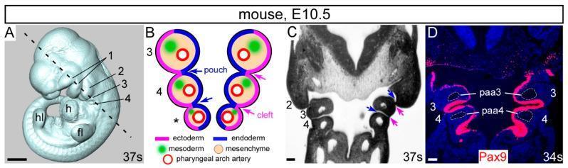
This composite educational image illustrates the developmental anatomy of the pharyngeal arches in a mouse embryo at embryonic day 10.5 (E10.5). (A) A 3D whole-mount embryo reconstruction showing anatomical landmarks including the forelimb bud (fl), hindlimb bud (hl), heart (h), and the sequential pharyngeal arches (1-4). (B) A schematic diagram of a coronal section detailing the embryological layers: outer ectoderm (pink), internal endoderm (blue) forming pouches, and a mesenchymal core containing mesoderm (green) and pharyngeal arch arteries (red circles). (C) A corresponding histological coronal section showing the physical arrangement of arches 2, 3, and 4. (D) An immunofluorescence-stained coronal section showing Pax9 expression (red signal) localized within the pharyngeal endoderm. The image highlights the specific positioning of the 3rd and 4th pharyngeal arch arteries (paa3, paa4) within their respective arches. This material demonstrates key concepts in embryology, specifically the rostral-caudal development of the pharyngeal apparatus and the molecular markers of the pharyngeal endoderm essential for cardiovascular and craniofacial morphogenesis.
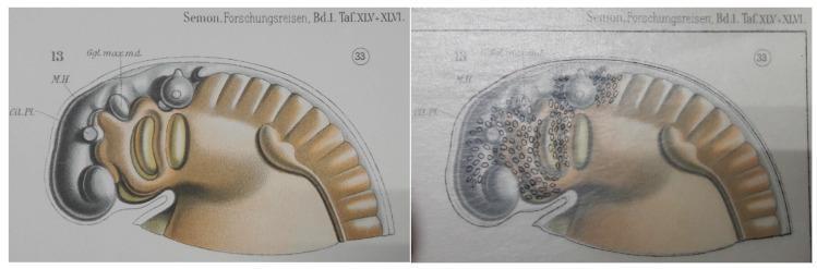
This comparative anatomical diagram illustrates the embryological development of the pharyngeal arches, using a stage 33 lungfish embryo as a model for vertebrate morphogenesis. The image is presented in two panels: the left displays a shaded specimen with key landmarks labeled, including 'Gl. Pl.' (Glandular Plate), 'M.H.' (Midbrain/Mittelhirn), and 'Ggl. max. md.' (Maxillomandibular ganglion). The right panel utilizes a historic transparent overlay method to demonstrate the migration of neural crest cells, represented by small black ovals. These cells are concentrated along the cephalic region and the dorsal-ventral axis of the pharyngeal arches, which appear as segmented bulges. The illustration highlights the pathways of migratory cells from the neural tube into the pharyngeal apparatus, which eventually contribute to the development of the aorticopulmonary septum, cardiac outflow tract, and craniofacial structures. This comparison is an educational resource for developmental biology and embryology, focusing on neural crest cell distribution and the evolution of vertebrate circulatory systems.
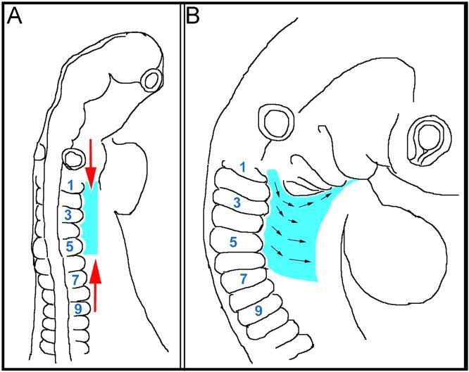
This medical illustration depicts an anatomical diagram of avian embryo development, specifically focused on the morphogenetic movements of the lateral mesoderm and ectoderm at the head-trunk interface. Panel A provides a dorsal view of the embryo, illustrating rostrocaudal compression of the lateral tissues (highlighted in cyan) indicated by opposing red arrows. Panel B provides a lateral view, showing mediolateral expansion of the same tissue layers. Somites are numbered 1 through 9 to serve as anatomical landmarks. Small black arrows in Panel B track the migration route of tissues originating from the level of rostral somites. These tissues move around the pharyngeal arches and extend along the floor of the pharynx toward the mandibular arch. This diagram serves to explain the 'conveyor belt' mechanism where the movement of lateral mesoderm and ectoderm aids in the assembly of the hypopharyngeal apparatus and facilitates the transport of non-migratory muscle precursors. Key structures shown include the neural tube, otic vesicle, somites, and pharyngeal arches within the context of vertebrate embryology and developmental anatomy.
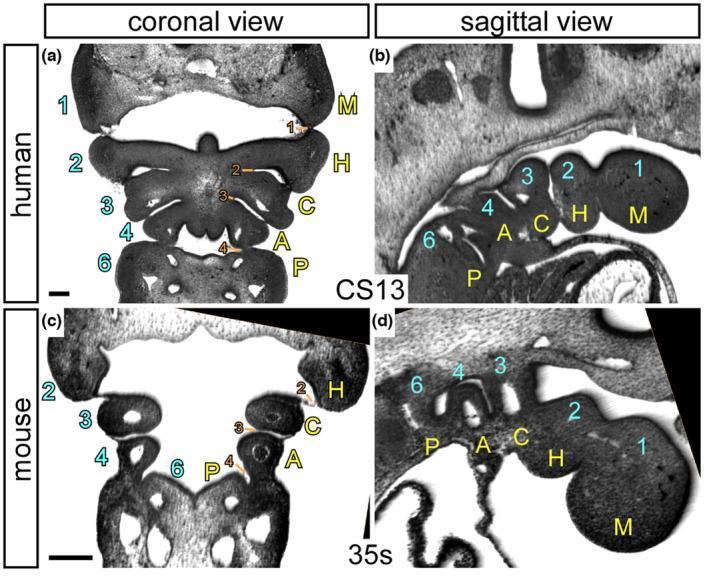
High-resolution episcopic microscopy (HREM) images illustrating the comparative embryological anatomy of the pharyngeal region in a human embryo (Carnegie Stage 13) and a mouse embryo (35 somites). The plate includes coronal (transverse) and sagittal sections, highlighting the segmental organization of pharyngeal arches and pouches. The images contrast the traditional numerical nomenclature (1, 2, 3, 4, 6) with a proposed descriptive naming system: Mandibular (M), Hyoid (H), Carotid (C), Aortic (A), and Pulmonary (P). In the human coronal view (a), bilateral symmetry of the arches is visible, while the sagittal view (b) clearly depicts the cranial-to-caudal sequence of these structures. Pharyngeal pouches are numerically labeled (1-4, 6) in orange, representing endodermal outpocketings between the arches. This visual resource is designed for developmental biology and embryology education, emphasizing that there are five distinct arches formed during normal amniote development, which has significant implications for understanding congenital cardiac malformations and the derivatives of the pharyngeal apparatus.
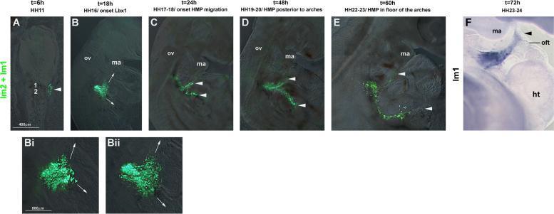
This composite diagnostic and pathophysiology diagram illustrates the migration of occipital lateral mesoderm in a developing avian embryo using GFP electroporation and in situ hybridization. Panels A through E display a temporal sequence from HH11 (t=6h) to HH22-23 (t=60h). Fluorescent GFP-labeled cells, initially localized near somites 1 and 2 (lm1+lm2), are shown organizing into two distinct migratory streams: a rostral stream projecting toward the pharyngeal arches and a lateral-caudal stream. Higher magnification panels (Bi, Bii) at HH16 highlight the onset of this bifurcation. Panel F shows GFP mRNA expression (blue staining) at HH23-24, demonstrating the accumulation of these lateral mesoderm-derived cells near the mandibular arch (ma) and the heart's outflow tract (oft). Anatomical landmarks include the otic vesicle (ov), heart (ht), and mandibular arch. This content serves as an educational resource for embryology and developmental biology, specifically regarding circumpharyngeal cell movements and the origins of cranial and cardiac mesodermal structures.
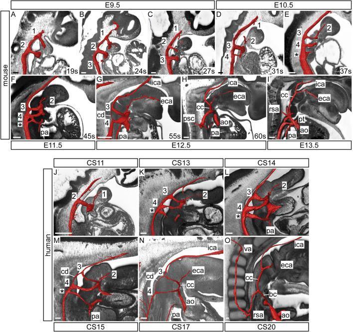
This comparative anatomical diagram displays three-dimensional reconstructions of pharyngeal arch artery development in mouse (E9.5 to E13.5) and human (CS11 to CS20) embryos, utilizing high-resolution episcopic microscopy (HREM) datasets. The reconstructions, highlighted in red, demonstrate the systematic transition from embryonic arch arteries to the definitive adult arterial configuration. Key developmental milestones illustrated include the formation of the initial first and second arches, the subsequent appearance of the third, fourth, and sixth (ultimate, denoted by *) arch arteries, and the involution of the carotid duct (cd). The images track the transformation of the third arch into the common carotid (cc) and proximal internal carotid arteries (ica), and the second arch into the external carotid artery (eca). Visible cardiovascular landmarks include the aorta (ao), pulmonary trunk (pt), and pulmonary arteries (pa). This series highlights the dramatic elongation of the common carotid arteries as the heart descends and the embryo grows along the anterior-posterior axis, providing a comprehensive visual of pharyngeal arch remodeling and outflow tract septation across mammalian species.
1. Pharyngeal Arch Structure - Embryological Layers
2. Human vs. Mouse Pharyngeal Arch Anatomy (Carnegie Stage 13)
3. Pharyngeal Arch Artery Remodeling (Mouse E9.5-E13.5 / Human CS11-CS20)
| Arch | Nerve | Cartilage | Muscle | Artery |
|---|---|---|---|---|
| 1 (Mandibular) | CN V2/V3 | Meckel's + malleus/incus | Muscles of mastication, tensor tympani | Maxillary a. |
| 2 (Hyoid) | CN VII | Stapes, styloid process, lesser cornu hyoid | Muscles of facial expression | Stapedial a. |
| 3 | CN IX | Greater cornu hyoid | Stylopharyngeus | Common/internal carotid |
| 4 | CN X (sup. laryngeal) | Thyroid cartilage | Pharyngeal constrictors | Aortic arch (L), subclavian (R) |
| 6 | CN X (recurrent laryngeal) | Cricoid/arytenoid cartilages | Intrinsic laryngeal muscles | Pulmonary arteries/ductus arteriosus |
Create a pharyngeal arch reference guide or study sheet
What format would you like for the study sheet? · What level of detail?
mkdir -p /tmp/workspace/pharyngeal-arches && ls /tmp/workspace/pharyngeal-arches/
~/pharyngeal-arches/build_study_sheet.py
from reportlab.lib.pagesizes import A4
from reportlab.lib import colors
from reportlab.lib.styles import getSampleStyleSheet, ParagraphStyle
from reportlab.lib.units import cm, mm
from reportlab.platypus import (
    SimpleDocTemplate, Paragraph, Spacer, Table, TableStyle,
    HRFlowable, KeepTogether
)
from reportlab.lib.enums import TA_CENTER, TA_LEFT, TA_JUSTIFY
from reportlab.pdfbase import pdfmetrics
from reportlab.pdfbase.ttfonts import TTFont
import os

OUTPUT = "/tmp/workspace/pharyngeal-arches/Pharyngeal_Arches_Study_Sheet.pdf"

doc = SimpleDocTemplate(
    OUTPUT,
    pagesize=A4,
    leftMargin=1.5*cm, rightMargin=1.5*cm,
    topMargin=1.5*cm, bottomMargin=1.5*cm
)

W, H = A4

# ─── Color palette ────────────────────────────────────────────────────────────
C_NAVY   = colors.HexColor("#1B3A6B")
C_TEAL   = colors.HexColor("#0D7377")
C_GOLD   = colors.HexColor("#F0A500")
C_LIGHT  = colors.HexColor("#EAF4F4")
C_PALE   = colors.HexColor("#FFF8E7")
C_WHITE  = colors.white
C_GRAY   = colors.HexColor("#555555")
C_LGRAY  = colors.HexColor("#E8E8E8")
C_RED    = colors.HexColor("#C0392B")
C_GREEN  = colors.HexColor("#1A7A4A")
C_PURPLE = colors.HexColor("#6C3483")
C_ORANGE = colors.HexColor("#D35400")
C_BROWN  = colors.HexColor("#7B4F2E")
C_PINK   = colors.HexColor("#A93226")

# Arch colours for the main table rows
ARCH_COLORS = {
    1: colors.HexColor("#FDEBD0"),  # warm orange
    2: colors.HexColor("#D5F5E3"),  # green
    3: colors.HexColor("#D6EAF8"),  # blue
    4: colors.HexColor("#E8DAEF"),  # purple
    6: colors.HexColor("#FDFEFE"),  # white/pale
}
ARCH_HDR_COLORS = {
    1: colors.HexColor("#E59866"),
    2: colors.HexColor("#58D68D"),
    3: colors.HexColor("#5DADE2"),
    4: colors.HexColor("#AF7AC5"),
    6: colors.HexColor("#AAB7B8"),
}

# ─── Styles ───────────────────────────────────────────────────────────────────
styles = getSampleStyleSheet()

def S(name, **kw):
    return ParagraphStyle(name, **kw)

title_style = S("Title_",
    fontName="Helvetica-Bold", fontSize=20, textColor=C_WHITE,
    alignment=TA_CENTER, spaceAfter=2)

subtitle_style = S("Subtitle_",
    fontName="Helvetica", fontSize=11, textColor=C_GOLD,
    alignment=TA_CENTER, spaceAfter=4)

section_style = S("Section_",
    fontName="Helvetica-Bold", fontSize=12, textColor=C_WHITE,
    alignment=TA_LEFT, spaceBefore=8, spaceAfter=4,
    leftIndent=4, leading=16)

body_style = S("Body_",
    fontName="Helvetica", fontSize=8.5, textColor=colors.black,
    alignment=TA_LEFT, spaceAfter=2, leading=12)

small_style = S("Small_",
    fontName="Helvetica", fontSize=7.5, textColor=C_GRAY,
    alignment=TA_LEFT, spaceAfter=1, leading=11)

bold_style = S("Bold_",
    fontName="Helvetica-Bold", fontSize=8.5, textColor=colors.black,
    alignment=TA_LEFT, spaceAfter=2)

italic_style = S("Italic_",
    fontName="Helvetica-Oblique", fontSize=8.5, textColor=C_GRAY,
    alignment=TA_LEFT, spaceAfter=2)

th_style = S("TH_",
    fontName="Helvetica-Bold", fontSize=8, textColor=C_WHITE,
    alignment=TA_CENTER, leading=11)

td_style = S("TD_",
    fontName="Helvetica", fontSize=7.5, textColor=colors.black,
    alignment=TA_LEFT, leading=11)

td_bold = S("TD_Bold",
    fontName="Helvetica-Bold", fontSize=7.5, textColor=colors.black,
    alignment=TA_LEFT, leading=11)

td_center = S("TD_Cen",
    fontName="Helvetica", fontSize=7.5, textColor=colors.black,
    alignment=TA_CENTER, leading=11)

note_style = S("Note_",
    fontName="Helvetica-Oblique", fontSize=7, textColor=C_GRAY,
    alignment=TA_LEFT, spaceAfter=2, leading=10)

mnemonic_style = S("Mnemonic_",
    fontName="Helvetica-Bold", fontSize=9, textColor=C_NAVY,
    alignment=TA_CENTER, spaceAfter=2, leading=13)

warn_style = S("Warn_",
    fontName="Helvetica-Bold", fontSize=8, textColor=C_RED,
    alignment=TA_LEFT, spaceAfter=2)

# ─── Helper: section header ───────────────────────────────────────────────────
def section_header(text, bg=C_NAVY):
    t = Table([[Paragraph(text, section_style)]], colWidths=[doc.width])
    t.setStyle(TableStyle([
        ("BACKGROUND", (0,0), (-1,-1), bg),
        ("TOPPADDING",    (0,0), (-1,-1), 5),
        ("BOTTOMPADDING", (0,0), (-1,-1), 5),
        ("LEFTPADDING",   (0,0), (-1,-1), 8),
        ("ROUNDEDCORNERS", [4,4,4,4]),
    ]))
    return t

def subsection_header(text, bg=C_TEAL):
    t = Table([[Paragraph(text, S("sub_h",
        fontName="Helvetica-Bold", fontSize=10, textColor=C_WHITE,
        alignment=TA_LEFT, leading=14)
    )]], colWidths=[doc.width])
    t.setStyle(TableStyle([
        ("BACKGROUND", (0,0), (-1,-1), bg),
        ("TOPPADDING",    (0,0), (-1,-1), 4),
        ("BOTTOMPADDING", (0,0), (-1,-1), 4),
        ("LEFTPADDING",   (0,0), (-1,-1), 8),
    ]))
    return t

def colored_box(content_paras, bg=C_LIGHT, border=C_TEAL):
    inner = [[p] for p in content_paras]
    t = Table(inner, colWidths=[doc.width - 1.2*cm])
    t.setStyle(TableStyle([
        ("BACKGROUND", (0,0), (-1,-1), bg),
        ("BOX",        (0,0), (-1,-1), 1.2, border),
        ("TOPPADDING",    (0,0), (-1,-1), 4),
        ("BOTTOMPADDING", (0,0), (-1,-1), 3),
        ("LEFTPADDING",   (0,0), (-1,-1), 8),
        ("RIGHTPADDING",  (0,0), (-1,-1), 8),
    ]))
    return t

# ─── Title block ──────────────────────────────────────────────────────────────
def make_title_block():
    title_para = Paragraph("PHARYNGEAL ARCHES", title_style)
    sub_para   = Paragraph("Complete Reference & Study Sheet  •  Embryology", subtitle_style)
    source_para = Paragraph(
        "Sources: The Developing Human (Moore & Persaud) | Langman's Medical Embryology",
        S("src", fontName="Helvetica-Oblique", fontSize=8, textColor=colors.HexColor("#CCDDDD"),
          alignment=TA_CENTER))
    block = Table([
        [title_para],
        [sub_para],
        [source_para],
    ], colWidths=[doc.width])
    block.setStyle(TableStyle([
        ("BACKGROUND",    (0,0), (-1,-1), C_NAVY),
        ("TOPPADDING",    (0,0), (-1,-1), 10),
        ("BOTTOMPADDING", (0,0), (-1,-1), 8),
        ("LEFTPADDING",   (0,0), (-1,-1), 10),
    ]))
    return block

# ─── Overview box ─────────────────────────────────────────────────────────────
def make_overview():
    items = [
        Paragraph("<b>What are Pharyngeal Arches?</b>", bold_style),
        Paragraph(
            "5 pairs of mesodermal arches (1-4, 6) separated by clefts (ectoderm) and pouches (endoderm). "
            "Develop in week 4-5. Each arch has: <b>artery</b> (from truncus arteriosus), "
            "<b>cartilage/bone</b>, <b>muscle</b>, and <b>cranial nerve</b>.",
            body_style),
        Spacer(1, 3),
        Paragraph("<b>Composition of each arch:</b>  Ectoderm (outer) | Mesenchyme core (neural crest cells + paraxial mesoderm) | Endoderm (inner)", body_style),
        Paragraph(
            "<b>Key concept:</b> Even when arch muscles migrate away, their cranial nerve follows — so nerve supply identifies arch of origin.",
            italic_style),
    ]
    return colored_box(items, bg=C_LIGHT, border=C_TEAL)

# ─── Main derivatives table ───────────────────────────────────────────────────
def make_main_table():
    col_w = [doc.width * x for x in [0.08, 0.11, 0.13, 0.21, 0.20, 0.16, 0.11]]

    header = [
        Paragraph("Arch", th_style),
        Paragraph("Name", th_style),
        Paragraph("Nerve (CN)", th_style),
        Paragraph("Muscles", th_style),
        Paragraph("Cartilage / Bone", th_style),
        Paragraph("Artery", th_style),
        Paragraph("Ligament", th_style),
    ]

    data = [header]

    arch_data = [
        {
            "num": "1",
            "name": "Mandibular",
            "nerve": "V2 (maxillary)\nV3 (mandibular)",
            "muscles": "• Muscles of mastication\n  (temporalis, masseter,\n  pterygoids)\n• Mylohyoid\n• Ant. belly of digastric\n• Tensor tympani\n• Tensor veli palatini",
            "cart": "• Meckel's cartilage\n• Malleus\n• Incus\n• Mandible (membranous)\n• Maxilla, zygoma\n• Part of temporal bone",
            "artery": "Maxillary a.\n(from ext. carotid)",
            "lig": "Ant. lig. of malleus\nSphenomandibular lig.",
            "color": ARCH_COLORS[1],
            "hdr": ARCH_HDR_COLORS[1],
        },
        {
            "num": "2",
            "name": "Hyoid\n(Reichert)",
            "nerve": "VII (Facial)",
            "muscles": "• Muscles of facial\n  expression\n• Stapedius\n• Stylohyoid\n• Post. belly of digastric\n• Auricular muscles",
            "cart": "• Stapes\n• Styloid process\n• Stylohyoid lig.\n• Lesser cornu of hyoid\n• Upper body of hyoid",
            "artery": "Stapedial a.\n(involutes)",
            "lig": "Stylohyoid lig.",
            "color": ARCH_COLORS[2],
            "hdr": ARCH_HDR_COLORS[2],
        },
        {
            "num": "3",
            "name": "Third",
            "nerve": "IX (Glosso-\npharyngeal)",
            "muscles": "• Stylopharyngeus\n  (only muscle!)",
            "cart": "• Greater cornu of hyoid\n• Lower body of hyoid",
            "artery": "Common carotid a.\nProximal int. carotid a.",
            "lig": "—",
            "color": ARCH_COLORS[3],
            "hdr": ARCH_HDR_COLORS[3],
        },
        {
            "num": "4",
            "name": "Fourth",
            "nerve": "X – Superior\nlaryngeal br.",
            "muscles": "• Cricothyroid\n• Levator veli palatini\n• Pharyngeal\n  constrictors",
            "cart": "• Thyroid cartilage\n• Superior cornu\n  of thyroid",
            "artery": "L: Aortic arch\nR: Subclavian a.",
            "lig": "—",
            "color": ARCH_COLORS[4],
            "hdr": ARCH_HDR_COLORS[4],
        },
        {
            "num": "6",
            "name": "Sixth\n(5 absent)",
            "nerve": "X – Recurrent\nlaryngeal br.",
            "muscles": "• Intrinsic muscles\n  of larynx\n• Striated muscles\n  of esophagus",
            "cart": "• Cricoid cartilage\n• Arytenoid cartilage\n• Corniculate cartilage\n• Cuneiform cartilage",
            "artery": "L: Ductus arteriosus\n  → lig. arteriosum\nR: Pulmonary a. (prox.)",
            "lig": "—",
            "color": ARCH_COLORS[6],
            "hdr": ARCH_HDR_COLORS[6],
        },
    ]

    def p(txt, style=td_style):
        return Paragraph(txt.replace("\n", "<br/>"), style)

    for a in arch_data:
        row = [
            p(f"<b>{a['num']}</b>", S("arch_num", fontName="Helvetica-Bold", fontSize=11,
               textColor=C_NAVY, alignment=TA_CENTER, leading=14)),
            p(f"<b>{a['name']}</b>", td_bold),
            p(a["nerve"]),
            p(a["muscles"]),
            p(a["cart"]),
            p(a["artery"]),
            p(a["lig"]),
        ]
        data.append(row)

    t = Table(data, colWidths=col_w, repeatRows=1)

    ts = TableStyle([
        # Header
        ("BACKGROUND",    (0,0), (-1,0), C_NAVY),
        ("TEXTCOLOR",     (0,0), (-1,0), C_WHITE),
        ("FONTNAME",      (0,0), (-1,0), "Helvetica-Bold"),
        ("FONTSIZE",      (0,0), (-1,0), 8),
        ("ALIGN",         (0,0), (-1,0), "CENTER"),
        ("TOPPADDING",    (0,0), (-1,0), 5),
        ("BOTTOMPADDING", (0,0), (-1,0), 5),
        # All cells
        ("FONTSIZE",      (0,1), (-1,-1), 7.5),
        ("TOPPADDING",    (0,1), (-1,-1), 4),
        ("BOTTOMPADDING", (0,1), (-1,-1), 4),
        ("LEFTPADDING",   (0,0), (-1,-1), 4),
        ("RIGHTPADDING",  (0,0), (-1,-1), 4),
        ("VALIGN",        (0,0), (-1,-1), "TOP"),
        ("GRID",          (0,0), (-1,-1), 0.5, colors.HexColor("#CCCCCC")),
        ("LINEABOVE",     (0,0), (-1,0), 1.5, C_NAVY),
        ("LINEBELOW",     (0,-1), (-1,-1), 1.5, C_NAVY),
    ])

    # Row colors
    for i, a in enumerate(arch_data):
        row_idx = i + 1
        ts.add("BACKGROUND", (0, row_idx), (-1, row_idx), a["color"])
        ts.add("BACKGROUND", (0, row_idx), (0, row_idx), a["hdr"])
        ts.add("TEXTCOLOR",  (0, row_idx), (0, row_idx), C_WHITE)

    t.setStyle(ts)
    return t

# ─── Pharyngeal Pouches ───────────────────────────────────────────────────────
def make_pouches_table():
    col_w = [doc.width * x for x in [0.10, 0.30, 0.30, 0.30]]
    header = [
        Paragraph("Pouch", th_style),
        Paragraph("Derivatives", th_style),
        Paragraph("Cleft (Ectoderm)", th_style),
        Paragraph("Clinical note", th_style),
    ]
    pouch_data = [
        ("1", "Tubotympanic recess\n• Tympanic cavity (middle ear)\n• Auditory (Eustachian) tube",
              "1st cleft → External auditory meatus\n(+ auricle formation)",
              "Branchial cyst/fistula if 1st cleft persists"),
        ("2", "• Palatine tonsils\n• Tonsillar fossa",
              "2nd–4th clefts → buried by 2nd arch\novergrowth → cervical sinus",
              "Branchial (cervical) cyst/fistula from\npersistent cervical sinus"),
        ("3", "• Inferior parathyroid glands (DORSAL)\n• Thymus (VENTRAL)",
              "(obliterated)",
              "Accessory thymic tissue possible;\nmigrates caudally with thymus"),
        ("4", "• Superior parathyroid glands (DORSAL)\n• Ultimobranchial body → C cells of thyroid\n  (parafollicular; make calcitonin)",
              "(obliterated)",
              "DiGeorge: 3rd+4th pouch failure →\nabsent thymus, absent parathyroids,\ncardiac outflow defects (22q11 del.)"),
        ("5", "Rudimentary — merges with 4th pouch", "(absent)", "—"),
    ]

    data = [header]
    row_bg = [colors.HexColor("#FDEBD0"), colors.HexColor("#D5F5E3"),
              colors.HexColor("#D6EAF8"), colors.HexColor("#E8DAEF"),
              C_LGRAY]

    def p(txt, style=td_style):
        return Paragraph(txt.replace("\n", "<br/>"), style)

    for i, (num, deriv, cleft, note) in enumerate(pouch_data):
        data.append([
            p(f"<b>{num}</b>", S("pn", fontName="Helvetica-Bold", fontSize=10,
              textColor=C_NAVY, alignment=TA_CENTER, leading=14)),
            p(deriv),
            p(cleft),
            p(note, note_style),
        ])

    t = Table(data, colWidths=col_w, repeatRows=1)
    ts = TableStyle([
        ("BACKGROUND",    (0,0), (-1,0), C_TEAL),
        ("TEXTCOLOR",     (0,0), (-1,0), C_WHITE),
        ("FONTNAME",      (0,0), (-1,0), "Helvetica-Bold"),
        ("FONTSIZE",      (0,0), (-1,0), 8),
        ("ALIGN",         (0,0), (-1,0), "CENTER"),
        ("TOPPADDING",    (0,0), (-1,-1), 4),
        ("BOTTOMPADDING", (0,0), (-1,-1), 4),
        ("LEFTPADDING",   (0,0), (-1,-1), 4),
        ("RIGHTPADDING",  (0,0), (-1,-1), 4),
        ("VALIGN",        (0,0), (-1,-1), "TOP"),
        ("GRID",          (0,0), (-1,-1), 0.5, colors.HexColor("#CCCCCC")),
        ("LINEABOVE",     (0,0), (-1,0), 1.5, C_TEAL),
        ("LINEBELOW",     (0,-1), (-1,-1), 1.5, C_TEAL),
    ])
    for i, bg in enumerate(row_bg):
        ts.add("BACKGROUND", (0, i+1), (-1, i+1), bg)
    t.setStyle(ts)
    return t

# ─── Arch Arteries / Vascular fate ───────────────────────────────────────────
def make_arteries_table():
    col_w = [doc.width * x for x in [0.08, 0.46, 0.46]]
    header = [
        Paragraph("Arch", th_style),
        Paragraph("Left-sided fate", th_style),
        Paragraph("Right-sided fate", th_style),
    ]
    art_data = [
        ("1", "Most involutes; maxillary a. (remnant)", "Same"),
        ("2", "Stapedial a. → involutes", "Same"),
        ("3", "Common carotid a. + proximal internal carotid a.", "Common carotid a. + proximal internal carotid a."),
        ("4", "Definitive aortic arch (between L. common carotid & L. subclavian)", "Right subclavian a. (proximal)"),
        ("5", "Transient / absent", "Transient / absent"),
        ("6", "Pulmonary trunk; ductus arteriosus (patent → lig. arteriosum)", "Right pulmonary a. (proximal portion)"),
    ]
    data = [header]
    for num, l, r in art_data:
        data.append([
            Paragraph(f"<b>{num}</b>", S("an", fontName="Helvetica-Bold", fontSize=10,
              textColor=C_NAVY, alignment=TA_CENTER, leading=14)),
            Paragraph(l.replace("\n","<br/>"), td_style),
            Paragraph(r.replace("\n","<br/>"), td_style),
        ])
    t = Table(data, colWidths=col_w, repeatRows=1)
    row_bgs = [colors.HexColor("#FDEBD0"), colors.HexColor("#D5F5E3"),
               colors.HexColor("#D6EAF8"), colors.HexColor("#E8DAEF"),
               C_LGRAY, colors.HexColor("#FDFEFE")]
    ts = TableStyle([
        ("BACKGROUND",    (0,0), (-1,0), colors.HexColor("#C0392B")),
        ("TEXTCOLOR",     (0,0), (-1,0), C_WHITE),
        ("FONTNAME",      (0,0), (-1,0), "Helvetica-Bold"),
        ("FONTSIZE",      (0,0), (-1,0), 8),
        ("ALIGN",         (0,0), (-1,0), "CENTER"),
        ("TOPPADDING",    (0,0), (-1,-1), 4),
        ("BOTTOMPADDING", (0,0), (-1,-1), 4),
        ("LEFTPADDING",   (0,0), (-1,-1), 5),
        ("RIGHTPADDING",  (0,0), (-1,-1), 5),
        ("VALIGN",        (0,0), (-1,-1), "TOP"),
        ("GRID",          (0,0), (-1,-1), 0.5, colors.HexColor("#CCCCCC")),
        ("LINEABOVE",     (0,0), (-1,0), 1.5, colors.HexColor("#C0392B")),
    ])
    for i, bg in enumerate(row_bgs):
        ts.add("BACKGROUND", (0, i+1), (-1, i+1), bg)
    t.setStyle(ts)
    return t

# ─── Mnemonics ────────────────────────────────────────────────────────────────
def make_mnemonics():
    items = [
        ("Arch nerves (1→6):", "V, VII, IX, X (sup), X (rec)", "CNN: V – VII – IX – X"),
        ("Pouches 3 & 4:", "3 = inferior PTH + thymus\n4 = superior PTH + C cells",
         "3 is INFERIOR (Thymus Travels); 4 is SUPERIOR (Stays)"),
        ("1st arch muscles:", "Mastication, Mylohyoid, ant. Digastric, tensor Tympani, tensor Veli palatini",
         "\"My Mouth Deserves Two Very Tasty Veggies\""),
        ("2nd arch muscles:", "Facial expression, Stapedius, Stylohyoid, Post. Digastric, Auricular",
         "\"FSSPA\" – Facial, Stapedius, Stylohyoid, Post-Dig, Auricular"),
        ("Arch 3 muscle:", "Only: Stylopharyngeus (CN IX)", "\"3 = Stylopharyngeus = Sole CN-IX muscle\""),
        ("Meckel's cartilage:", "Malleus + Incus (ossicles); mandible formed around it by membranous ossification",
         "\"Meckel Makes Middle-ear ossicles\""),
        ("Arch 6 artery:", "Ductus arteriosus (L side) — remember: RECURRENT laryngeal wraps around it!",
         "\"6 = ductus = recurrent nerve hook\""),
    ]
    rows = []
    for label, fact, tip in items:
        rows.append([
            Paragraph(f"<b>{label}</b>", S("ml", fontName="Helvetica-Bold", fontSize=8,
              textColor=C_NAVY, leading=11)),
            Paragraph(fact.replace("\n","<br/>"), td_style),
            Paragraph(f"<i>{tip}</i>", S("mt", fontName="Helvetica-Oblique", fontSize=7.5,
              textColor=C_PURPLE, leading=11)),
        ])
    t = Table(rows, colWidths=[doc.width*0.22, doc.width*0.38, doc.width*0.40])
    t.setStyle(TableStyle([
        ("BACKGROUND",    (0,0), (-1,-1), C_PALE),
        ("BOX",           (0,0), (-1,-1), 1.2, C_GOLD),
        ("LINEBELOW",     (0,0), (-1,-2), 0.5, colors.HexColor("#DDDDAA")),
        ("TOPPADDING",    (0,0), (-1,-1), 4),
        ("BOTTOMPADDING", (0,0), (-1,-1), 3),
        ("LEFTPADDING",   (0,0), (-1,-1), 5),
        ("RIGHTPADDING",  (0,0), (-1,-1), 5),
        ("VALIGN",        (0,0), (-1,-1), "TOP"),
    ]))
    return t

# ─── Clinical conditions ──────────────────────────────────────────────────────
def make_clinical():
    clin = [
        ("Treacher Collins Syndrome",
         "1st arch syndrome",
         "Autosomal dominant (TCOF1 / treacle protein). Malar hypoplasia, down-slanting palpebral fissures, "
         "deformed external ears, lower eyelid defects. Cause: insufficient neural crest migration into arch 1."),
        ("Pierre Robin Sequence",
         "1st arch syndrome",
         "Micrognathia (small mandible) → posterior tongue displacement → bilateral cleft palate. "
         "Most cases sporadic; rarely AD."),
        ("DiGeorge Syndrome\n(22q11.2 deletion)",
         "3rd & 4th pouch failure",
         "Absent thymus + parathyroids, cardiac outflow defects (VSD, TOF, truncus arteriosus), "
         "hypoparathyroidism (hypocalcaemia), T-cell immunodeficiency. CATCH-22 mnemonic."),
        ("Branchial (Lateral Cervical)\nCyst / Fistula / Sinus",
         "2nd arch / cervical sinus remnant",
         "Persistent cervical sinus → cyst along ant. border of SCM at level of angle of jaw. "
         "Fistula opens externally. Common presentation: painless lateral neck mass in young adult."),
        ("Patent Ductus Arteriosus\n(PDA)",
         "Arch 6 artery",
         "Ductus arteriosus (arch 6 L) fails to close → L→R shunt. "
         "Continuous machinery murmur. Treat: indomethacin (premature) or surgical ligation."),
        ("Right-sided Aortic Arch /\nVascular Ring",
         "Arch 4 anomaly",
         "Double aortic arch or aberrant R subclavian (arch 4 R) → vascular ring compressing trachea/esophagus. "
         "Stridor, dysphagia. Dx: barium swallow / CT angiography."),
        ("Preauricular Sinus/Tag",
         "1st arch cleft remnant",
         "Pit or tag anterior to helix. Often benign; can get recurrent infections."),
    ]
    rows = []
    for name, arch, desc in clin:
        rows.append([
            Paragraph(f"<b>{name}</b>", S("cn", fontName="Helvetica-Bold", fontSize=8,
              textColor=C_RED, leading=11)),
            Paragraph(arch.replace("\n","<br/>"), S("ca", fontName="Helvetica-Oblique", fontSize=7.5,
              textColor=C_NAVY, leading=11)),
            Paragraph(desc, td_style),
        ])
    t = Table(rows, colWidths=[doc.width*0.24, doc.width*0.20, doc.width*0.56])
    t.setStyle(TableStyle([
        ("BACKGROUND",    (0,0), (-1,-1), colors.HexColor("#FEF9F9")),
        ("BOX",           (0,0), (-1,-1), 1.2, C_RED),
        ("LINEBELOW",     (0,0), (-1,-2), 0.5, colors.HexColor("#DDCCCC")),
        ("TOPPADDING",    (0,0), (-1,-1), 4),
        ("BOTTOMPADDING", (0,0), (-1,-1), 3),
        ("LEFTPADDING",   (0,0), (-1,-1), 5),
        ("RIGHTPADDING",  (0,0), (-1,-1), 5),
        ("VALIGN",        (0,0), (-1,-1), "TOP"),
    ]))
    return t

# ─── Quick recall boxes ───────────────────────────────────────────────────────
def make_quick_boxes():
    boxes = [
        ("Arch 1 nerve – CN V", "#FDEBD0",
         ["V2 sensory: face (maxillary)",
          "V3 motor: mastication muscles",
          "V3 sensory: lower face/chin"]),
        ("Arch 2 nerve – CN VII", "#D5F5E3",
         ["Motor: facial expression",
          "Stapedius (dampens loud sounds)",
          "Chorda tympani (taste ant. 2/3 tongue)"]),
        ("Arch 3 nerve – CN IX", "#D6EAF8",
         ["Motor: stylopharyngeus only",
          "Sensory: post. 1/3 tongue taste",
          "Carotid body/sinus afferents"]),
        ("Arch 4 nerve – CN X (sup.)", "#E8DAEF",
         ["Motor: cricothyroid (pitch)",
          "Levator veli palatini",
          "Pharyngeal constrictors"]),
        ("Arch 6 nerve – CN X (rec.)", "#EAF4F4",
         ["All other intrinsic laryngeal muscles",
          "L recurrent loops under arch 6 artery\n(ductus arteriosus)",
          "R recurrent loops under arch 4 artery\n(subclavian)"]),
    ]
    cell_w = doc.width / len(boxes)
    cells = []
    for title, bg_hex, points in boxes:
        bg = colors.HexColor(bg_hex)
        content = Paragraph(f"<b>{title}</b>",
            S("bh", fontName="Helvetica-Bold", fontSize=7.5, textColor=C_NAVY,
              alignment=TA_CENTER, leading=11))
        pts = [Paragraph(f"• {p.replace(chr(10),'<br/>  ')}", small_style) for p in points]
        inner = [[content]] + [[p] for p in pts]
        inner_t = Table(inner, colWidths=[cell_w - 0.6*cm])
        inner_t.setStyle(TableStyle([
            ("BACKGROUND",    (0,0), (-1,-1), bg),
            ("TOPPADDING",    (0,0), (-1,-1), 3),
            ("BOTTOMPADDING", (0,0), (-1,-1), 2),
            ("LEFTPADDING",   (0,0), (-1,-1), 4),
            ("RIGHTPADDING",  (0,0), (-1,-1), 4),
            ("LINEBELOW",     (0,0), (0,0), 0.8, C_NAVY),
        ]))
        cells.append(inner_t)
    t = Table([cells], colWidths=[cell_w]*len(boxes))
    t.setStyle(TableStyle([
        ("VALIGN",       (0,0), (-1,-1), "TOP"),
        ("LEFTPADDING",  (0,0), (-1,-1), 2),
        ("RIGHTPADDING", (0,0), (-1,-1), 2),
        ("BOX",          (0,0), (-1,-1), 1, C_NAVY),
    ]))
    return t

# ─── High-yield USMLE facts ───────────────────────────────────────────────────
def make_usmle_facts():
    facts = [
        "Arch 5 is absent/vestigial in humans — skip from 4 to 6.",
        "The RECURRENT laryngeal nerve hooks around arch 6 artery (L=ductus; R=subclavian derived from arch 4).",
        "Arch 3 gives stylopharyngeus — the ONLY muscle innervated by CN IX.",
        "Thymus + inferior PTH from pouch 3 (migrate together; PTH ends lower = inferior).",
        "Superior PTH from pouch 4 (migrates less; stays superior).",
        "C cells (parafollicular; calcitonin) come from ultimobranchial body — pouch 4/5.",
        "DiGeorge = 22q11.2 deletion → CATCH-22: Cardiac defects, Abnormal facies, T-cell deficit, Cleft palate, Hypocalcaemia.",
        "Meckel's cartilage (arch 1) → malleus + incus; DOES NOT form mandible directly (membranous ossification around it).",
        "Second arch overgrows to cover arches 2-4 clefts → cervical sinus; failure to obliterate = branchial cyst.",
        "Left recurrent laryngeal nerve = longest cranial nerve course due to looping under ductus arteriosus.",
    ]
    rows = []
    for i, f in enumerate(facts):
        rows.append([
            Paragraph(f"<b>{'★' if i < 5 else '◆'}</b>",
                S("fc", fontName="Helvetica-Bold", fontSize=10,
                  textColor=C_GOLD if i<5 else C_TEAL,
                  alignment=TA_CENTER, leading=13)),
            Paragraph(f, td_style),
        ])
    t = Table(rows, colWidths=[doc.width*0.06, doc.width*0.94])
    t.setStyle(TableStyle([
        ("BACKGROUND",    (0,0), (-1,-1), colors.HexColor("#F0F8FF")),
        ("BOX",           (0,0), (-1,-1), 1.2, C_NAVY),
        ("LINEBELOW",     (0,0), (-1,-2), 0.4, colors.HexColor("#CCDDEE")),
        ("TOPPADDING",    (0,0), (-1,-1), 3),
        ("BOTTOMPADDING", (0,0), (-1,-1), 3),
        ("LEFTPADDING",   (0,0), (-1,-1), 5),
        ("RIGHTPADDING",  (0,0), (-1,-1), 5),
        ("VALIGN",        (0,0), (-1,-1), "MIDDLE"),
    ]))
    return t

# ─── Assemble document ────────────────────────────────────────────────────────
story = []

story.append(make_title_block())
story.append(Spacer(1, 6))

story.append(make_overview())
story.append(Spacer(1, 8))

story.append(section_header("ARCH DERIVATIVES — COMPLETE REFERENCE TABLE"))
story.append(Spacer(1, 3))
story.append(make_main_table())
story.append(Spacer(1, 8))

story.append(section_header("CRANIAL NERVE QUICK REFERENCE — PER ARCH", bg=C_TEAL))
story.append(Spacer(1, 3))
story.append(make_quick_boxes())
story.append(Spacer(1, 8))

story.append(section_header("PHARYNGEAL POUCHES & CLEFTS", bg=colors.HexColor("#1A7A4A")))
story.append(Spacer(1, 3))
story.append(make_pouches_table())
story.append(Spacer(1, 8))

story.append(section_header("ARCH ARTERIES & VASCULAR FATE", bg=colors.HexColor("#C0392B")))
story.append(Spacer(1, 3))
story.append(make_arteries_table())
story.append(Spacer(1, 8))

story.append(section_header("MNEMONICS & MEMORY AIDS", bg=C_GOLD))
story.append(Spacer(1, 3))
story.append(make_mnemonics())
story.append(Spacer(1, 8))

story.append(section_header("CLINICAL CONDITIONS & CONGENITAL ANOMALIES", bg=C_RED))
story.append(Spacer(1, 3))
story.append(make_clinical())
story.append(Spacer(1, 8))

story.append(section_header("HIGH-YIELD USMLE / EXAM FACTS", bg=C_NAVY))
story.append(Spacer(1, 3))
story.append(make_usmle_facts())
story.append(Spacer(1, 6))

# Footer note
story.append(HRFlowable(width="100%", thickness=0.5, color=C_GRAY))
story.append(Paragraph(
    "Sources: Moore & Persaud — The Developing Human: Clinically Oriented Embryology (10th ed.) | "
    "Sadler — Langman's Medical Embryology (14th ed.)  •  Compiled for educational use.",
    S("ft", fontName="Helvetica-Oblique", fontSize=7, textColor=C_GRAY,
      alignment=TA_CENTER, spaceAfter=0)))

doc.build(story)
print(f"PDF saved: {OUTPUT}")
cd /tmp/workspace/pharyngeal-arches && python build_study_sheet.py
ls -lh /tmp/workspace/pharyngeal-arches/Pharyngeal_Arches_Study_Sheet.pdf
Pharyngeal Arches Study Sheet
PDF Document · PDF
1. Overview Box
- Arch composition (ectoderm / neural crest mesenchyme / endoderm)
- Key principle: nerve supply always identifies arch of origin
2. Complete Derivatives Table (color-coded per arch)
| Arch | Nerve | Muscles | Cartilage/Bone | Artery | Ligament |
|---|---|---|---|---|---|
| 1 | CN V2/V3 | Mastication + mylohyoid + tensor tympani/palatini | Meckel's, malleus, incus, mandible, maxilla | Maxillary a. | Ant. lig. of malleus, sphenomandibular lig. |
| 2 | CN VII | Facial expression, stapedius, stylohyoid, post. digastric | Stapes, styloid, lesser cornu hyoid | Stapedial a. | Stylohyoid lig. |
| 3 | CN IX | Stylopharyngeus (only!) | Greater cornu + lower body of hyoid | Common/int. carotid | — |
| 4 | CN X (sup. laryngeal) | Cricothyroid, levator palatini, constrictors | Thyroid cartilage | Aortic arch (L) / subclavian (R) | — |
| 6 | CN X (recurrent) | All intrinsic laryngeal + esophagus | Cricoid, arytenoid, corniculate, cuneiform | Ductus arteriosus (L) / pulm. a. | — |
3. Cranial Nerve Quick Reference - mini cards per arch
4. Pharyngeal Pouches & Clefts table
- Pouch 1 → middle ear + Eustachian tube
- Pouch 2 → palatine tonsils
- Pouch 3 → inferior PTH + thymus (migrate together)
- Pouch 4 → superior PTH + C cells (calcitonin)
5. Arch Arteries - left vs. right vascular fate
6. Mnemonics & Memory Aids
- Nerve sequence, muscle memory aids, "3 = inferior, 4 = superior" PTH rule
7. Clinical Conditions
10 High-Yield USMLE Facts highlighted at the bottom
Diagram for pharyngeal arches
Detailed medical embryology diagram of pharyngeal arches. Show a lateral view of a 4-week human embryo head and neck region with all 5 pharyngeal arches (1, 2, 3, 4, 6) clearly labeled and color-coded. Each arch should be a distinct color. Include labeled arrows pointing to: the cranial nerve for each arch (CN V for arch 1, CN VII for arch 2, CN IX for arch 3, CN X superior laryngeal for arch 4, CN X recurrent laryngeal for arch 6), the pharyngeal pouches (1-4) between arches labeled with their derivatives (pouch 1 = middle ear/Eustachian tube, pouch 2 = palatine tonsil, pouch 3 = thymus/inferior PTH, pouch 4 = superior PTH/C cells), and pharyngeal clefts on the outer surface. Also show a small inset cross-section of a single arch showing ectoderm outer layer, mesenchyme core with neural crest cells, arch artery in center, and endoderm inner layer. Include a legend/key at the bottom. Clean, professional, textbook-quality medical illustration style with white background.
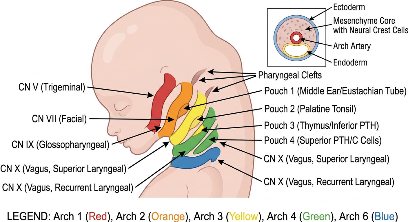
- 5 color-coded arches (1, 2, 3, 4, 6) in lateral embryo view
- Cranial nerve labels for each arch (CN V, VII, IX, X sup., X rec.)
- Pharyngeal pouches (1-4) with their key derivatives
- Pharyngeal clefts on the outer ectoderm surface
- Cross-section inset of a single arch showing ectoderm, mesenchyme core, arch artery, and endoderm layers
To draw in exam
Simple clean hand-drawn style diagram of pharyngeal arches suitable for reproducing in a medical exam. Black lines on white background, no shading or complex art. Show a lateral profile outline of an embryo head and neck (simple oval/teardrop shape). Draw 5 arch bars (numbered 1, 2, 3, 4, 6) as curved ridges on the neck, separated by clefts. Between arches on the inside draw pouches numbered 1-4. Use simple straight labeled arrows pointing outward for: Arch 1 = CN V (mandibular), Arch 2 = CN VII (facial), Arch 3 = CN IX, Arch 4 = CN X sup. laryngeal, Arch 6 = CN X rec. laryngeal. Below each arch number write the key muscle in small text. Add a small separate box showing cross-section of arch with concentric labels: ectoderm outside, mesoderm/neural crest middle, arch artery dot in center, endoderm inside. At bottom add a compact 2-column table: Pouch | Derivative. Style: simple pencil-sketch outline, easy to copy in 5 minutes in exam, clear labels, nothing decorative.
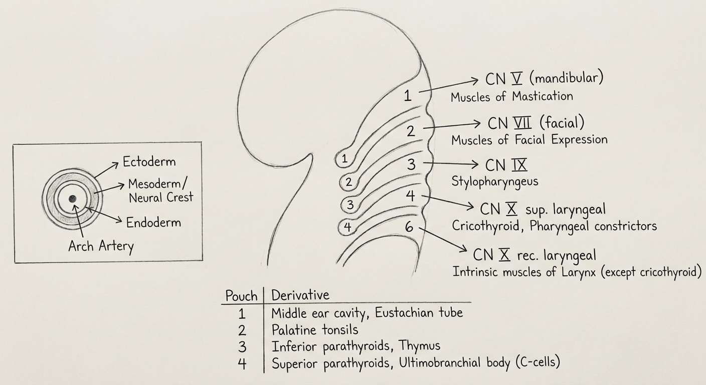
- Simple teardrop/oval for the head, curving neck below
- 5 curved ridges labeled 1, 2, 3, 4, 6 (skip 5!)
- Clefts (ectoderm grooves) between them on the outside
- 4 internal pockets labeled P1, P2, P3, P4 between the arches
| Arch | Nerve | Key muscle/structure |
|---|---|---|
| 1 | CN V (V2/V3) | Mastication muscles |
| 2 | CN VII | Facial expression |
| 3 | CN IX | Stylopharyngeus |
| 4 | CN X (sup. laryngeal) | Cricothyroid |
| 6 | CN X (rec. laryngeal) | Intrinsic laryngeal |
- Outer ring = Ectoderm
- Middle = Neural crest/mesenchyme
- Inner ring = Endoderm
- Central dot = Arch artery
| Pouch | Derivative |
|---|---|
| 1 | Middle ear + Eustachian tube |
| 2 | Palatine tonsil |
| 3 | Thymus + inf. PTH |
| 4 | Sup. PTH + C cells |
Cranial nerve
What would you like about cranial nerves?
all 12 cranial nerves overview diagram base of brain
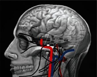
This medical illustration presents a lateral projection of the human cranial region, serving as an anatomical diagram of the skull base, brain, and neurovascular structures. The image overlays a detailed skull rendering with the cerebral hemispheres, emphasizing cortical gyri and sulci. A key feature is the schematic mapping of the twelve cranial nerves (labeled I–XII in Roman numerals), showing their approximate pathways as they exit the brainstem and navigate through skull base foramina. Major vascular structures are highlighted in red (arterial, including the internal carotid artery) and blue (venous), demonstrating their close spatial proximity to the cranial nerves near the zygomatic arch and mandible. The diagram includes orientation labels for 'Anterior' and 'Lateral' perspectives. It is designed for clinical education in neuroanatomy and skull base surgery, illustrating the complex topographic relationships essential for diagnostic imaging and surgical planning in procedures such as microvascular decompression or tumor resection.
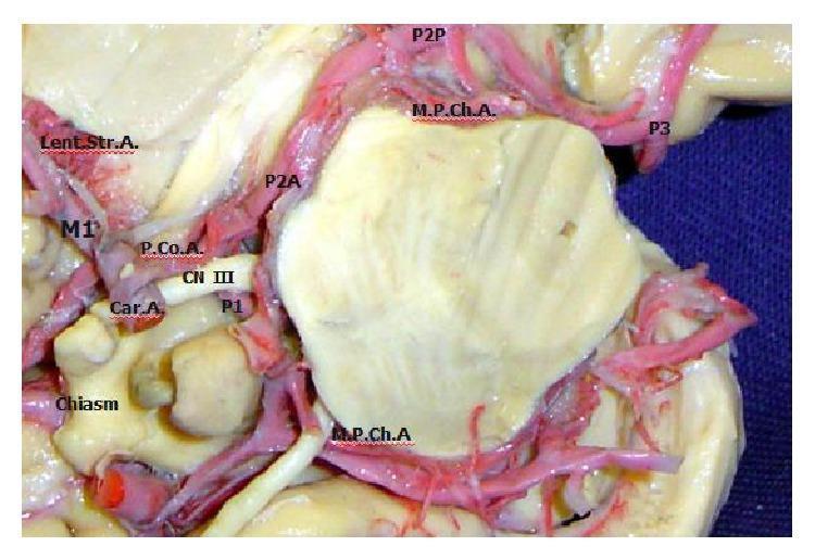
An anatomical diagram featuring a high-magnification photograph of a cadaveric brain dissection, illustrating the neurovascular relationships at the base of the brain. The image highlights the Circle of Willis and associated cranial nerves. Key vascular structures labeled include the internal carotid artery (Car. A.), which branches into the first segment of the middle cerebral artery (M1) and the posterior communicating artery (P.Co.A.). The posterior cerebral artery (PCA) is shown in its various segments: the precommunicating (P1), crural (P2A), ambient (P2P), and quadrigeminal (P3) segments. Also visible are the lateral lenticulostriate arteries (Lent. Str. A.) originating from M1, and the medial posterior choroidal artery (M.P.Ch.A.). Neurological landmarks include the optic chiasm and the oculomotor nerve (CN III), which is positioned between the P.Co.A. and the P1 segment of the PCA. This visual is designed for advanced neuroanatomical education, demonstrating the spatial orientation and arterial supply pathways critical for neurosurgery and vascular neurology.
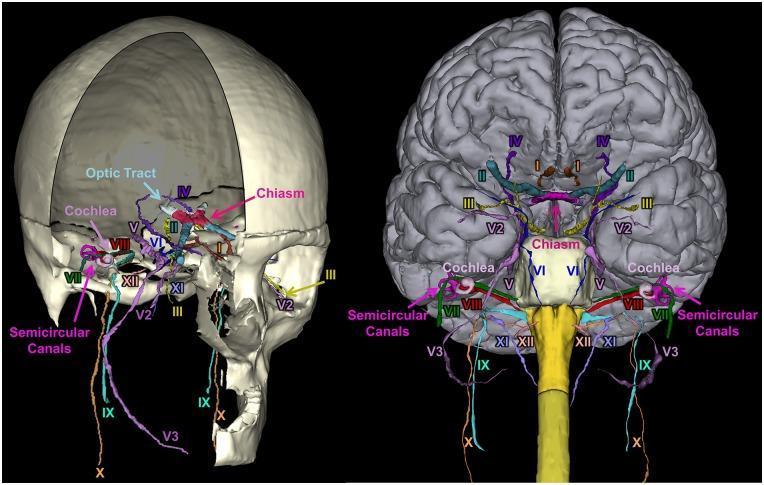
A 3D anatomical reconstruction showcasing the segmentation and spatial orientation of the 12 pairs of human cranial nerves (CN I–XII). The image presents two views: a lateral perspective within a partially transparent skull (left) and an anterior-inferior view relative to the brain and brainstem (right). Each nerve is color-coded for identification: CN I (Olfactory), CN II (Optic) with the optic chiasm and tract, CN III (Oculomotor), CN IV (Trochlear), CN V (Trigeminal) including V2 (maxillary) and V3 (mandibular) divisions, CN VI (Abducens), CN VII (Facial), CN VIII (Vestibulocochlear), CN IX (Glossopharyngeal), CN X (Vagus), CN XI (Accessory), and CN XII (Hypoglossal). Key inner ear structures, including the snail-shaped cochlea and the loop-like semicircular canals, are visualized in relation to the facial and vestibulocochlear nerves. This diagnostic-quality illustration demonstrates the complex neuroanatomical pathways through the skull base and their origins at the brainstem, serving as a pedagogical tool for neurology, radiology, and neurosurgery training.
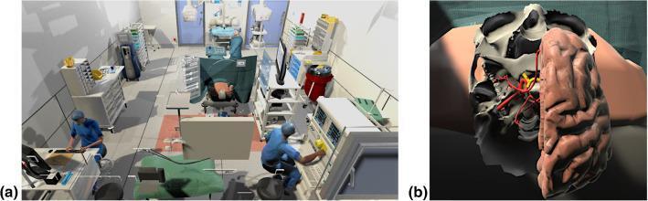
This composite image illustrates a neurosurgical virtual reality (VR) simulation environment. Panel (a) presents an Overview of a Virtual Operating Room, featuring 3D models of surgical staff, anesthesia equipment, and a patient positioned for a procedure. Panel (b) provides a detailed Anatomical Diagram within the simulator, showing a rendered model of the skull base and brain. Key structures include a segmented right cerebral hemisphere with visible gyri and sulci, the Circle of Willis (CoW) highlighted in red to demonstrate cerebrovascular landmarks, and the optic nerve protruding from the optic canal highlighted in yellow. The model is designed for educational training in neurosurgical approaches, such as craniotomy and Sylvian fissure dissection. The visualization emphasizes the spatial relationships between the sphenoid bone, cranial nerves, and major intracranial vessels, providing a high-fidelity environment for pre-operative planning and anatomical study.
All 12 Cranial Nerves — Complete Overview
Functional Components at a Glance
| Type | Abbrev. | Function | CNs |
|---|---|---|---|
| General Somatic Afferent | GSA | Touch, pain, temperature | V, VII, IX, X |
| General Visceral Afferent | GVA | Sensory from viscera | IX, X |
| Special Afferent | SA | Smell, taste, vision, hearing, balance | I, II, VII, VIII, IX, X |
| General Somatic Efferent | GSE | Motor to voluntary muscles | III, IV, VI, XII |
| General Visceral Efferent | GVE | Motor to smooth muscle/glands (parasympathetic) | III, VII, IX, X |
| Branchial Efferent | BE | Motor to pharyngeal arch muscles | V, VII, IX, X, XI |
Parasympathetic outflow in the head travels via CN III, VII, IX, X only.
The 12 Cranial Nerves
CN I - Olfactory
| Type | Special sensory (smell) |
| Origin | Olfactory epithelium (nasal mucosa) → cribriform plate → olfactory bulb |
| Foramen | Cribriform plate (ethmoid) |
| Function | Smell (olfaction) |
| Lesion | Anosmia - loss of smell (head trauma, meningioma, COVID-19) |
| Exam tip | Only CN that does NOT relay in thalamus first |
CN II - Optic
| Type | Special sensory (vision) - technically part of CNS, not PNS |
| Origin | Retinal ganglion cells → optic disc → optic nerve → chiasm → tracts |
| Foramen | Optic canal |
| Function | Vision; pupillary light reflex (afferent limb) |
| Lesion | Bitemporal hemianopia (chiasm lesion = pituitary adenoma); homonymous hemianopia (tract lesion) |
CN III - Oculomotor
| Type | GSE + GVE (parasympathetic) |
| Origin | Midbrain (anterior to cerebral aqueduct) |
| Foramen | Superior orbital fissure |
| Function | Most eye movements (SR, IR, MR, IO); levator palpebrae; pupil constriction + accommodation (via ciliary ganglion) |
| Lesion | "Down and out" eye; ptosis; FIXED DILATED pupil (surgical CN III palsy — e.g. PComm aneurysm). Medical CN III palsy (diabetes) = pupil spared |
| Mnemonic | LR6SO4 — the REST is CN III |
CN IV - Trochlear
| Type | GSE |
| Origin | Midbrain (posterior - only CN to exit dorsal brainstem) |
| Foramen | Superior orbital fissure |
| Function | Superior oblique muscle → depresses eye when adducted |
| Lesion | Vertical diplopia; head tilt away from lesion. Patient tilts head to compensate ("reading palsy") |
| Exam tip | Longest intracranial course; thinnest CN; most common CN injured in closed head trauma |
CN V - Trigeminal
| Type | GSA + BE (branchial motor) |
| Origin | Pons (motor nucleus + main sensory nucleus) |
| Divisions | V1 (ophthalmic) - superior orbital fissure; V2 (maxillary) - foramen rotundum; V3 (mandibular) - foramen ovale |
| Function | Largest CN. Sensory: entire face, scalp, sinuses, teeth, meninges. Motor: muscles of mastication (arch 1) + tensor tympani + tensor veli palatini |
| Reflexes | Corneal reflex (afferent); jaw jerk (both limbs = V3) |
| Lesion | Trigeminal neuralgia (tic douloureux) - electric shock pain V2/V3; numbness in face |
| Ganglia | Trigeminal (Gasserian) ganglion - sensory; no parasympathetic ganglion of its own |
CN VI - Abducens
| Type | GSE |
| Origin | Pons (floor of 4th ventricle) |
| Foramen | Superior orbital fissure |
| Function | Lateral rectus muscle → abducts eye |
| Lesion | Failure to abduct; medial deviation (convergent squint). Most common CN palsy due to raised ICP (false localizing sign - long course) |
CN VII - Facial
| Type | BE + GVE + SA (taste) + GSA |
| Origin | Pons (cerebellopontine angle) |
| Foramen | Internal acoustic meatus → facial canal → stylomastoid foramen |
| Function | Motor: muscles of facial expression (arch 2), stapedius, stylohyoid, post. digastric. Taste: anterior 2/3 of tongue (chorda tympani). Parasympathetic: lacrimal + submandibular + sublingual glands |
| Branches (TEMPORAL) | To Five Zones Be Ready: Temporal, Zygomatic (Frontal), Buccal, Marginal mandibular, Cervical |
| Lesion | UMN lesion = contralateral lower face only (forehead spared). LMN (Bell's palsy) = ipsilateral entire face + hyperacusis + taste loss |
| Ganglia | Geniculate (sensory); pterygopalatine; submandibular |
CN VIII - Vestibulocochlear
| Type | Special afferent (hearing + balance) |
| Origin | Cochlea (cochlear division) + semicircular canals/utricle/saccule (vestibular division) |
| Foramen | Internal acoustic meatus |
| Function | Hearing; balance; nystagmus reflex |
| Lesion | Sensorineural deafness; tinnitus; vertigo; nystagmus. Acoustic neuroma (CN VIII schwannoma) at CP angle → also affects CN VII |
CN IX - Glossopharyngeal
| Type | BE + GVE + GVA + SA |
| Origin | Medulla |
| Foramen | Jugular foramen |
| Function | Motor: stylopharyngeus (arch 3 - only muscle!). Taste: posterior 1/3 of tongue. Sensory: pharynx, carotid sinus/body (baroreceptor + chemoreceptor). Parasympathetic: parotid gland (via otic ganglion) |
| Reflex | Gag reflex (afferent limb) |
| Lesion | Loss of gag reflex afferent; glossopharyngeal neuralgia (rare) |
CN X - Vagus
| Type | BE + GVE + GVA + SA |
| Origin | Medulla (dorsal motor nucleus = parasympathetic; nucleus ambiguus = motor) |
| Foramen | Jugular foramen |
| Function | Motor: pharynx/larynx (arch 4 + 6 muscles via sup. + recurrent laryngeal branches). Parasympathetic: heart, lungs, gut to splenic flexure. Sensory: larynx, trachea, esophagus, thoracic/abdominal viscera |
| Key branches | Recurrent laryngeal (loops under arch 6 L = ductus; arch 4 R = subclavian); superior laryngeal |
| Lesion | Hoarseness (recurrent laryngeal); dysphagia; absent gag reflex efferent; uvula deviates AWAY from lesion |
| Exam tip | Only CN that leaves head/neck to supply thorax + abdomen |
CN XI - Accessory
| Type | BE (branchial efferent) |
| Origin | Cranial root (medulla) + spinal root (C1-C5 spinal cord) |
| Foramen | Jugular foramen |
| Function | Sternocleidomastoid (turns head away) + trapezius (shrugs shoulder) |
| Lesion | Weak shoulder shrug; failure to turn head to opposite side; drooping shoulder |
| Exam tip | Only CN to originate partly from spinal cord |
CN XII - Hypoglossal
| Type | GSE |
| Origin | Medulla |
| Foramen | Hypoglossal canal |
| Function | All intrinsic + extrinsic tongue muscles (except palatoglossus = CN X) |
| Lesion | Tongue deviates TOWARD lesion (LMN); wasting + fasciculations. Away from lesion (UMN). "Lick your wound" - tongue goes to the injured side |
Summary Table
| CN | Name | Type | Foramen | Key Function | Key Lesion |
|---|---|---|---|---|---|
| I | Olfactory | Sensory | Cribriform plate | Smell | Anosmia |
| II | Optic | Sensory | Optic canal | Vision | Hemianopia |
| III | Oculomotor | Motor + Para | SOF | Eye movement, pupil constriction | Down & out, dilated pupil |
| IV | Trochlear | Motor | SOF | Superior oblique | Vertical diplopia, head tilt |
| V | Trigeminal | Both | SOF/FR/FO | Face sensation, mastication | Trigeminal neuralgia |
| VI | Abducens | Motor | SOF | Lateral rectus | Medial deviation |
| VII | Facial | Both + Para | IAM/Stylomastoid | Facial expression, taste ant. 2/3 | Bell's palsy |
| VIII | Vestibulocochlear | Sensory | IAM | Hearing, balance | Deafness, vertigo |
| IX | Glossopharyngeal | Both + Para | Jugular | Stylopharyngeus, taste post. 1/3 | Loss of gag (afferent) |
| X | Vagus | Both + Para | Jugular | Larynx/pharynx, viscera | Hoarseness, uvula deviation |
| XI | Accessory | Motor | Jugular | SCM, trapezius | Weak shoulder shrug |
| XII | Hypoglossal | Motor | Hypoglossal canal | Tongue | Tongue deviation toward lesion |
Classic Mnemonics
- Names: Oh Oh Oh To Touch And Feel Very Good Velvet. Such Heaven → Olfactory, Optic, Oculomotor, Trochlear, Trigeminal, Abducens, Facial, Vestibulocochlear, Glossopharyngeal, Vagus, Spinal accessory, Hypoglossal
- Type (S/M/Both): Some Say Marry Money But My Brother Says Bad Business Marry Money → Sensory, Sensory, Motor, Motor, Both, Motor, Both, Sensory, Both, Both, Motor, Motor
- Parasympathetic CNs: 3, 7, 9, 10 only
- Eye movement CNs: LR6(SO4) rest = 3