
In reference of bailey and love and s das compare pain , vomiting, fever, decubitus of appendicitis, pancreatitis, cholecystitis
Comparison: Appendicitis vs. Pancreatitis vs. Cholecystitis
1. PAIN
| Feature | Acute Appendicitis | Acute Pancreatitis | Acute Cholecystitis |
|---|---|---|---|
| Onset | Gradual (catarrhal) or sudden (obstructive) | Rapid, reaching maximum intensity within minutes | Sudden, often nocturnal onset |
| Initial character | Visceral - periumbilical colic (central/midline) regardless of appendix position (as midgut is a median organ - S. Das) | Severe, constant, refractory to usual analgesic doses | Dull, continuous, severe - "colicky" but not truly colicky |
| Progression | Shifts and localises to right iliac fossa (RIF) as parietal peritoneum becomes irritated | Remains epigastric but may spread diffusely; sometimes localises to either upper quadrant | Localises to right upper quadrant (RUQ) or epigastric region |
| Radiation | None typical; pelvic appendix causes suprapubic pain + tenesmus; retrocaecal causes right flank/posterior pain | To the back in ~50%; relief by leaning forward; can radiate to chest (mimicking MI/pneumonia) | Radiates to the back or right shoulder tip |
| Aggravation | Coughing, sudden movement exacerbate RIF pain | Movement; no specific relief position gives consistent benefit | Movement; fatty food intake can precipitate/worsen episodes |
| Notes | Classic visceral-somatic sequence present in only ~50% (Bailey & Love). Non-obstructive: dull aching; obstructive: colicky (S. Das) | Can mimic most causes of the acute abdomen (Bailey & Love). The suddenness may simulate perforated peptic ulcer | Worsened by deep inspiration during palpation in right subcostal region - Murphy's sign (Bailey & Love) |
2. VOMITING
| Feature | Acute Appendicitis | Acute Pancreatitis | Acute Cholecystitis |
|---|---|---|---|
| Occurrence | Nausea and vomiting present; in obstructive type vomiting is common and early | Nausea, repeated vomiting and retching - usually marked | Present, with associated nausea |
| Severity | Moderate; related to degree of appendix distension (S. Das) | Severe, persistent retching that may continue even with an empty stomach / nasogastric aspiration (Bailey & Love) | Moderate; associated with biliary pain episodes |
| Relation to pain | Follows pain - S. Das emphasises "Pain → Vomiting → Fever" (Murphy's syndrome) | Accompanies pain from early on; hiccoughs also troublesome | Accompanies pain; food - especially fatty food - can trigger both |
| Notes | In catarrhal appendicitis: nausea/vomiting accompany discomfort. In obstructive appendicitis: sudden severe vomiting (S. Das) | Hiccoughs due to gastric distension or diaphragmatic irritation. NGT aspiration may not relieve retching (Bailey & Love) | Dislike for fatty foods is characteristic of gallbladder disease (S. Das: "qualitative dyspepsia") |
3. FEVER
| Feature | Acute Appendicitis | Acute Pancreatitis | Acute Cholecystitis |
|---|---|---|---|
| Timing | Comes last in the sequence (after pain and vomiting) - Murphy's syndrome (S. Das) | Initially body temperature may be normal or even subnormal; rises as inflammation develops (Bailey & Love) | Develops with acute cholecystitis; fever moderate in degree (S. Das) |
| Degree | Mild pyrexia 37.2-37.7°C in early stages; 20% of patients show no pyrexia initially (Bailey & Love). Quite high in children (S. Das). Temperature >38.5°C in children suggests other causes such as mesenteric adenitis (Bailey & Love) | Temperature part of SIRS criteria (>38°C or <36°C). Swinging pyrexia suggests cholangitis complicating gallstone pancreatitis (Bailey & Love) | Raised to a moderate degree - less dramatic than appendicitis in children (S. Das). Fever is a systemic sign in Tokyo Guidelines diagnostic criteria for acute cholecystitis (Bailey & Love) |
| Sequence | Pain → Vomiting → Fever last (Murphy's syndrome - S. Das) | Fever is not an early cardinal feature; part of SIRS; "never an early sign" in acute conditions (S. Das principle) | Fever moderate, part of systemic inflammatory response; accompanies local RUQ signs |
| Notes | After perforation: initially pain subsides temporarily, then peritonitis ensues with subnormal temperature and rising pulse (S. Das) | Cyanosis may be visible in haemorrhagic pancreatitis. Mild icterus seen in gallstone pancreatitis (Bailey & Love) | Intermittent fever (Charcot's triad: fever + rigor + jaundice) suggests calculus in common bile duct, not simple cholecystitis (S. Das) |
4. DECUBITUS (Position of Comfort / Posture)
| Feature | Acute Appendicitis | Acute Pancreatitis | Acute Cholecystitis |
|---|---|---|---|
| Preferred position | Patient lies still, typically on back or slightly flexed on the right side; reluctant to move as movement exacerbates pain | Patient may gain relief by sitting up or leaning forwards - reduces traction on inflamed retroperitoneal pancreas (Bailey & Love) | Patient lies still (movement worsens pain); no specific relief posture documented |
| Movements | Avoids movement; coughing or sudden movement exacerbates RIF pain (Bailey & Love) | Restless due to severity; leaning forward or assuming the knee-chest position may reduce pain | Avoids deep inspiration (Murphy's sign is based on this - pain on inspiring during RUQ palpation) |
| Abdominal wall | Guarding and rebound tenderness over RIF; hyperaesthesia in Sherren's triangle (S. Das) | Abdomen may be distended (ileus); epigastric mass may develop; tenderness with guarding | Tenderness RUQ; Murphy's sign positive; omentum may wall off gallbladder (mass palpable) |
| Notes | Retrocaecal appendicitis: pain and tenderness in right flank or posterior - patient may be rolled to the left side to elicit tenderness (S. Das) | "At the other extreme, gravely ill with profound shock, toxicity and confusion" - prostrated (Bailey & Love) | Contour of abdomen remains normal in uncomplicated cholecystitis and biliary colic (S. Das) |
Summary at a Glance
| Parameter | Appendicitis | Pancreatitis | Cholecystitis |
|---|---|---|---|
| Pain site | Periumbilical → RIF | Epigastric → diffuse | RUQ / epigastric |
| Pain radiation | None (varies by position) | Back (50%), chest | Back / right shoulder |
| Vomiting | Moderate; after pain | Severe, persistent, unrelieved by NGT | Moderate; with fatty food |
| Fever | Mild (37.2-37.7°C); last in sequence | Normal/subnormal early; rises later; SIRS | Moderate degree |
| Sequence | Pain → Vomiting → Fever (Murphy's syndrome) | Pain dominant; fever late | Pain + vomiting together; fever moderate |
| Decubitus | Lies still, reluctant to move | Leans forward / knee-chest for relief | Lies still; avoids deep inspiration |
- Murphy's syndrome (S. Das): In acute appendicitis, the invariable sequence is pain first, then vomiting, then fever. This sequence is diagnostically important.
- Pancreatitis pain is uniquely refractory to standard analgesics and is relieved (partially) by leaning forward - a posture not seen in the other two conditions (Bailey & Love).
- Cholecystitis fever is only moderate, distinguished from the higher fever of appendicitis (especially in children) and the SIRS-range fever of severe pancreatitis (S. Das).
- Vomiting is most severe and persistent in pancreatitis; it can persist despite nasogastric aspiration due to ongoing retching (Bailey & Love).
- The contour of the abdomen remains normal in biliary colic and acute cholecystitis, unlike the distension seen in pancreatitis (with ileus) (S. Das).
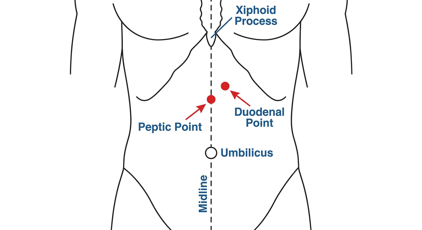
Create a image of human body showing peptic point and duodenal point
Medical diagram of the front of a human body (anatomical illustration, clean clinical style) showing the abdomen with clearly labeled tender points. Show two specific points marked with red dots and arrows: 1) "Peptic Point" - located in the epigastric region, on the midline between the xiphisternum and the umbilicus; 2) "Duodenal Point" - located just to the right of the midline in the epigastric region, slightly above and right of center. Include visible anatomical landmarks: xiphoid process, umbilicus, midline. Label both points clearly with text annotations and arrows. The body outline should be a simple clean anterior view of the torso from chest to pelvis. Use a white or light background, professional medical illustration style.
-
Peptic Point - located on the midline in the epigastric region, midway between the xiphisternum and the umbilicus. Tenderness here is elicited in gastric ulcer.
-
Duodenal Point - located just to the right of the midline in the epigastric region, at roughly the same level as the peptic point. Tenderness here is characteristic of duodenal ulcer (also called the pyloroduodenal point in some texts).
Uti patient history in mbbs 3 rd year
UTI History Taking - 3rd Year MBBS
PRESENTING COMPLAINTS (with duration)
- Burning/pain on passing urine (dysuria) - since ___ days
- Increased frequency of urination - since ___ days
- Urgency (sudden, uncontrollable desire to pass urine) - since ___ days
- Passage of blood in urine (haematuria) - if present
- Suprapubic pain / lower abdominal pain - if present
- Loin pain / flank pain - if upper UTI (pyelonephritis)
- Fever with chills - if present (suggests upper UTI)
Dysuria alone is the single most reliable symptom of UTI. Presence of dysuria + frequency predicts UTI in >90% of cases when vaginal symptoms are absent. - Campbell Walsh Wein Urology
HISTORY OF PRESENTING ILLNESS (HPI)
For Dysuria:
- Onset: Sudden or gradual?
- Character: Burning, stinging, or pain? At the start, end, or throughout micturition?
- Burning throughout urination - typical of cystitis
- Pain at the end of urination (strangury) - suggests bladder neck/trigone involvement
- Severity: How much is it bothering you?
- Progression: Is it getting worse or better?
- Relieving/aggravating factors: Does drinking water help? Does holding urine worsen it?
For Frequency:
- How many times are you urinating during the day? At night (nocturia)?
- Normal is 4-6 times/day; nocturia: 0-1 times at night
- Is the amount passed each time normal, or are you passing small amounts?
For Urgency:
- Do you feel a sudden urge that you cannot control?
- Have you had any leakage of urine before reaching the toilet? (urge incontinence)
For Urine Appearance:
- Is the urine cloudy or foul-smelling?
- Is there blood in the urine? (gross haematuria)
- If yes: is it throughout the stream (total haematuria - suggests bladder/kidney) or at the end (terminal haematuria - suggests bladder neck/trigone)?
- Is blood clots present?
For Loin/Flank Pain (upper UTI):
- Site: unilateral or bilateral?
- Character: dull ache or severe colicky?
- Radiation: does it radiate to the groin? (suggests ureteric calculus as differential)
- Associated with fever, chills, rigors?
For Fever:
- Low grade or high grade?
- Associated with chills and rigors? (rigors suggest bacteraemia/pyelonephritis)
- Time of fever: continuous, intermittent, or remittent?
ASSOCIATED SYMPTOMS (to ask systematically)
| Symptom | Significance |
|---|---|
| Nausea / vomiting | Pyelonephritis; systemic illness |
| Loss of appetite | Systemic infection |
| Flank pain + fever + lower UTI symptoms | Classic triad of pyelonephritis |
| Vaginal discharge / irritation | Suggests vaginitis / STI rather than UTI (reduces UTI probability) |
| Urethral discharge | Suggests urethritis / STI (Chlamydia, Gonorrhoea) |
| Suprapubic fullness / inability to pass urine | Acute urinary retention |
| Backache | Upper UTI, renal pathology |
| Swelling of feet | Renal involvement, nephrotic syndrome |
| Weight loss | Malignancy, TB of urinary tract |
NEGATIVE HISTORY (symptoms to ask about and note as absent)
- No vaginal discharge / No abnormal per vaginal symptoms
- No urethral discharge
- No obstructive symptoms (poor stream, hesitancy, dribbling) - if absent, less likely to be BPH/stricture
- No history of renal stones
- No jaundice
- No joint pains / skin rash (to rule out systemic diseases like SLE causing nephritis)
PAST HISTORY
- Previous episodes of UTI - How many times? How treated? Did they require admission?
- Previous investigations: Any urine culture done before? What organism? What antibiotic was given? Did it resolve?
- History of renal stones / kidney disease
- History of urinary tract surgery / instrumentation - catheterisation, cystoscopy, urethral dilation
- History of diabetes mellitus - major predisposing factor for complicated/recurrent UTI
- History of tuberculosis - sterile pyuria can be due to renal TB
- History of sickle cell disease / anaemia - associated with UTI
- History of spinal cord injury / neurological disease - neurogenic bladder predisposes to UTI
- History of STI (sexually transmitted infection) - Chlamydia, Gonorrhoea can mimic UTI
- History of benign prostatic hyperplasia (BPH) - in elderly males
MENSTRUAL AND OBSTETRIC HISTORY (in female patients - IMPORTANT)
- Last menstrual period (LMP) - Is the patient possibly pregnant? (Pregnancy is a major risk factor and changes management completely)
- Any missed period / pregnancy symptoms?
- Menstrual cycle: Regular or irregular?
- Post-menopausal status - oestrogen deficiency causes atrophic urethritis, increases UTI risk
- Number of pregnancies (gravida/para): Pregnancy increases UTI risk due to ureteric dilatation and vesicoureteric reflux
- Any UTI during previous pregnancies?
Asymptomatic bacteriuria in pregnancy must be treated (unlike in non-pregnant women) as it can progress to pyelonephritis in 20-30% of cases. - Harrison's Principles of Internal Medicine 22e
SEXUAL HISTORY (ask sensitively, with appropriate privacy)
- Is the patient sexually active?
- Recent change in sexual partner or new sexual partner - increases UTI risk (post-coital UTI/"honeymoon cystitis")
- History of unprotected intercourse - to rule out STI as cause of dysuria
- Use of contraceptives: Diaphragm/spermicide use increases UTI risk; condom use is protective
- In men: any penile discharge? Any partners with known STI?
DRUG HISTORY
- Current medications - any antibiotics already started? (affects culture results)
- Cyclophosphamide - causes haemorrhagic cystitis
- Immunosuppressants - increase infection risk
- NSAIDs / analgesics - can cause interstitial nephritis mimicking UTI
- Any known drug allergies? (Especially to sulfa drugs, penicillins, fluoroquinolones - important before prescribing)
- Oral contraceptive pill use - slightly increases UTI risk
PERSONAL HISTORY
- Diet: Fluid intake - is the patient drinking adequate water? (dehydration predisposes)
- Bowel habits: Constipation? (faecal flora close to urethra in women increases ascending infection risk)
- Hygiene practices: Wiping direction after defecation (back to front = risk in females)
- Bladder habits: Does the patient delay urination frequently? (urinary stasis predisposes)
- Occupation: Sedentary work, truck drivers (hold urine for long periods)
- Smoking/alcohol: Note for general history; less directly relevant but ask routinely
- Travel history: Geographic exposure (TB-endemic areas for renal TB differential)
FAMILY HISTORY
- Any family history of renal disease, polycystic kidney disease?
- Family history of diabetes mellitus?
- Family history of recurrent UTI (some genetic predispositions exist - P-blood group antigen receptors for E. coli)
- Family history of urinary tract anomalies or vesicoureteric reflux
SOCIOECONOMIC HISTORY
- Standard of living; water source (contaminated water?)
- Toilet facilities available? (lack of access to toilets leads to urine holding)
- Occupation
SUMMARY OF HISTORY (how to present in viva/ward)
"Mrs. [Name], a [age]-year-old female, presented with a [X]-day history of burning micturition, increased frequency of urination [__ times/day], urgency, and passage of cloudy urine. She also gives a history of lower abdominal pain. There is no vaginal discharge. She has had [X] previous similar episodes, last treated with [antibiotic]. She is a known diabetic on metformin. LMP was [date], she is not pregnant. There is no history of fever, loin pain, or rigors to suggest upper urinary tract involvement."
KEY DISTINGUISHING FEATURES IN HISTORY
| Feature | Lower UTI (Cystitis) | Upper UTI (Pyelonephritis) |
|---|---|---|
| Dysuria | Present | May be present |
| Frequency/urgency | Prominent | Less prominent |
| Fever | Absent or low grade | High fever with rigors |
| Loin/flank pain | Absent | Present (unilateral usually) |
| Nausea/vomiting | Absent | Present |
| Systemic illness | Absent | Present |
| CVA tenderness | Absent | Present |
IMPORTANT RISK FACTORS TO ELICIT IN HISTORY
| Risk Factor | Mechanism |
|---|---|
| Female sex | Short urethra, proximity to rectum |
| Sexual activity | "Milking" of bacteria into bladder |
| Pregnancy | Ureteric dilatation, urinary stasis |
| Diabetes mellitus | Impaired immunity, glucosuria (bacterial growth medium) |
| Urinary catheter | Direct inoculation, biofilm formation |
| Urinary tract obstruction (stone, BPH, stricture) | Urinary stasis |
| Vesicoureteric reflux (VUR) | Ascending infection to kidney |
| Immunosuppression | Decreased host defence |
| Post-menopause | Oestrogen deficiency, vaginal flora change |
| Recent antibiotic use | Disrupts protective flora |
MNEMONICS FOR 3RD YEAR VIVA
- Frequency
- Urgency
- Nocturia
- Dysuria
- Fever (high grade with rigors)
- Loin pain (CVA tenderness)
- Lower UTI symptoms (dysuria, frequency)
- Sexual activity
- Trauma/instrumentation
- Obstruction (stones, BPH)
- Diabetes
- Dehydration
- Anomalies (VUR, horseshoe kidney)
- Renal calculi
Abdominal examination in uti
Abdominal Examination in UTI - MBBS 3rd Year
BEFORE YOU BEGIN
- Position: Patient supine, relaxed, arms by the sides, knees slightly flexed (to relax abdominal muscles)
- Exposure: Nipples to pubic symphysis (fully expose the abdomen)
- Warm hands, stand on the right side of the patient
- Ask about pain site before palpating - always begin away from the painful area
I. GENERAL SURVEY (Before touching the patient)
| Finding | Significance in UTI |
|---|---|
| Ill-looking / flushed / sweating | Suggests systemic infection / pyelonephritis |
| Pallor | Anaemia from chronic UTI, haematuria |
| Jaundice | Associated hepatic dysfunction in severe sepsis |
| Dehydration (dry lips, sunken eyes) | Inadequate fluid intake, vomiting with pyelonephritis |
| Fever | Low-grade: cystitis; High-grade with rigors: pyelonephritis |
| Tachycardia / tachypnoea | Systemic inflammatory response, early sepsis |
| Patient lying still vs. writhing | Peritonitis (lies still) vs. renal/ureteric colic (writhes) |
II. INSPECTION OF THE ABDOMEN
1. Shape / Contour
- Normal contour - typical in uncomplicated cystitis (lower UTI)
- Suprapubic fullness / bulge - suggests bladder distension (urinary retention due to obstruction e.g. BPH, urethral stricture)
- Flank fullness - hydronephrosis, perinephric abscess
2. Skin
- Note any scars (previous surgery - nephrectomy, cystoscopy, catheterisation)
- Herpes zoster vesicles in flank/loin - can mimic UTI pain; prodromal hyperaesthesia before rash
- Surgical stoma - increases UTI risk
3. Movements with Respiration
- Normal in uncomplicated UTI
- Restricted abdominal wall movement with deep inspiration suggests peritoneal irritation (e.g. perinephric abscess)
4. Visible Pulsations / Peristalsis
- Not typically relevant to UTI; note if present to consider other diagnoses
III. PALPATION
A. Superficial Palpation (all 9 regions)
- Guarding / rigidity - absent in simple cystitis; may be present in perinephric abscess or if UTI complicated by peritonitis
- Tenderness map:
| Area | Finding | Significance |
|---|---|---|
| Suprapubic / hypogastric region | Tenderness on light palpation | Cystitis (bladder inflammation) |
| Right / left iliac fossa | Tenderness | Consider appendicitis (right), ovarian pathology (differential) |
| Right / left lumbar (flank) region | Tenderness | Upper UTI, pyelonephritis, perinephric abscess |
| Epigastric region | Usually non-tender in UTI | If tender - consider differential diagnoses |
B. Deep Palpation
- Normally the bladder is not palpable when empty
- If the bladder is palpable as a smooth, rounded, midline suprapubic mass - indicates urinary retention (>300 mL of urine retained)
- The mass arises from the pelvis into the abdomen, is dull to percussion, and the patient may feel urge to urinate on palpation
"Bimanual examination, or renal ballottement, can be performed by placing the non-examining hand posteriorly at the costophrenic angle and palpating for the kidney with the examining hand through the anterior abdominal wall. Deep inspiration can help inferiorly displace the kidney to aid in exposure." - Campbell Walsh Wein Urology
- Left hand: placed posteriorly under the loin at the costovertebral angle (CVA) - between the 12th rib and the lateral border of erector spinae
- Right hand: placed anteriorly in the corresponding flank
- Ask patient to take a deep breath - kidney descends
- Ballot the kidney between the two hands
| Findings | Significance |
|---|---|
| Normal kidney - not palpable (or just the lower pole of right kidney in thin adults) | Normal |
| Enlarged, tender kidney | Pyelonephritis, perinephric abscess, hydronephrosis |
| Smooth, ballottable kidney | Hydronephrosis, polycystic kidney |
| Irregular, hard mass | Renal tumour (differential) |
IV. PERCUSSION
1. Suprapubic Percussion
- Percuss from the umbilicus downward toward the pubis
- Dullness above the pubic symphysis - confirms bladder distension / urinary retention
- Normal finding: resonant (when bladder is empty)
2. Renal / Flank Percussion
- Percuss the flanks in all four quadrants
3. Murphy's Kidney Punch / Fist Percussion (CVA Tenderness) - ★ KEY SIGN ★
"The classic presentation (of pyelonephritis) is an abrupt onset of chills, fever, and unilateral or bilateral flank or costovertebral angle pain and/or tenderness." - Campbell Walsh Wein Urology
- Ask patient to sit upright (or lean forward)
- Place one hand flat over the costovertebral angle (CVA) - angle between the 12th rib and lateral border of erector spinae muscle, posteriorly
- Strike the back of your placed hand with the ulnar border of your other fist
- Elicit from both sides; compare
- Ball of the fist directly percussed over the CVA from behind
| Finding | Interpretation |
|---|---|
| Positive CVA tenderness (pain elicited) | Pyelonephritis, perinephric abscess, renal calculus |
| Bilateral CVA tenderness | Bilateral pyelonephritis |
| Negative (no pain) | Favours lower UTI (cystitis) |
Important: CVA tenderness is absent in uncomplicated cystitis. Its presence immediately suggests upper urinary tract involvement.
V. AUSCULTATION
- Bowel sounds: Normal in UTI; reduced / absent in paralytic ileus (suggests complicated peritonitis)
- Renal bruit: Over the renal angles / epigastrium - a bruit here suggests renal artery stenosis (relevant as a cause of recurrent/resistant hypertension + UTI)
- Absent bowel sounds in a febrile patient with flank pain - may indicate retroperitoneal or peritoneal extension of infection
VI. SPECIAL MANOEUVRES / ADDITIONAL EXAMINATION
1. Deep Palpation for Ureteric Tenderness
- Upper ureteric point: At the junction of lateral border of rectus abdominis and the umbilical horizontal plane (at the umbilical level, 2 cm lateral to midline)
- Middle ureteric point: At the junction of a line joining the ASIS and the pubic tubercle, at the lateral border of rectus abdominis
- Lower ureteric point: Only accessible on per rectal / per vaginal examination
2. Psoas Stretch Test (in suspected perinephric abscess / retroperitoneal irritation)
- Patient in lateral decubitus
- Extend the hip passively - pain suggests irritation of psoas (perinephric collection)
3. Per Rectal Examination (in male patients)
- Assess the prostate gland: size, consistency, tenderness
- Exquisitely tender, swollen prostate = acute prostatitis (UTI variant in males)
- Enlarged, smooth, non-tender prostate = BPH (predisposing factor for UTI in males)
4. Per Vaginal Examination (in female patients)
- Vaginal discharge? (suggests vaginitis / STI as differential, reduces UTI probability)
- Cervical excitation tenderness? (suggests PID - important differential)
- Assess for pelvic organ prolapse (can cause incomplete bladder emptying → recurrent UTI)
- Urethral palpation: urethral diverticulum, Skene's gland abscess
VII. FINDINGS SUMMARY TABLE
| Sign | Lower UTI (Cystitis) | Upper UTI (Pyelonephritis) | Urinary Retention |
|---|---|---|---|
| General appearance | Looks well / mildly ill | Looks ill, flushed, toxic | Distressed, in pain |
| Fever | Absent / low-grade | High-grade with rigors | Absent |
| Abdominal contour | Normal | Normal / flank fullness | Suprapubic fullness |
| Suprapubic tenderness | Present | May be present | Present (tense) |
| Flank/loin tenderness | Absent | Present | Absent |
| Bladder palpable | No | No | Yes (midline, dull) |
| Kidney ballottement | Not enlarged | Enlarged / tender | Not enlarged |
| CVA tenderness (fist) | Negative | Positive (hallmark) | Negative |
| Bowel sounds | Normal | Normal / reduced | Normal |
HOW TO PRESENT IN VIVA
"On abdominal examination, the patient was febrile and appeared ill. The abdomen was flat with no visible distension. On palpation, there was deep tenderness in the right lumbar and right iliac fossa regions. The kidney was ballottable and tender on the right side. Bladder was not palpable. On percussion, there was positive right-sided costovertebral angle tenderness (Murphy's kidney punch positive on the right). Bowel sounds were present. These findings are consistent with right-sided acute pyelonephritis."