
Angioplasty
"angioplasty"[MeSH Terms] AND "percutaneous coronary intervention"
balloon angioplasty and stent placement diagram
coronary angioplasty balloon stent
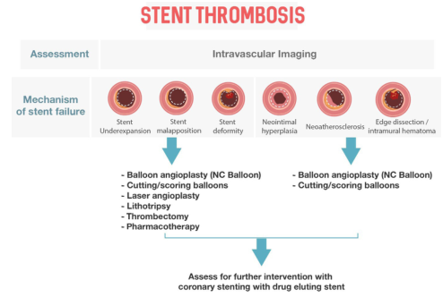
Summary : This flowchart outlines the assessment and management of stent thrombosis, focusing on mechanisms of stent failure identified by intravascular imaging and corresponding interventional strategies.
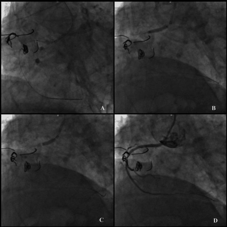
A four-panel (A-D) coronary angiogram documenting a complex percutaneous coronary intervention (PCI) for a right coronary artery (RCA) ostial lesion involving in-stent restenosis with stent protrusion. Panel A: Initial phase showing a guiding catheter attempting to engage the RCA ostium, with a coronary guidewire successfully crossing into the vessel through a side strut of an existing, overhanging stent. Panel B: Sequential balloon dilation showing an inflated balloon catheter positioned within the ostial lesion to expand the narrowed segment. Panel C: Demonstrates post-dilation using a non-compliant balloon to optimize stent apposition and luminal diameter. Panel D: The final angiographic result showing a patent RCA with restored blood flow and a more uniform vessel diameter, indicating successful lesion treatment. The images highlight the technical challenge of managing protruding ostial stents and the use of balloon angioplasty and stenting to re-establish coronary perfusion in the setting of acute coronary syndrome.
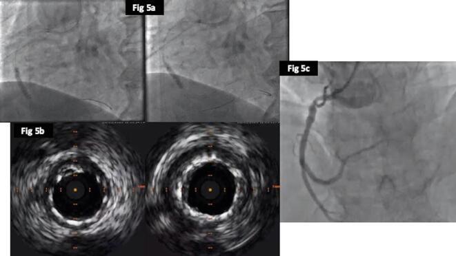
This composite figure illustrates a percutaneous coronary intervention (PCI) procedure for in-stent restenosis (ISR). Panel 5a consists of two fluoroscopic frames showing balloon angioplasty (POBA) being performed to optimize a previously deployed stent in the Right Coronary Artery (RCA); a radiopaque balloon catheter is visible within the vessel lumen. Panel 5b presents cross-sectional Intravascular Ultrasound (IVUS) images, showing a circular, widely patent lumen with improved stent expansion and wall apposition following the intervention. The bright, circumferential echoes represent the expanded stent struts against the vessel wall. Panel 5c displays the final coronary angiogram, revealing an excellent procedural result with a smooth, unobstructed lumen in the RCA and successful restoration of distal blood flow without signs of residual stenosis or dissection. This sequence demonstrates the use of intravascular imaging to guide and verify successful balloon optimization in complex coronary revascularization cases.
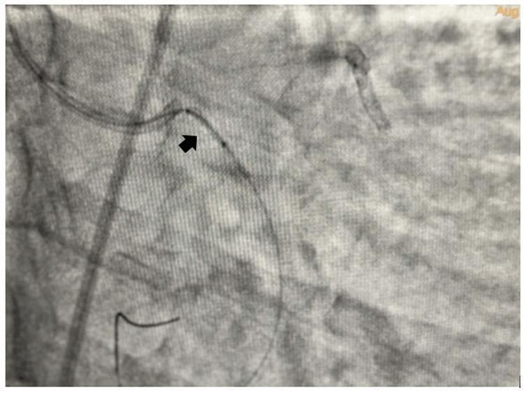
This diagnostic image is a coronary angiogram captured during an interventional cardiology procedure. The grayscale fluoroscopic view demonstrates the left circumflex coronary artery (LCX) anatomy. Visible is a radiopaque 0.014-inch guidewire navigated through the vessel lumen. A small (1.25x8 mm) semicompliant angioplasty balloon is shown inflated within the proximal segment of a dislodged coronary stent, indicated by a black arrow. The radiopaque markers of the balloon catheter are visible, delineating its position relative to the stent and the vessel wall. This visual illustrates a bail-out maneuver for stent dislodgement, where a small balloon is used to re-engage or track through a displaced stent when a full-sized catheter cannot cross. The background shows characteristic pulmonary and cardiac tissue mottling typical of invasive angiography. This material is intended for advanced medical education in percutaneous coronary intervention (PCI) and complication management.
Angioplasty (percutaneous transluminal angioplasty)
Basic technique
- Access is gained through the radial or femoral artery.
- A guide catheter is engaged at the vessel origin. A thin guidewire (0.014 inch in the coronaries) is passed across the lesion.
- A balloon is advanced over the wire and inflated under fluoroscopy. This compresses and remodels the plaque and stretches the vessel wall.
- A stent is usually deployed, then post-dilated if needed. Intravascular imaging (IVUS or OCT) can check expansion and apposition.
Coronary indications
- Stable angina: PCI is for symptoms that persist despite medical therapy, with ischemia on stress testing. PCI relieves angina better than medical therapy. In stable exertional angina, trials show it does not reduce death or MI compared with optimal medical therapy (Harrison's Principles of Internal Medicine 22E, p. 2147).
- Unstable angina and MI: PCI improves outcomes, including early in MI with or without cardiogenic shock (Harrison's, p. 2147).
- STEMI: Guidelines favor primary PCI over fibrinolysis if first medical contact to balloon time is under 90 to 120 minutes (Tintinalli's Emergency Medicine, p. 385).
- Left main or three-vessel disease: CABG is generally best, especially with diabetes or impaired LV function. PCI can be considered in selected patients, such as those with prohibitive surgical risk (Harrison's, p. 2147).
Outcomes and complications
- Success: Primary success with angina relief is above 95%.
- Stents: Current drug-eluting stents have restenosis rates under 5%, lower than bare metal stents, and are the standard of care.
- Complications: These are mainly dissection, thrombosis with vessel occlusion, uncontrolled ischemia and ventricular failure.
- Stent thrombosis: Delayed endothelial healing around drug-eluting stents extends the period of risk for subacute stent thrombosis.
- Vein grafts: An embolic protection device improves success in diseased vein grafts.
Antithrombotic therapy
- Aspirin, a P2Y12 inhibitor and an antithrombin (heparin or bivalirudin) are given to reduce thrombus formation.
- After a drug-eluting stent, continue aspirin indefinitely. Give a P2Y12 antagonist ideally for 1 year (dual antiplatelet therapy, DAPT).
- Longer DAPT, up to 30 months, may add benefit but increases bleeding.
- Shorter DAPT is used in patients with high bleeding risk or a long-term indication for oral anticoagulation.
- Newer approaches under study include dropping aspirin after 1 to 3 months, de-escalating prasugrel or ticagrelor to clopidogrel, and low-dose DOAC plus an antiplatelet (Harrison's, p. 2147).
Peripheral angioplasty (e.g., superficial femoral artery)
- Plain balloon angioplasty (POBA) is inferior to other SFA treatments except in short TASC A lesions away from the adductor canal.
- Paclitaxel drug-coated balloons reduce restenosis compared with POBA. They approach 1-year SFA stent patency.
- Balloon diameter is chosen 5 to 10% larger than the vessel. Typical sizes are 5 to 7 mm for the SFA, 4 to 6 mm for the popliteal and 2 to 3 mm for tibial arteries.
- Lesion preparation (POBA, atherectomy, balloon lithotripsy) improves results.
- Cutting or scoring balloons are mostly reserved for neointimal hyperplasia at bypass anastomoses.
- Source: Fischer's Mastery of Surgery, 8th ed., p. 6599-6600.