
Assalaam o alykum! Dear students,, Here is the syllabus for upcoming Class test of Pathology on Monday, 27th July Insha.ALLAH: Topic : Muscle Remodeling Code: *MS-Pa-001* Specific LO: Describe the Hyperplasia, hypertrophy, and atrophy of muscle fibre Topic: Diseases of Bone Code: *MS-Pa-003* Specific LO: Describe the clinical presentation and histological justification for osteoporosis, osteopetrosis ° Briefly enlist the steps of repair Topic: Disease of Cartilage Code: *MS-Pa-004* Specific LO: Describe the histological basis for bone repair after fracture and cartilage growth and repair
Reading File
Wa Alaikum Assalam! I'll build comprehensive notes for all three topics right away. Let me search the medical library in parallel.
Finding Sources
Finding Sources
Finding Sources
Finding Sources
Finding Sources
Finding Sources
Good - I have key file paths. Now let me read the most relevant sections in parallel.
Reading File
Reading File
Reading File
Finding Sources
Finding Sources
Excellent - Robbins Pathology has exactly what I need. Let me read those sections now.
Reading File
Reading File
Reading File
Reading File
Reading File
Finding Sources
Reading File
Reading File
I now have comprehensive material from Guyton (muscle), Robbins Pathology 10e & Basic Path (osteoporosis, osteopetrosis), Miller's Orthopaedics (fracture repair), and Rockwood & Green (cartilage). Let me now compile the full study notes.Here are your complete, exam-ready notes for Monday's Pathology test. Insha'Allah they serve you well!
📚 Pathology Class Test Notes
Monday, 27th July | Musculoskeletal Pathology
MS-Pa-001 | Muscle Remodeling
Learning Objective: Hyperplasia, Hypertrophy, and Atrophy of Muscle Fibre
🔴 Muscle Hypertrophy
Definition: An increase in the size (not number) of individual muscle fibres.
Mechanism:
- Occurs in response to increased work demand (e.g., exercise, chronic loading)
- The myofibrils within each fibre increase in number and size
- New sarcomeres are added at the ends of muscle fibres when a muscle is stretched to greater than normal length - new sarcomeres can be added at a rate of several per minute
- Enzyme systems for energy production also increase, especially glycolytic enzymes, to supply rapid energy during forceful contraction
Key mediators:
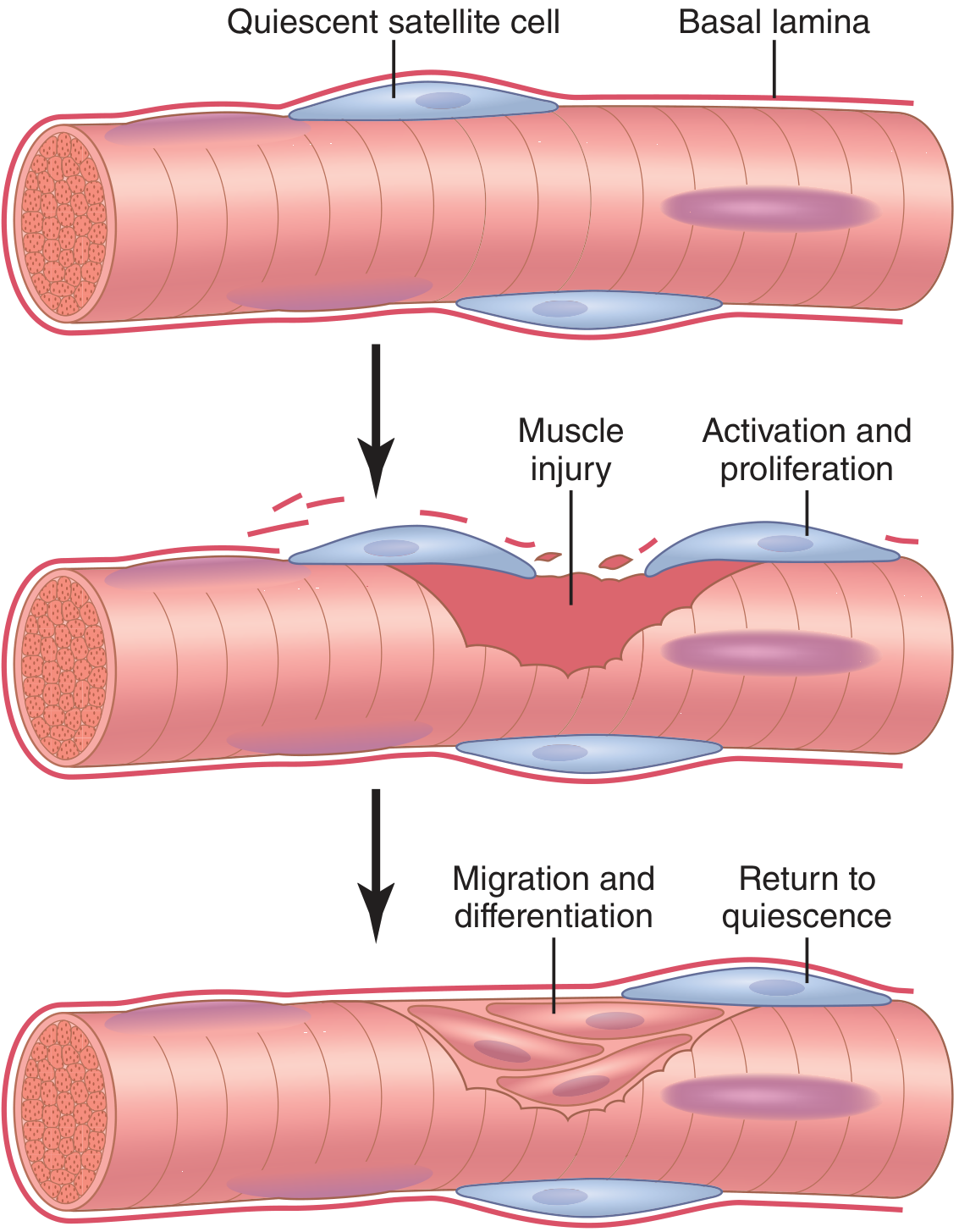
- Satellite cells - adult stem cells that lie quiescent beneath the basal lamina above the sarcolemma
- Upon activation (by exercise or injury), they proliferate, differentiate, migrate through the sarcolemma, and fuse with existing fibres to contribute to repair and hypertrophy
- Some satellite cells self-renew to ensure continued repair capacity
Satellite cells: quiescent beneath basal lamina → activated → proliferate → differentiate → fuse with fibre or self-renew (Guyton & Hall, Medical Physiology)
🔴 Muscle Hyperplasia
Definition: An actual increase in the number of muscle fibres (rare).
Key facts:
- Occurs only under extreme conditions of muscle force generation
- Increase is only by a few percent - a minor contributor compared to hypertrophy
- Primary mechanism: Linear splitting of previously enlarged fibres (not satellite cell division producing new fibres)
- Occurs in addition to fibre hypertrophy, not instead of it
Source: Guyton & Hall Textbook of Medical Physiology
🔴 Muscle Atrophy
Definition: A decrease in muscle fibre size due to degradation exceeding synthesis.
Two main causes:
1. Disuse Atrophy
- When a muscle remains unused for many weeks, the rate of degradation of contractile proteins exceeds replacement
- The key pathway: ATP-dependent Ubiquitin-Proteasome Pathway
- Proteasomes = large protein complexes that degrade damaged or unneeded proteins by proteolysis
- Ubiquitin = regulatory protein that labels cells targeted for proteosomal degradation
- Conversely: when a muscle remains shortened to less than normal length, sarcomeres at the ends disappear
2. Denervation Atrophy
- When a muscle loses its nerve supply, it no longer receives signals to maintain normal size
- Atrophy begins almost immediately after denervation
- After ~2 months: degenerative changes appear in fibres
- If nerve regrows rapidly: full return of function possible in as little as 3 months
- No further functional return after 1-2 years of denervation
Sarcopenia: Age-related reduction in satellite cell populations and repair capacity contributes to progressive loss of muscle mass. Exercise activates satellite cells and may prevent or reverse some effects of aging on muscle mass.
MS-Pa-003 | Diseases of Bone
Learning Objective 1: Osteoporosis - Clinical Presentation & Histological Justification
Definition
Osteoporosis = osteopenia severe enough to significantly increase the risk of fracture.
- Radiographically: bone mass ≥ 2.5 SD below mean peak bone mass in young adults (T-score ≤ -2.5)
- Osteopenia = 1 to 2.5 SD below the mean
Types
| Type | Mechanism |
|---|---|
| Postmenopausal | Estrogen loss → ↑ RANKL → ↑ osteoclast activity → excess resorption |
| Senile | Osteoblasts show reduced proliferative & biosynthetic capacity; "low-turnover" form |
| Secondary | Endocrine (Cushing's, hyperthyroidism), GI malabsorption, drugs (corticosteroids) |
Pathogenesis
Peak bone mass is achieved in early adulthood. From the 4th decade onwards, resorption exceeds formation. Key factors:
- Reduced estrogen (postmenopausal) - estrogen normally suppresses RANKL and promotes OPG (decoy receptor); its loss → unchecked osteoclast activity
- Age-related changes - osteoblasts have reduced biosynthetic response to growth factors
- Reduced physical activity - osteocytes detect mechanical stress and signal bone formation; reduced activity reduces this stimulus
- Calcium/Vitamin D deficiency - impairs mineralization and increases PTH
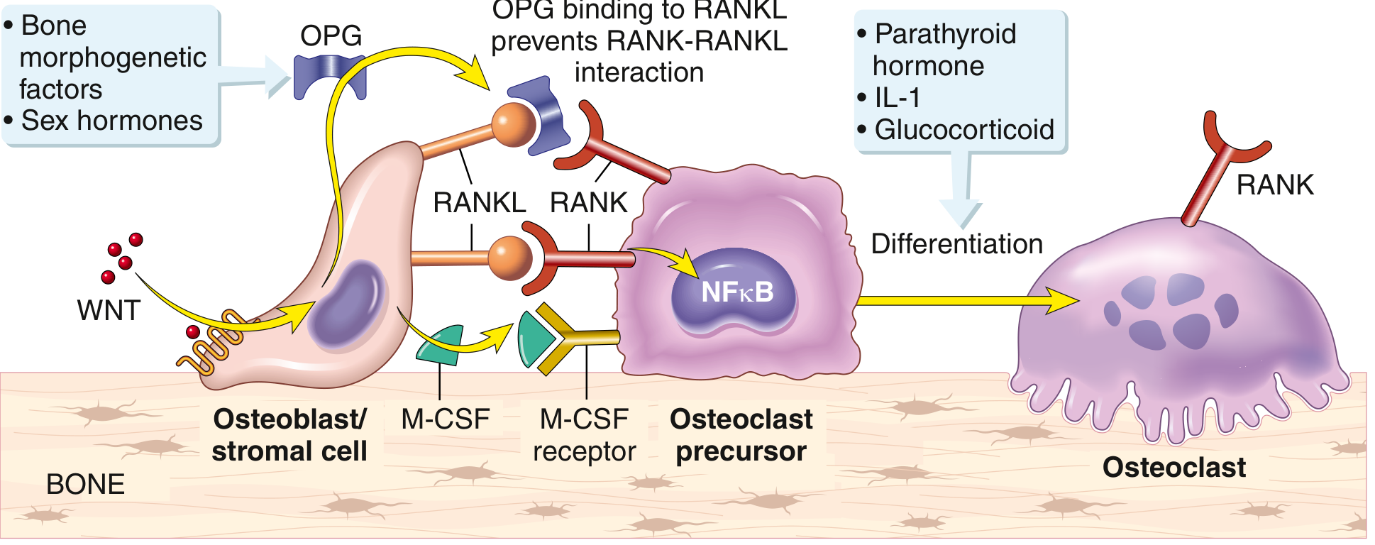
RANKL-OPG Axis (Core Mechanism):
- RANKL (on osteoblasts/stromal cells) binds RANK on osteoclast precursors → osteoclast differentiation and bone resorption
- OPG (osteoprotegerin) = decoy receptor secreted by osteoblasts in response to WNT signaling → blocks RANKL-RANK binding → inhibits resorption
- Estrogen loss tips the balance toward RANKL dominance
Paracrine regulation of osteoclast formation via RANKL/RANK/OPG (Robbins Basic Pathology)
Histological Features (Justification)
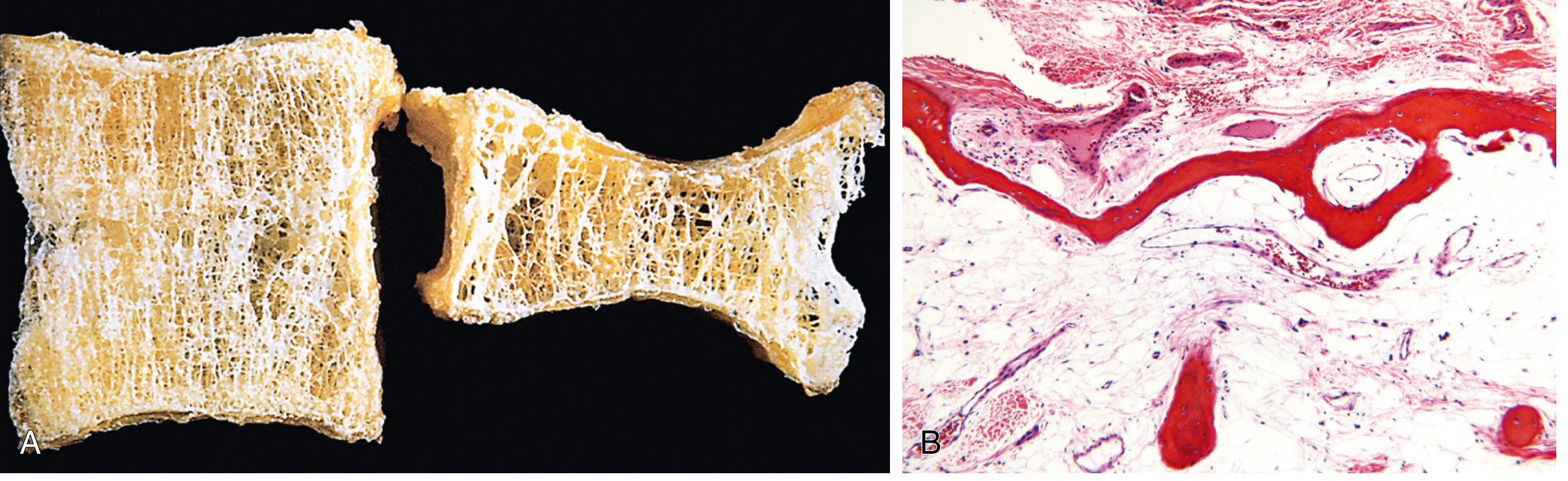
The hallmark: histologically NORMAL bone that is DECREASED in quantity.
- Bone that is present is normally mineralized (this distinguishes it from osteomalacia)
Postmenopausal osteoporosis - affects mainly cancellous bone (high surface area):
- Trabecular plates become thinned and perforated
- Loss of interconnections between trabeculae
- Leads to microfractures → eventually vertebral body collapse
- Characteristic: loss of horizontal trabeculae with relative preservation of vertical trabeculae
Senile osteoporosis - affects both cortical and cancellous bone:
- Cortex is thinned by subperiosteal and endosteal resorption
- Haversian systems are widened
(A) Osteoporotic vertebral body - loss of horizontal trabeculae, compression fractures. (B) Advanced osteoporosis - thinning of both trabecular and cortical bone (Robbins Pathologic Basis of Disease 10e)
Clinical Features
- Vertebral fractures (thoracic & lumbar) - painful; multiple fractures → loss of height, kyphoscoliosis, lumbar lordosis
- Femoral neck, pelvis, spine fractures → immobilization → pulmonary embolism, pneumonia → >50,000 deaths/year
- Plain X-rays: osteoporosis NOT detectable until 30-40% of bone mass is lost
- Blood tests (Ca, PO4, Alk Phos) are usually normal
- Diagnosis: DEXA scan (dual-energy X-ray absorptiometry) or quantitative CT
Treatment
- Exercise, calcium & Vitamin D supplementation
- Bisphosphonates - reduce bone resorption (first-line)
- Denosumab - anti-RANKL antibody (promising for postmenopausal osteoporosis)
- Menopausal hormone therapy (estrogen) - slows bone loss but limited by DVT/stroke risks
Learning Objective 2: Osteopetrosis - Clinical Presentation & Histological Justification
Definition
Osteopetrosis (= Marble Bone Disease / Albers-Schönberg disease) = a group of rare genetic diseases characterized by:
- Reduced bone resorption due to impaired osteoclast formation or function
- Diffuse, symmetric skeletal sclerosis
- Bones appear stone-like on X-ray but are abnormally brittle and fracture easily
Pathogenesis
Core defect = Failure to acidify the osteoclast resorption pit
Osteoclasts normally create a sealed extracellular trench and secrete acid + neutral proteases (MMPs) to dissolve calcium hydroxyapatite. Mutations disrupt this:
| Mutation | Effect |
|---|---|
| CLCN7 (proton-chloride exchanger) | Mild autosomal dominant form - cannot acidify resorption pit |
| TCIRG1 (vacuolar H⁺-ATPase subunit) | Most common autosomal recessive form - acid production fails |
| Carbonic anhydrase 2 deficiency | Autosomal recessive form |
Result: Osteoclasts are present but non-functional → bone matrix accumulates without being resorbed → dense but structurally weak bone
Classification
| Type | Inheritance | Onset |
|---|---|---|
| Autosomal recessive ("malignant") | AR | Infancy - severe, often fatal if untreated |
| Autosomal dominant ("benign") | AD (Albers-Schönberg) | Adulthood - milder |
Histological Features
- Bones are hyperdense and sclerotic - medullary canal is obliterated by unresorbed bone
- Osteoclasts may be increased in number but are non-functional (resorption pits absent)
- Woven bone persists - normal remodeling to lamellar bone does not occur
- Cartilage remnants within bone (in growth plate region) - endochondral ossification occurs but chondroclasts cannot resorb the primary spongiosa
- On X-ray: characteristic "Erlenmeyer flask" deformity of metaphyses; "bone within bone" appearance
Clinical Features
| Feature | Explanation |
|---|---|
| Pathological fractures | Dense but brittle bone (chalk-stick fractures) |
| Anemia, thrombocytopenia, extramedullary hematopoiesis | Obliteration of medullary cavity → no space for marrow → liver/spleen take over hematopoiesis |
| Hepatosplenomegaly | Extramedullary hematopoiesis |
| Cranial nerve palsies (CN II, VII most common) | Foraminal narrowing compresses cranial nerves → blindness, facial palsy |
| Recurrent infections | Reduced marrow space → leukopenia |
| Hypocalcemia | Osteoclasts cannot release calcium from bone |
| Dental abnormalities | Failure of tooth eruption, osteomyelitis of jaw |
Learning Objective 3: Steps of Bone Repair (Briefly)
Three Stages of Fracture Repair
Stage 1: INFLAMMATION (Days 1-7)
- Fracture disrupts periosteum and blood vessels → Fracture hematoma forms
- Hematoma provides hematopoietic cells that secrete growth factors (TGF-β, PDGF, BMP)
- Neutrophils and monocytes arrive → phagocytosis of debris
- Fibroblasts, mesenchymal cells, and osteoprogenitor cells migrate to site
- Granulation tissue forms around fracture ends
Stage 2: REPAIR (Weeks 2-12)
- Primary callus response within 2 weeks
- For fracture ends not in continuity: soft (bridging) callus forms - consists of fibrocartilage and fibrous tissue (type II collagen expressed early)
- Soft callus undergoes endochondral ossification → replaced by woven bone (hard callus)
- Intramedullary (medullary) callus forms more slowly to supplement bridging callus
- Amount of callus is inversely proportional to degree of immobilization (more motion = more callus)
- Type II collagen (cartilage) → Type I collagen (bone) transition
Stage 3: REMODELING (Months to years - up to 7 years)
- Begins in the middle of the repair phase
- Woven bone is replaced by lamellar bone (organized parallel collagen)
- Bone remodels according to Wolff's Law - assumes normal configuration according to mechanical stress and electric charges (piezoelectric)
- Fracture healing is complete when the marrow space is repopulated
- Extramedullary callus provides greater structural strength than intramedullary callus (bending stiffness increases with the 4th power as callus is deposited further from the neutral axis)
Source: Miller's Review of Orthopaedics 9e; Rockwood & Green's Fractures in Adults 10e
MS-Pa-004 | Disease of Cartilage
Learning Objective: Histological Basis for Bone Repair After Fracture AND Cartilage Growth and Repair
Part A: Histological Basis of Bone Repair
Two types of bone matrix relevant to healing:
| Bone Type | Features | Significance |
|---|---|---|
| Woven bone | Haphazard collagen arrangement, more cellular, disorganized | Formed rapidly during fetal development AND fracture repair; abnormal in adult at rest |
| Lamellar bone | Parallel collagen fibers, organized Haversian systems | Normal mature bone; structural stability |
The histological sequence during fracture repair:
- Hematoma - clot provides scaffold and growth factor reservoir (TGF-β, BMP, PDGF)
- Fibrovascular granulation tissue - fibroblasts synthesize collagen type III initially
- Soft callus (fibrocartilage) - chondrocytes differentiate from MSCs; type II collagen deposited; resembles growth plate
- Hard callus (woven bone) - endochondral ossification converts fibrocartilage to woven bone via osteoblast activity; type I collagen replaces type II
- Remodeling to lamellar bone - osteoclasts resorb woven bone; osteoblasts deposit organized lamellar bone along stress lines
Key histological marker: Woven bone in an adult skeleton ALWAYS indicates active bone repair or pathological process.
Part B: Cartilage Growth and Repair
Normal Cartilage Growth
Cartilage grows by two mechanisms:
- Appositional growth - new cartilage added at the periphery by chondroblasts from the perichondrium, which differentiate into chondrocytes
- Interstitial growth - chondrocytes within lacunae divide and produce new matrix from within (possible because cartilage matrix is flexible) - this is the main mechanism of long bone elongation at growth plates
Growth Plate (Physeal) Zones (endochondral ossification):
| Zone | Histology |
|---|---|
| Resting zone | Small, scattered chondrocytes; reserve cells |
| Proliferative zone | Chondrocytes rapidly dividing, forming columns |
| Hypertrophic zone | Enlarged chondrocytes; matrix mineralizes |
| Zone of provisional calcification | Matrix calcified; blood vessels invade; osteoblasts deposit woven bone |
Cartilage Repair - Why It Fails
Cartilage has very limited intrinsic repair capacity due to:
- Avascular - no blood supply to initiate or support repair
- Low cell density - few chondrocytes per unit volume
- Low migratory capacity - chondrocytes do not migrate readily through the dense matrix to the injury site
- Limited stem cell population - insufficient progenitors to repopulate defects
- Chondrocyte death in tissue surrounding the injury impairs integration
Response to Isolated Chondral Injury (No subchondral bone involvement)
- Viable chondrocytes near injury may proliferate and form clusters (chondrocyte clones)
- Synthesize new matrix - but insufficient to recapitulate native cartilage
- Synovial mesenchymal progenitor cells may migrate to defect via:
- Direct contact with synovium, or
- Pannus-like migration of synovial membrane onto articular surface
- These progenitors (marked by DPP4 and PI16 expression; derived from GDF-5-expressing joint interzone lineage) can differentiate into chondrocyte-like cells and deposit repair matrix
- Mediated in part by YAP1 (Yes-associated protein-1)
- Result: Functionally inferior fibrocartilage - not true hyaline cartilage
Response to Osteochondral Injury (Subchondral bone also involved)
This triggers a much better healing response because blood vessels are accessible:
- Subchondral vessels rupture → hematoma fills both bony and chondral defect
- Inflammatory phase: neutrophils/monocytes → phagocytosis of debris
- Mesenchymal progenitors from bone marrow migrate into clot → synthesize collagenous matrix
- Some cells assume rounded morphology → synthesize type III and type I collagen (not native type II + proteoglycans)
- Within weeks: differentiation into fibrocartilaginous and osseous tissues → fibrocartilage repair tissue
- The repair matrix has some chondrocyte-like properties but is type I/III collagen-rich rather than type II collagen + proteoglycans (as in native hyaline cartilage)
- Subsequent fate varies - some defects improve, others deteriorate
Bottom line: Osteochondral injuries heal with fibrocartilage (not hyaline cartilage). Fibrocartilage is biomechanically inferior and lacks the compressive resistance of hyaline cartilage, predisposing to secondary osteoarthritis.
📝 Quick Revision Summary Table
| Topic | Key "1-liner" for MCQs |
|---|---|
| Muscle hypertrophy | ↑ myofibril size + new sarcomeres added; driven by satellite cells |
| Muscle hyperplasia | ↑ fibre number by linear splitting; rare, only under extreme load |
| Disuse atrophy | Ubiquitin-proteasome pathway degrades contractile proteins |
| Denervation atrophy | Immediate; irreversible after 1-2 years |
| Osteoporosis histology | Normal bone, decreased quantity; trabecular perforation; thinned cortex |
| Osteoporosis clinical | Vertebral crush fractures; femoral neck #; normal Ca/PO4/ALP; diagnosed by DEXA |
| Osteopetrosis | Marble bone; ↓ osteoclast function; dense but brittle; marrow obliteration → pancytopenia |
| Fracture repair | Hematoma → granulation tissue → soft callus → hard callus → remodeling (woven → lamellar) |
| Cartilage repair | Poor; avascular; fibrocartilage (type I/III) replaces hyaline (type II); osteochondral > chondral healing |
Sources: Guyton & Hall Medical Physiology | Robbins Pathologic Basis of Disease 10e | Robbins & Kumar Basic Pathology | Miller's Review of Orthopaedics 9e | Rockwood & Green's Fractures in Adults 10e
Best of luck for your test tomorrow, Insha'Allah! 🤍
This is a shared conversation. Sign in to Orris to start your own chat.