
Make notes on pitrosporum ovale for md derma theory exam
Malassezia furfur seborrheic dermatitis
KOH preparation Malassezia spaghetti and meatballs yeast and hyphae
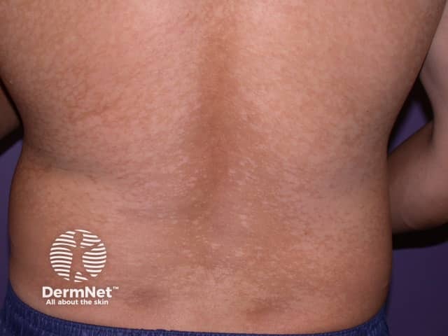
Clinical photography of the back using a high‑resolution digital camera under standard white‑light illumination was obtained to document cutaneous skin findings. The view is a posterior trunk (dorsal back) with bilateral, well‑circumscribed hypopigmented macules and patches showing fine surface scaling. Lesions are diffuse across the mid to lower back with mild contrast against surrounding tan skin, consistent with pityriasis versicolor (tinea versicolor). The textural appearance is smooth, with less erythema than inflammatory dermatoses; scaling is subtle and best appreciated on oblique lighting. Pityriasis versicolor is a superficial fungal infection caused by Malassezia species that disrupt epidermal pigmentation, producing hypo‑ or hyperpigmented macules and mild pruritus in some patients. The condition commonly affects the trunk and proximal limbs in warm, humid climates and may be asymptomatic. In the differential diagnosis, vitiligo, pityriasis alba, and postinflammatory hypopigmentation are considerations. If tested, KOH preparation or fungal culture can reveal yeast with short hyphae (“spaghetti and meatballs”) compatible with Malassezia species. Management typically involves topical antifungals; extensive cases may require oral therapy. This photograph serves educational and reference purposes for pattern recognition, trunk involvement, and characterization of pigmented skin lesions in dermatology practice and training.
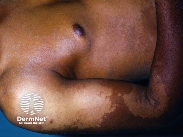
Clinical photograph of in vivo human forearm skin showing hypopigmented, confluent patches compatible with pityriasis versicolor (tinea versicolor). The image demonstrates localized involvement of the volar forearm with pale, irregularly shaped macules that coalesce into larger hypopigmented patches. The lesions display subtle surface scaling and well-defined but variable borders; surrounding skin is darker, giving a contrast typical for this condition. The distribution is on sun-exposed or humid skin regions and may appear more prominent after tanning. Pathophysiology involves superficial Malassezia yeast colonization of the stratum corneum with production of acids that disrupt melanin synthesis, leading to pigmentary heterogeneity. Clinically, pityriasis versicolor presents as nonpruritic hypo- or hyperpigmented patches that fluoresce under Wood's lamp. Differential diagnoses include vitiligo, postinflammatory hypopigmentation, and pityriasis alba; however, tinea versicolor generally preserves skin texture and shows minimal scale on closer inspection. Diagnostic confirmation is often via KOH preparation revealing the spaghetti-and-meatballs pattern of yeast and hyphae. Management is antifungal (topical azoles, selenium sulfide, or oral therapy for extensive disease). This image is valuable for dermatology education, pigmentary disorder assessment, and search-indexing for clinical skin disease repositories, aiding learners in recognition and differential diagnosis of superficial fungal hypopigmentation.
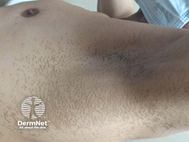
The image depicts a clinical photograph of axillary and proximal chest skin showing pigmented follicular lesions consistent with pityriasis versicolor. Imaging modality is clinical photography using a standard digital color camera to document cutaneous involvement. Visual features include multiple small brown, slightly scaly papules and patches centered on hair follicles, with fine surface scaling and patchy distribution. Lesions appear hypo- or hyperpigmented relative to adjacent skin, with preserved contour and no overt inflammation. The follicular localization suggests involvement of the follicular ostia and sebaceous-rich zones of the upper trunk. Histopathology (if sampled) would typically reveal Malassezia yeast and hyphae within the stratum corneum; KOH preparation may demonstrate the classic spaghetti-and-meatballs pattern. Clinically, pityriasis versicolor is a superficial fungal infection that can mimic other pigmentary disorders such as seborrheic dermatitis or vitiligo; differential diagnoses include post-inflammatory hyperpigmentation and tinea corporis. This image is valuable for dermatology education, telemedicine, and patient counseling, and supports planning of antifungal therapy (azoles, selenium sulfide) with attention to recurrence in hot, humid environments. For educational indexing, include commonly used synonyms such as tinea versicolor, Malassezia infection, pityriasis versicolor; this enhances recognition in dermatology databases, textbooks, and image banks used by clinicians, students, and researchers.
Pityrosporum ovale (Malassezia furfur) - Exam Notes
Nomenclature
- Pityrosporum ovale = old name for Malassezia furfur (also called P. orbiculare historically). Currently classified under genus Malassezia (14 species, 11 detected as human skin commensals).
- Other clinically important species: M. globosa, M. sympodialis, M. restricta.
- First suspected as a pathogen by Unna, who linked it to seborrheic dermatitis.
Microbiology
- Lipophilic, dimorphic yeast - part of normal resident cutaneous flora, especially in sebum-rich ("seborrheic") areas: scalp, face, upper trunk.
- Requires exogenous lipid (fatty acids) to grow - hence cannot easily be cultured on routine media; needs lipid-supplemented (e.g., Dixon's) media.
- Colonization begins by age 3-6 months, coinciding with maturation of sebaceous glands; earlier colonization noted in neonates with prolonged NICU stay.
- On KOH mount: classic "spaghetti and meatballs" appearance - short curved hyphae (spaghetti) + round budding yeast cells (meatballs).
Clinical Conditions Caused/Associated
1. Pityriasis (Tinea) Versicolor
- Overgrowth of M. furfur/M. globosa in stratum corneum.
- Predisposing factors: heat, humidity, sweating, oily skin, corticosteroid use, immunosuppression - highest sebaceous activity age group (adolescents/young adults).
- Clinical: asymptomatic (occasionally pruritic) hypo- or hyperpigmented, finely scaly macules that coalesce, mainly upper trunk/chest/back; face involved more in infants/immunocompromised.
- Mechanism of pigment change: yeast produces dicarboxylic acids (e.g., azelaic acid) that inhibit tyrosinase -> hypopigmentation; hyperpigmented lesions from thicker stratum corneum/inflammatory response.
- Diagnosis: KOH scraping - spaghetti and meatballs pattern; Wood's lamp - yellow-green to golden fluorescence.
- Treatment: topical azoles (ketoconazole 2% shampoo daily x1-2 weeks), selenium sulfide 2.5%; extensive disease - oral fluconazole/itraconazole. High recurrence - prophylactic monthly antifungal shampoo/therapy.
2. Malassezia (Pityrosporum) Folliculitis
- Chronic, moderately pruritic, monomorphic dome-shaped follicular papules and pustules on upper back, chest, shoulders, sometimes face/scalp.
- Risk factors: organ/marrow transplant recipients, antibiotic therapy (esp. tetracyclines for acne, which suppress P. acnes allowing Malassezia overgrowth), occlusion, sweating, immunosuppression, warm weather.
- Often coexists with tinea versicolor or seborrheic dermatitis.
- Diagnosis: characteristic morphology + yellow-green Wood's lamp fluorescence + KOH/biopsy showing yeast in follicles + response to antifungals confirms diagnosis (a somewhat controversial entity historically).
- Treatment: oral fluconazole 400 mg single dose, or itraconazole 200 mg/day x5-7 days; topical selenium sulfide 2.5% overnight, propylene glycol 30-50%, topical imidazoles. PDT for refractory cases. Prophylaxis needed - relapses common.
3. Seborrheic Dermatitis
- Chronic eczematous eruption of sebum-rich areas (scalp, eyebrows, nasolabial folds, retroauricular, presternal).
- Pathogenesis: not a true infection but an inflammatory/immune response to Malassezia metabolites and lipase/lipoxygenase products releasing irritant free fatty acids, in a susceptible host (barrier dysfunction, altered immune response) rather than simple organism overload.
- No direct quantitative correlation between yeast load and disease severity - unaffected skin can carry similar organism counts.
- Confirmed link: 1984 study showed systemic ketoconazole suppresses seborrheic dermatitis - corroborated by later studies.
- Infantile form (self-limited, first 3-4 months) vs adult form (chronic, peaks 4th-6th decade); more common in men.
- Associated with HIV (extensive, treatment-resistant SD is a cutaneous marker), Parkinson disease, stroke, immunosuppression, and drugs (neuroleptics, dupilumab, TKIs).
4. Other Associations
- Neonatal cephalic pustulosis (neonatal acne-like eruption).
- Possible role in atopic dermatitis exacerbation (IgE sensitization to M. sympodialis/furfur, especially head-and-neck dermatitis).
- Rare systemic infection: catheter-related fungemia in neonates on IV lipid emulsions (since organism is lipid-dependent).
Diagnosis Summary
- KOH mount: yeast + short hyphae ("spaghetti and meatballs").
- Wood's lamp: yellow-green/golden fluorescence in versicolor and folliculitis.
- Culture not routinely done (needs lipid supplementation).
- Skin biopsy in folliculitis - yeast forms within dilated follicles.
Treatment Overview (exam-relevant table)
| Condition | Topical | Systemic |
|---|---|---|
| Pityriasis versicolor | Ketoconazole 2% shampoo, selenium sulfide 2.5% | Fluconazole/itraconazole (extensive disease) |
| Malassezia folliculitis | Selenium sulfide, topical imidazoles | Fluconazole 400 mg single dose or itraconazole 200 mg/day x5-7d |
| Seborrheic dermatitis | Ketoconazole/zinc pyrithione shampoo, mild topical steroids, calcineurin inhibitors | Oral itraconazole/ketoconazole in resistant cases |