
Supraclavicular Brachial plexus usg anatoy
ultrasound anatomy supraclavicular brachial plexus transverse scan subclavian artery first rib pleura
"brachial plexus" AND ultrasound
supraclavicular brachial plexus ultrasound anatomy subclavian artery first rib pleura authoritative
https://nysora.com/regional-anesthesia/topics/regional-anest…
Supraclavicular brachial plexus: ultrasound anatomy
Probe position
- Patient supine, head turned to the opposite side, ipsilateral arm adducted.
- Use a high-frequency linear probe in the supraclavicular fossa, just superior/posterior to the midpoint of the clavicle.
- Obtain a transverse or slightly oblique transverse view.
Structures on the ultrasound image
| Structure | Typical appearance | Location |
|---|---|---|
| Subclavian artery (SCA) | Round, pulsatile, anechoic structure. Confirm with color Doppler. | Central to inferomedial landmark |
| Brachial plexus | Cluster of round/oval hypoechoic nodules, described as a “bunch of grapes.” | Usually posterolateral/superficial to the SCA |
| First rib | Bright curved hyperechoic line with a clean posterior acoustic shadow | Deep and lateral/posterior to SCA and plexus |
| Pleura | Bright sliding line deep to the first rib, moving with respiration | Deepest important safety structure |
| Anterior scalene muscle | Muscle medial/anterior to plexus at more cephalad levels | Helps trace plexus cranially |
| Middle scalene muscle | Muscle lateral/posterior to plexus | Plexus emerges between anterior and middle scalenes |
| Subclavian vein | Compressible anechoic vessel, usually medial and anterior to artery | Identify before needling |
Key spatial relationship
Superficial
   Skin / subcutaneous tissue

      Brachial plexus
     (trunks/divisions)
            ○ ○ ○
              \
               \  posterolateral to
                [ Subclavian artery ]

                  First rib  ─────────  bright cortex + shadow
                  Pleura     ─────────  sliding echogenic line
Deep
How to recognize the plexus
- Find the subclavian artery first.
- Look lateral and posterior/superficial to it.
- The plexus appears as several tightly packed hypoechoic circular or oval structures.
- Identify the first rib deep to the plexus. Its acoustic shadow helps distinguish it from pleura.
- Locate the pleural sliding line below the rib before any needle advancement.
The “corner pocket”
- the inferolateral subclavian artery, and
- the first rib.
Safety anatomy
- Pleura is close, commonly only 1-2 cm deep to the plexus. Do not advance a needle if the tip is not clearly seen.
- First rib is the protective backstop. Aim to keep the needle trajectory such that the rib remains between needle tip and pleura.
- Use color Doppler to identify vessels. The dorsal scapular and transverse cervical arteries may pass through or near plexus elements.
- A lateral-to-medial in-plane approach is commonly used, but technique should only be performed by clinicians trained in ultrasound-guided regional anesthesia.
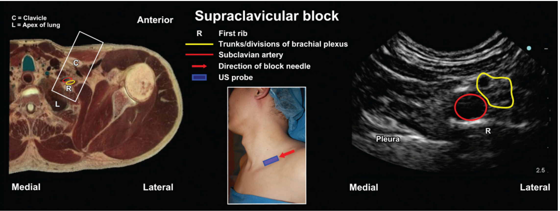
Supraclavicular Brachial plexus usg anatomy
Supraclavicular brachial plexus: USG anatomy
Probe position
- Supine patient, head turned away from the block side, arm adducted.
- High-frequency linear probe in the supraclavicular fossa, just above the midpoint of the clavicle.
- Start in a transverse/slightly oblique transverse orientation.
Structures to identify, superficial to deep
| USG structure | Appearance | Relationship |
|---|---|---|
| Brachial plexus | Compact cluster of round/oval hypoechoic nodules, the “bunch of grapes” appearance | Usually posterolateral and superficial to the subclavian artery |
| Subclavian artery | Round, pulsatile, anechoic lumen. Use color Doppler if needed. | Main sonographic landmark, inferomedial to plexus |
| First rib | Bright hyperechoic curved line with posterior acoustic shadow | Deep to the artery and plexus, especially laterally |
| Pleura | Echogenic sliding line with respiratory movement | Deep to the first rib, toward the lung apex |
| Subclavian vein | Compressible anechoic vessel | Usually medial and anterior to the artery |
| Anterior and middle scalene muscles | Hypoechoic muscle with striations | More cephalad, the plexus can be followed between them |
Key relation
                 Superficial
                      Skin

          Brachial plexus: ○ ○ ○
            posterolateral/superficial
                       to
              Subclavian artery: ●

                  First rib: ─────
                    Pleura: ~~~~~
                    Deep
Scan sequence
- Identify the subclavian artery.
- Find the first rib deep to it. Look for its bright cortex and acoustic shadow.
- Confirm the pleural sliding deep to the rib.
- Sweep lateral/posterior to the artery to identify the clustered plexus trunks/divisions.
- Trace cephalad if needed: upper trunk = C5-C6, middle trunk = C7, lower trunk = C8-T1.
Important practical points
- The lower trunk is closest to the subclavian artery and pleura.
- The corner pocket lies between the inferolateral subclavian artery and the first rib, near the lower trunk.
- Always identify the first rib and pleura before needle insertion. The rib is the protective bony backstop; loss of visible needle-tip control risks pleural injury and pneumothorax.
- Color Doppler helps detect vessels, including vessels that may run among plexus elements.
Show labeled ultrasound images of the supraclavicular view
labeled ultrasound image supraclavicular brachial plexus subclavian artery first rib pleura
"brachial plexus" AND ultrasound
labeled ultrasound image supraclavicular brachial plexus first rib pleura subclavian artery
https://asra.com/news-publications/asra-updates/blog-landing…
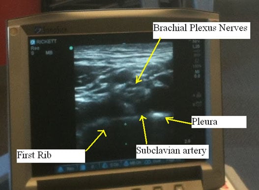
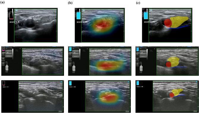
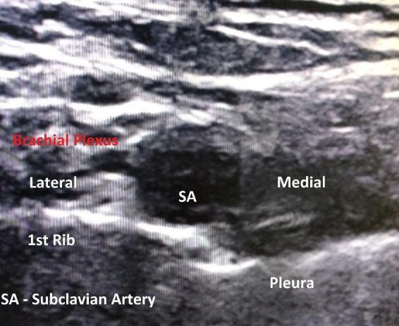
Labeled ultrasound images: supraclavicular brachial plexus view
Labels include the brachial plexus, subclavian artery, first rib, and pleura.
Read the image in this order
- SA: subclavian artery, round and anechoic.
- BP: brachial plexus, a cluster of hypoechoic round fascicles lateral/posterolateral to the artery.
- 1st rib: bright curved line with acoustic shadow.
- Pleura: sliding echogenic line deep to the rib.
Create a one-page PDF of supraclavicular anatomy