Can you show 3d images of quadrangular membrane,cricovocal membrane and muscles more clearly enlarged different images
3D anatomy of laryngeal membranes quadrangular membrane cricovocal membrane and intrinsic laryngeal muscles enlarged diagram
laryngeal anatomy
3D larynx anatomy quadrangular membrane cricovocal membrane intrinsic laryngeal muscles
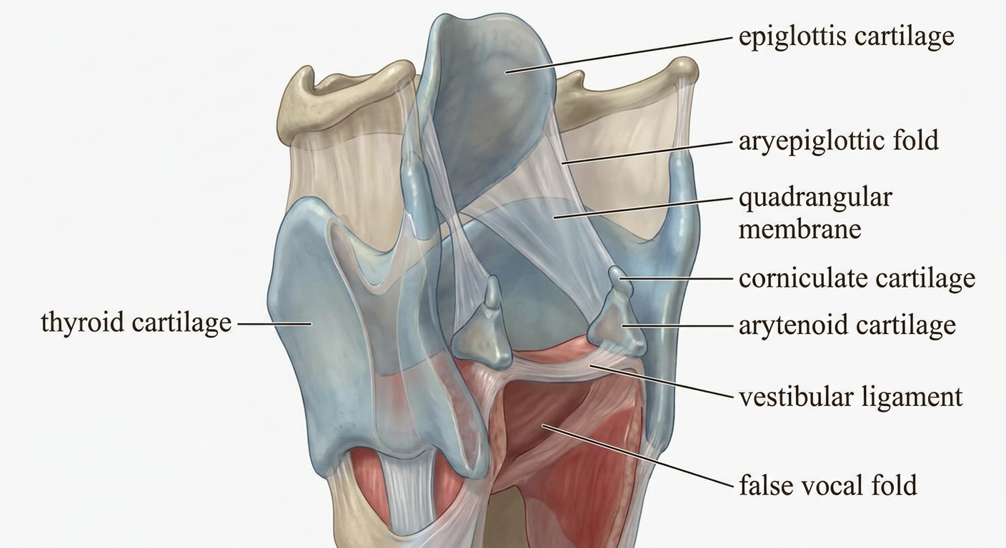
An enlarged anatomically accurate 3D educational cutaway of the human larynx focused only on the quadrangular membrane. The thyroid cartilage is partly removed to expose the epiglottis, arytenoid and corniculate cartilages. Show the paired thin quadrangular membranes extending from lateral epiglottis to arytenoid cartilages, with their upper free edge forming the aryepiglottic fold and lower free edge thickened as the vestibular ligament. Include clean readable labels: epiglottis, quadrangular membrane, aryepiglottic fold, vestibular ligament, arytenoid cartilage, false vocal fold.
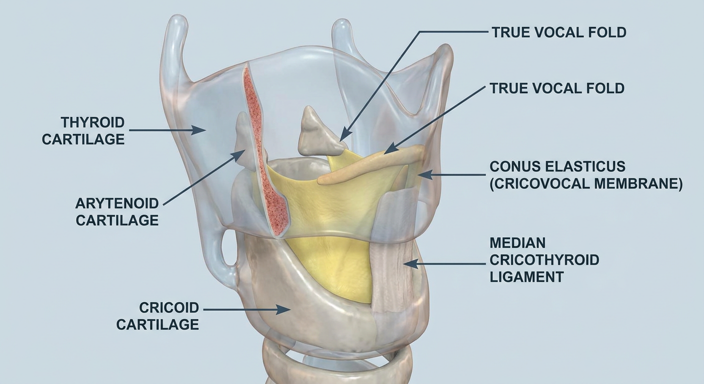
An enlarged anatomically accurate 3D educational cutaway of the human larynx focused only on the cricovocal membrane, also called conus elasticus. The thyroid cartilage is partly removed. Show the elastic membrane rising from the superior border of cricoid cartilage to the vocal processes of arytenoid cartilages and the anterior thyroid cartilage, with its free upper margin forming the vocal ligament. Include clean readable labels: cricoid cartilage, conus elasticus (cricovocal membrane), vocal ligament, true vocal fold, arytenoid cartilage, median cricothyroid ligament.
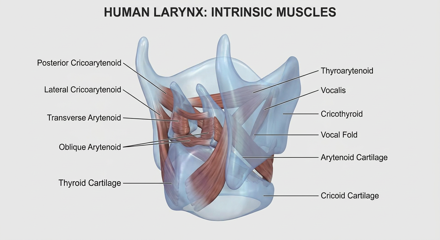
An enlarged anatomically accurate 3D posterior and lateral cutaway of the human larynx showing the intrinsic muscles clearly. Render the cartilages semi-transparent where helpful. Show and label posterior cricoarytenoid, lateral cricoarytenoid, transverse arytenoid, oblique arytenoid, thyroarytenoid, vocalis, and cricothyroid muscles, plus arytenoid, cricoid, thyroid cartilage and vocal fold. Educational anatomy illustration.
1. Quadrangular membrane
2. Cricovocal membrane / conus elasticus
3. Intrinsic muscles of the larynx
- Posterior cricoarytenoid: abducts the vocal folds, the only abductor
- Lateral cricoarytenoid and interarytenoid muscles: adduct the vocal folds
- Cricothyroid: lengthens and tenses vocal folds
- Thyroarytenoid/vocalis: fine control of vocal-fold tension