
Why should we do CT proximal humerus in Proximal humerus fracture
Reading File
Now let me search the medical library for CT in proximal humerus fractures.
Finding Sources
Finding Sources
Searching the Web
CT scan proximal humerus fracture indications importance classification
Good results. Now let me read the key textbook sections in detail.
Reading File
Reading File
Excellent - I have comprehensive textbook content. Let me also fetch the 3D CT reconstruction image to include visually.
Loading Image
Finding Sources
I now have very rich content. Here is the comprehensive answer:
Why CT is Done in Proximal Humerus Fractures
CT of the proximal humerus is not just a supplementary imaging step - it addresses several key limitations of plain radiographs and directly influences management. Here is a systematic breakdown:
1. Better Understanding of Fracture Pattern
Plain radiographs give a 2D picture of a 3D injury. CT overcomes this by providing:
- Axial cuts - show displacement of lesser and greater tuberosity fragments in the transverse plane
- Coronal reconstructions - assess alignment of humeral head, comminution at the humeral calcar, integrity of the inferomedial hinge, and size of metaphyseal fracture extension
- Sagittal reconstructions - determine flexion or extension deformity of the proximal humerus relative to the shaft
"CT of proximal humeral fractures is extremely helpful for providing further understanding of the fracture pattern." - Rockwood and Green's Fractures in Adults, 10th ed.
2. Glenohumeral Relationship and Associated Injuries
Simple axial CT images provide critical information that plain X-rays frequently miss:
- Confirm the spatial relationship between the humeral head and the glenoid (especially for subtle fracture-dislocations or posterior dislocations)
- Demonstrate glenoid rim fractures
- Reveal whether the tuberosities are fractured
- Identify occult coracoid process and lesser tuberosity fractures that are easily missed on radiographs
3. Assessment of Bone Quality and Impaction
CT allows:
- Quantification of the degree of osteopenia - important in elderly patients where bone quality affects implant selection
- Detection of bone impaction patterns (e.g., valgus-impacted fractures, calcar comminution)
- Assessment of fracture comminution that determines fixation options
4. Medial Hinge Integrity (Vascular Implication)
Assessment of the inferomedial periosteal hinge on coronal CT images is a key step - integrity of this hinge is used as an indicator of vascular supply to the humeral head in multipart fractures. Loss of medial hinge increases the risk of avascular necrosis (AVN) of the humeral head post-fixation.
5. Head-Splitting Fractures
Head-splitting fractures are a specific subtype where the articular surface is cleaved. These are best characterized on CT by combining axial, coronal, and sagittal sequences. Missing a head-split on plain films can lead to an inappropriate attempt at fixation when arthroplasty is actually required.
6. 3D Reconstruction for Surgical Planning
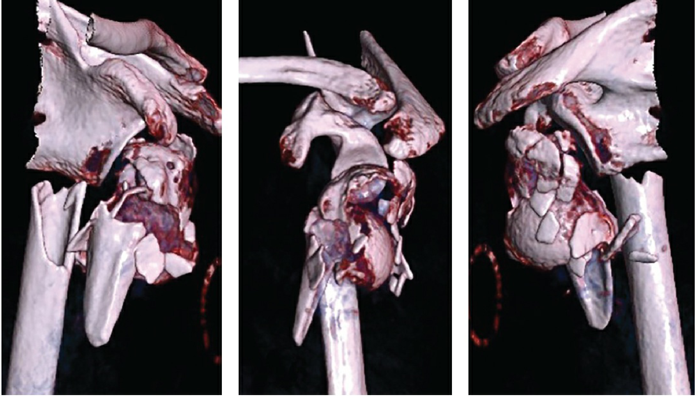
3D CT reconstruction of a proximal humerus fracture - anterior, lateral, and posterior views (Rockwood & Green's, 10th ed)
- 3D reconstructions (ideally with and without scapular subtraction) allow the proximal humerus to be analyzed from every angle
- Studies show that 3D imaging of proximal humerus fractures significantly increases the number of surgical indications compared to radiographs alone or radiographs + 2D CT
- Helps plan the reduction sequence, implant type, and surgical approach
"3D reconstruction images can be of great value to complete the understanding of various fracture patterns." - Rockwood and Green's, 10th ed.
Important caveat: 3D reconstructions provide only a surface view - they do not allow assessment of impaction and bony deficiencies within the fractured humerus. So 2D axial + reformats remain essential alongside 3D.
7. Soft Tissue Assessment
Using soft tissue windows on sagittal CT images:
- Fatty atrophy of the rotator cuff muscles can be analyzed - relevant in patients with suspected pre-injury rotator cuff pathology
- Assessment of potential injury to adjacent neurovascular structures
8. Improving Operative vs. Non-operative Decision-Making
Research comparing plain radiographs alone vs. CT for proximal humerus fractures showed:
- CT significantly improved interobserver reliability in fracture characterization and assessment
- Agreement on whether to operate improved from "fair" (κ = 0.38) to "good" (κ = 0.65) with the addition of CT scans
Summary Table
| CT Feature | Clinical Value |
|---|---|
| Axial cuts | Glenohumeral relationship, tuberosity displacement |
| Coronal reformat | Calcar comminution, medial hinge integrity |
| Sagittal reformat | Flexion/extension deformity, rotator cuff atrophy |
| 3D reconstruction | Surgical planning, fragment orientation, implant choice |
| Soft tissue window | Rotator cuff, neurovascular structures |
| All sequences | Head-split detection, occult fractures (coracoid, lesser tuberosity) |
Source: Rockwood and Green's Fractures in Adults, 10th ed. 2025, Chapter 36 (Proximal Humerus) and Chapter 7 (Imaging); Schwartz's Principles of Surgery, 11th ed.
Why will we do both bone nailing over both bone plating
Both bone nailing (intramedullary nailing) vs both bone plating for forearm fractures - nailing is preferred in certain situations due to biomechanical and biological advantages. Let me pull the textbook evidence.
Searching sources...
Finding Sources
Finding Sources
Searching the Web
both bone forearm fracture intramedullary nailing vs plating advantages indications
Reading File
Finding Sources
Reading File
Finding Sources
Reading File
Finding Sources
Reading File
Reading File
Reading File
I now have comprehensive data from both Campbell's and Rockwood & Green's. Here is the full answer:
Both Bone Nailing vs Both Bone Plating in Forearm Fractures
First, the important clinical context: plating (ORIF with compression plates) remains the gold standard for both-bone forearm fractures in adults. However, there are specific situations and advantages where intramedullary (IM) nailing is preferred or chosen over plating.
Why Nailing Over Plating? - The Advantages
1. Minimally Invasive - Less Soft Tissue Damage
Nailing can be done via small percutaneous incisions with closed or semi-open reduction, avoiding the extensive soft tissue stripping required for plate application. This is the single most important advantage.
"The use of intramedullary nails for injuries is often reserved for patients in whom the soft-tissue envelope is so traumatized that safe plate application is not possible." - Campbell's Operative Orthopaedics, 15th ed.
This applies particularly in:
- Severely traumatized soft tissue (crush injuries, degloving)
- Burns over the forearm
- Severely contaminated open fractures (Gustilo IIIB/IIIC)
2. Less Blood Loss
A randomized comparison (Ozkaya et al.) directly measured this:
- IM nailing (closed technique): zero blood loss
- ORIF with plate fixation: mean 60 mL (range 20-240 mL)
- Difference was statistically significant
This matters in polytrauma patients, patients on anticoagulants, or elderly patients with limited physiologic reserve.
3. Faster Union Time
- IM nailing: mean union ~10 weeks
- Plate fixation: mean union ~14 weeks
- This difference was statistically significant (Ozkaya et al., Köse et al.)
The biological reason is that nailing is an extraperiosteal technique - it does not strip the periosteum, preserving the endosteal and periosteal blood supply that drives callus formation.
4. Shorter Operative Time
Multiple comparative studies have shown statistically shorter operative times with IM nailing compared to plate fixation. This is relevant in:
- Polytrauma patients with limited anaesthetic tolerance
- Patients with significant medical comorbidities
5. Smaller Scars / Better Cosmesis
Especially relevant in paediatric patients and young adults, where cosmesis is a concern. Elastic Stable Intramedullary Nailing (ESIN) in children gives excellent cosmetic results through tiny stab incisions.
6. Avoids Risks Specific to Plating
| Plate-specific risk | How nailing avoids it |
|---|---|
| Refracture after plate removal | Smaller stress riser after nail removal |
| Wound dehiscence over plate | No bulky implant under tight skin |
| Plate irritation / prominence | Nail buried within medullary canal |
| Periosteal stripping causing delayed union | Nailing preserves periosteum |
Refracture after plate removal is a well-documented complication (~5.5% in paediatric patients, higher in adults), particularly after 3.5-mm plate removal. Nails avoid the cortical stress riser that plates leave behind.
7. Specific Scenario - Hybrid Fixation
Modern practice has evolved to a hybrid approach in difficult cases:
- IM nail for the ulna (straight bone, easier to nail)
- Plate fixation for the radius (complex bow, better restored with plate)
This limits soft tissue complications while preserving the radial bow anatomically.
"Commonly an intramedullary nail can be used for one bone (usually the ulna) with plate fixation of the other (usually the radius) to limit soft-tissue complications." - Campbell's Operative Orthopaedics, 15th ed.
Studies (Behnke et al.) showed equivalent union rates, range of motion, and functional outcomes with this hybrid approach vs. dual plating.
Why Plating Remains the Standard (Disadvantages of Nailing)
To be balanced - this is why plating is still routinely preferred:
| Issue with IM Nailing | Detail |
|---|---|
| Cannot restore radial bow reliably | The radial bow is critical for pronation/supination |
| Poor rotational stability in older designs | Modern interlocking nails have improved this |
| Length-unstable or comminuted fractures | Nails provide inadequate stability |
| Requires postoperative cast in some cases | Plates allow early mobilization |
| Increased compartment syndrome risk | Especially with prolonged attempts at closed nailing |
| Cannot achieve anatomic articular reduction | For any intra-articular component |
"Despite the satisfactory outcomes with modern intramedullary nailing, the outcomes of open reduction and plate fixation remain superior." - Campbell's Operative Orthopaedics, 15th ed.
Summary - When to Choose Nailing Over Plating
| Indication for IM Nailing | Reason |
|---|---|
| Severely compromised soft tissue envelope | Plating unsafe; wound complications too high |
| Grossly contaminated open fractures (Gustilo IIIB) | Plating risks deep infection; nail is biomechanically less demanding of soft tissue |
| Polytrauma / damage control orthopaedics | Shorter operative time, less blood loss |
| Paediatric both-bone forearm fractures (ESIN) | Minimal invasion, small incisions, good cosmesis |
| Refracture after prior plate removal | Medullary canal already compromised |
| Patient preference / cosmetic concern | Smaller scars |
Sources: Rockwood and Green's Fractures in Adults, 10th ed. 2025, Chapter 42 (Forearm Shaft Fractures); Campbell's Operative Orthopaedics, 15th ed. 2026, Chapter 62 (Fractures of the Shafts of the Radius and Ulna)
Malunion
Given the context of our conversation (both-bone forearm fractures), I'll look up malunion of forearm fractures specifically.
Searching sources...
Finding Sources
Finding Sources
Reading File
Reading File
Reading File
Reading File
Excellent content. Here is a comprehensive answer:
Malunion of Both-Bone Forearm Fractures
Why Malunion is Particularly Problematic in the Forearm
The forearm is often called a "functional joint" - the radius and ulna together function as a linked unit, and their precise 3D anatomical relationship is essential for pronation and supination. Even small degrees of malunion directly impair this rotation.
"Residual malalignment correlates directly to loss of motion and function. In addition, malunion can result in a decrease in grip and pinch strength." - Rockwood and Green's, 10th ed.
Types of Malunion in the Forearm
1. Angular Malunion
The bone heals with a bent angulation. This is the most common type.
- Mechanism of disability: Angular deformity encroaches on the interosseous space between radius and ulna, physically blocking rotation
- Critical thresholds (from cadaver studies):
- 10 degrees of deformity → insignificant functional loss (acceptable)
- 15 degrees → >27% loss of pronosupination (clinically significant)
- 20 degrees → ~30% deficit in pronosupination (functionally significant)
- Location matters: Middle-third deformities more significantly limit supination; deformities at the midshaft of the ulna maximally affect rotation
2. Rotational Malunion
The bone heals in a rotated position.
- Pure rotational deformity causes loss of rotation equal to the magnitude of the deformity (1:1 relationship)
- This is particularly difficult to detect on plain radiographs
3. Loss of Radial Bow
The radius has a specific lateral bow - this is not just a cosmetic feature, it is biomechanically essential for pronosupination.
- Maximum radial bow normally located at 60% of the proximal-to-distal distance from the biceps tuberosity to the ulnar aspect of the distal articular surface
- Normal magnitude: 15.3 ± 0.3 mm
- If restoration is within 4.3% of original location and within 1.5 mm of original magnitude → ≥80% normal rotation is preserved
- Loss of radial bow reduces both forearm rotation AND grip strength
4. Shortening
Leads to ulnar positive variance at the wrist, causing DRUJ dysfunction and TFCC problems.
Consequences of Forearm Malunion
| Consequence | Mechanism |
|---|---|
| Loss of pronation / supination | Interosseous space encroachment, loss of radial bow |
| Reduced grip and pinch strength | Altered muscle tension and lever arm |
| DRUJ instability / arthritis | Disturbance of bony geometry between ulnar head and sigmoid notch |
| Proximal RUJ arthritis | Develops after long-standing (>20 years) malunions |
| Cosmetic deformity | Visible bowing or shortening |
Treatment of Malunion
Decision to Operate
Surgery is based on functional limitations and physical demands, not radiographic deformity alone. Indications:
- Loss of pronosupination (primary indication)
- DRUJ instability
- Unacceptable cosmetic appearance
Important caveat: Restoring skeletal alignment will NOT improve functional deficits caused by soft tissue injury or prolonged immobilization - these must be assessed and discussed preoperatively.
Timing is Critical
- Surgery within 12 months of injury: average gain of 79 degrees of rotation (range 20-160°)
- Surgery after 12 months: average gain of only 30 degrees (range 25-95°)
- Complication rate: 15% if operated early vs. 48% if operated >1 year after injury
Surgical Technique
Corrective osteotomy + compression plating + bone grafting
Steps (Trousdale and Linscheid technique):
- Document preoperative ROM (pronation, supination, elbow flexion/extension)
- Assess DRUJ and PRUJ stability
- Full-length AP, lateral, and rotation radiographs of both forearms for comparison
- Osteotomy at the malunion site of the involved bone(s)
- Correct deformity in all planes (angular + rotational)
- Fix with 3.5-mm compression plates + iliac crest bone graft
- Single-bone osteotomy if only one bone is deformed; both bones if required
Modern adjuncts:
- 3D CT modeling and patient-specific cutting guides (PSI) are now used for complex multiplanar deformities - especially useful in cases where the deformity is oblique to both coronal and sagittal planes
- 3D printing of the affected bones allows full preoperative understanding of the deformity and correction required
Complications of Corrective Surgery
- Delayed union / nonunion
- Infection
- Loss of motion
- Radial nerve paresthesias (PIN at risk during Henry approach)
- Wrist pain
- DRUJ instability
- Heterotopic ossification along the interosseous membrane (late cases)
- Refracture after plate removal
Key Numbers to Remember
| Deformity | Functional consequence |
|---|---|
| 10° angulation | Acceptable - minimal deficit |
| 15° angulation | >27% loss of rotation |
| 20° angulation | ~30% deficit in pronosupination |
| Surgery <12 months | Average 79° rotation gained |
| Surgery >12 months | Average 30° rotation gained, 48% complication rate |
Sources: Rockwood and Green's Fractures in Adults, 10th ed. 2025, Chapter 42; Campbell's Operative Orthopaedics, 15th ed. 2026, Chapter 63
This is a shared conversation. Sign in to Orris to start your own chat.