
Hot water spill on hand .what's the management
hot water scald burn hand first aid management treatment 2024
burn degree classification skin diagram superficial partial full thickness

This clinical series demonstrates the progression of burn severity categorized by depth and tissue involvement. (a) First-degree burn (superficial): Presents as erythema of the epidermis without blistering, consistent with a sunburn. (b) Second-degree superficial partial-thickness burn (SPTB): Displays clear blistering (bullae) and moist, red tissue, indicating damage to the papillary dermis. (c) Second-degree deep partial-thickness burn (DPTB): Shows a mottled appearance with ruptured blisters and more significant exudate, extending into the reticular dermis. (d) Third-degree burn (full-thickness): Characterized by a central area of leathery, dark eschar formation surrounded by a zone of stasis, indicating total destruction of the epidermis and dermis into the subcutaneous fat. (e) Fourth-degree burn: Illustrates the most severe injury, extending through all skin layers to involve underlying fascia, skeletal muscle (rectus abdominis), and potentially bone, appearing charred and necrotic. This visual comparison serves as an educational tool for clinical triage and the classification of thermal injuries.
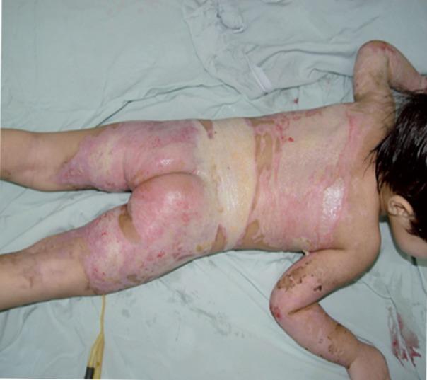
Clinical photograph of a pediatric patient in prone position demonstrating extensive partial-thickness and full-thickness burns across the posterior trunk, buttocks, and lower extremities. The burn wounds exhibit mixed characteristics: erythematous and weeping areas suggestive of superficial partial-thickness (second-degree) injury, interspersed with pale, whitish, and yellowish leathery patches indicative of deep partial-thickness or full-thickness (third-degree) tissue damage. Significant skin desquamation and raw dermal exposure are visible, particularly on the back and proximal thighs. The distribution is patchy but widespread, involving a substantial percentage of the total body surface area (TBSA). The presence of a moist appearance suggests the application of topical antimicrobial agents or wound exudate. This image serves as a clinical example of severe thermal trauma in a child, highlighting the necessity for intensive burn management, including fluid resuscitation, wound debridement, and potential surgical intervention.
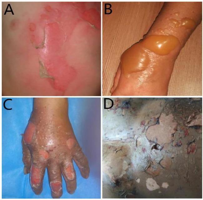
A composite of four clinical photographs demonstrating the progression and classification of burn wound severity. Image A illustrates a first-degree (superficial) burn, showing blanching erythema of the skin with areas of peeling epidermis. Image B depicts a superficial second-degree (partial-thickness) burn on an extremity, characterized by large, tense, fluid-filled bullae with translucent amber-colored serous fluid and surrounding erythematous inflammation. Image C shows a deep second-degree burn on the hand, featuring mottled red and white tissue with ruptured blisters and areas of exposed dermis. Image D represents a third-degree (full-thickness) burn, displaying a dry, leathery, and inelastic eschar with a variegated color palette of white, grey, and brown, indicating destruction of the epidermis, dermis, and subcutaneous tissue. This comparison chart serves as a diagnostic educational tool for identifying burn depth based on visual morphological features such as blistering, tissue color, and texture.
Management of a Hot Water Spill (Scald Burn) on the Hand
Step 1 - Immediate First Aid (First 20 minutes)
- Hold the burned hand under cool (not cold) running water for 20 minutes continuously. Do not stop early.
- Do NOT use ice, iced water, or ice packs - these cause vasoconstriction and can deepen the burn.
- Do NOT apply butter, oil, toothpaste, or any home remedy - these trap heat and promote infection.
- Remove any rings, watches, or tight clothing/jewelry near the burn before swelling begins, unless stuck to the skin (do not forcibly remove anything adhered to skin).
Step 2 - Assess Burn Depth and Severity
| Degree | Depth | Appearance | Pain | Healing |
|---|---|---|---|---|
| 1st degree (superficial) | Epidermis only | Red, dry, no blisters | Painful | 3-5 days, no scar |
| 2nd degree superficial partial-thickness | Papillary dermis | Red, blistered, moist, weeping | Very painful | 10-14 days, minimal scar |
| 2nd degree deep partial-thickness | Reticular dermis | Mottled red/white, ruptured blisters | Less painful (nerve damage) | >21 days, scarring likely |
| 3rd degree (full-thickness) | Through dermis | Leathery, white/brown/charred, dry | Little/no pain | Needs grafting |
Step 3 - Wound Care
- After cooling, gently pat dry.
- Cover with a non-adherent, sterile dressing (e.g. paraffin gauze / Mepitel / cling film as a temporary cover).
- Apply a topical agent:
- Silver sulfadiazine 1% cream - broad-spectrum, standard for partial-thickness burns
- Petroleum jelly (Vaseline) - acceptable for very superficial burns
- Aloe vera gel - soothing for 1st degree burns
- Change dressing every 2-3 days or when soaked.
- Do not burst blisters intentionally - they protect against infection. If a blister ruptures spontaneously, trim dead skin and re-dress.
- Do not apply home topical agents.
- Cover loosely with a clean cloth or cling film and proceed to the ED.
Step 4 - Pain Relief
- Paracetamol (acetaminophen) 500-1000 mg as first-line for mild pain.
- Ibuprofen 400 mg (if not contraindicated) for anti-inflammatory effect.
- Elevation of the hand above heart level reduces swelling and pain.
- For severe pain: ED assessment and IV opioids may be needed.
Step 5 - When to Seek Emergency Medical Care
- Is deep partial-thickness or full-thickness (white, charred, leathery, or painless)
- Covers a large area (>1% TBSA, roughly the size of the patient's palm)
- Involves the palms, fingers, or dorsum of the hand (special area - risk of functional impairment and contracture)
- Shows circumferential involvement around a finger/hand (risk of vascular compromise)
- Has suspected inhalation injury or other associated trauma
- Affects children, elderly, or immunocompromised patients
- Shows signs of infection (increasing redness, warmth, pus, fever, red streaking)
Step 6 - Follow-up and Monitoring
- Dressing changes every 48-72 hours.
- Watch for infection signs: increased pain, purulent discharge, fever, wound breakdown.
- Physiotherapy/occupational therapy referral for deep hand burns to prevent contracture.
- Tetanus prophylaxis if not up to date (for wounds with blistering/open skin).
- Consider burn center referral for deep partial or full-thickness burns.
Summary Algorithm
Hot water spill on hand
       |
Cool water 20 min → Remove jewelry
       |
Assess depth & size
       |
1st/Superficial 2nd degree   →   Clean, non-stick dressing, silver sulfadiazine, analgesia
(small area)                      Review in 48-72h
       |
Deep 2nd / 3rd degree           → ED / Burn center immediately
OR large area OR fingers/palm    → Analgesia, IV fluids if large, surgical consult