
What view will take to see fracture clavicle
clavicle fracture X-ray AP view radiograph
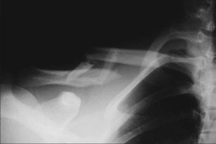
This diagnostic image is an anteroposterior (AP) X-ray radiograph of the shoulder and upper thoracic region, demonstrating a displaced mid-shaft clavicle fracture. The primary finding is a complete cortical disruption of the middle third of the clavicle with 100% displacement and significant shortening exceeding 2 cm. The fracture is comminuted, with at least one visible 'butterfly' fragment present at the fracture site. The medial fragment is superiorly displaced due to the pull of the sternocleidomastoid muscle, while the lateral fragment is depressed. The surrounding anatomy, including the visible upper ribs, coracoid process, and scapula, appears intact without obvious secondary trauma or pneumothorax in this view. This image is an educational example of severe clavicle injury typically requiring surgical intervention, such as intramedullary pinning or plate fixation, particularly in the context of high-grade shortening and displacement.
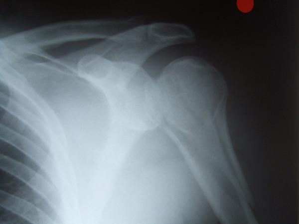
Anteroposterior (AP) view X-ray of the left shoulder demonstrating a complex, comminuted fracture of the proximal humerus. The radiograph reveals multiple fracture lines involving the anatomical neck and humeral head, extending into the proximal shaft. There is significant cortical disruption and misalignment of fragments, consistent with an iatrogenic fracture following a manipulation attempt. The humeral head shows loss of normal congruence with the glenoid fossa, suggesting an associated glenohumeral dislocation or subluxation. Surrounding structures visible include the clavicle, acromion, scapula, and superior ribs, which appear radiographically intact. This image serves as a clinical example of potential complications during the reduction of shoulder dislocations, illustrating the importance of accurate pre-procedural diagnosis of fracture-dislocations in orthopaedic trauma.
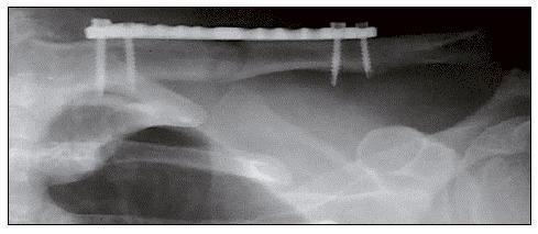
Postoperative anteroposterior (AP) X-ray radiography of the shoulder region, specifically focusing on the clavicle. The image demonstrates surgical management of a middle-third clavicle fracture using internal fixation. A radiopaque metallic locking plate is positioned along the superior aspect of the clavicular shaft. Four cortical/locking screws are visible, oriented perpendicularly to the plate and extending into the bone to provide stable fixation across the fracture site. The radiograph shows successful fracture reduction with the bone fragments aligned in a neutral anatomical position. This imaging illustrates standard orthopedic intervention for displaced clavicular fractures, likely utilizing a Minimally Invasive Plate Osteosynthesis (MIPO) technique, indicated by the distal and proximal screw clusters and the preserved central biological bridge over the primary fracture site.
Radiographic Views for Clavicle Fracture
Standard Views (Midshaft Fractures)
| View | Beam Angle | Notes |
|---|---|---|
| AP (Anteroposterior) | Standard 0° | Sufficient for initial diagnosis |
| AP Cephalad Tilt (Tilted view) | 15-40° superiorly (commonly 30°) | Preferred if only one view is taken; better profiles displacement |
Key principle: If only ONE view is taken, the cephalad-tilted AP view (15-40° superior angulation) is preferred over the standard AP, as it reduces superimposition and reveals true displacement.

Additional / Situational Views
| Situation | View Used |
|---|---|
| Assess shortening | AP Chest X-ray (compares both clavicles; shortening ≥2 cm is a relative indication for surgery) |
| Polytrauma / Trauma bay | Supine AP chest radiograph (first available film; note - this underestimates displacement vs. upright) |
| Medial third fractures | CT scan (better defines complex anatomy) |
| Distal clavicle / AC joint | Stress views (weighted AP) or Zanca view |
| Coracoid base fracture | Stryker notch view |
Why Upright Position Matters
CT Scan Role
- Medial third clavicle fractures
- "Floating shoulder" (associated glenoid neck fracture)
- Complex 3D deformity assessment