
I have chapter called coagulants and anti coagulants I want to be smart how to make notes for that with all classification and how to make all notes give me idea I am also adhd person I want to make it simple yest smart
coagulation cascade clotting factors intrinsic extrinsic pathway diagram
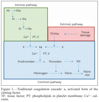
Summary : This diagram illustrates the traditional coagulation cascade, detailing the sequence of events and factors involved in blood clotting via the intrinsic, extrinsic, and common pathways. flowchart: # Pathways : • Intrinsic pathway (green box): Initiated by factor XII, proceeds through XI, IX, and VIII, with activation steps indicated by "a" (e.g., XIIa, XIa, IXa). • Extrinsic pathway (red box): Initiated by tissue damage, which activates tissue factor (TF) and factor VIIa. • Common pathway (blue box): Both intrinsic and extrinsic pathways converge at factor X, leading to the activation of prothrombin to thrombin, which then converts fibrinogen to fibrin, forming a fibrin clot. # Nodes : • XII → XIIa (Intrinsic) • XI → XIa (Intrinsic) • IX → IXa (Intrinsic) • Ca²⁺, PT, V (Intrinsic, Common) • TF/VIIa (Extrinsic) • Tissue damage (Extrinsic) • X → Xa (Common) • Prothrombin → Thrombin (Common) • Fibrinogen → Fibrin (Common) • XIII (Common) • Fibrin clot (Common) # Connectors : • Arrows indicate the direction of activation or conversion between factors. • Intrinsic and extrinsic pathways both feed into the activation of factor X in the common pathway. • Ca²⁺ and PT, V are required cofactors at specific steps. # Layout : • Three main colored sections: green (intrinsic, top left), red (extrinsic, top right), blue (common, bottom). • Flow proceeds from top (initiation) to bottom (clot formation). • Arrows show sequential activation and convergence of pathways. # Analysis : • The diagram clearly separates the intrinsic and extrinsic initiation mechanisms, both converging on the common pathway at factor X. • The cascade is highly sequential, with each activated factor catalyzing the next step. • Calcium ions (Ca²⁺) and phospholipids (PT) are essential cofactors at multiple steps. • The final outcome is the formation of a stable fibrin clot, highlighting the coordinated nature of the coagulation process.
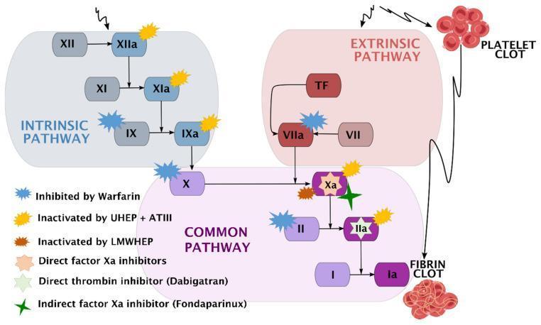
A comprehensive pathophysiology diagram illustrating the human coagulation cascade and the specific pharmacodynamic sites of action for various anticoagulants. The diagram is divided into three functional zones: the Intrinsic Pathway (blue), starting with Factor XII; the Extrinsic Pathway (red), initiated by Tissue Factor (TF) and Factor VII; and the Common Pathway (purple), where the pathways converge at Factor X. The cascade terminates in the conversion of Fibrinogen (I) to Fibrin (Ia), forming a fibrin clot. A secondary visual element depicts a platelet clot associated with the extrinsic pathway. Key clinical pharmacotherapeutic targets are marked with color-coded symbols: Warfarin inhibits Factors IX, VII, X, and II; Unfractionated Heparin (UHEP) + Antithrombin III (ATIII) inactivates Factors XIIa, XIa, IXa, Xa, and IIa; Low-Molecular-Weight Heparin (LMWHEP) and Fondaparinux target Factor Xa; Direct Factor Xa inhibitors act on Factor Xa; and Dabigatran acts as a direct thrombin inhibitor (Factor IIa). This schematic is designed for medical education regarding hemostasis and thromboprophylaxis.
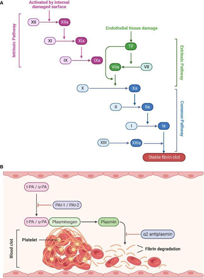
This medical schematic illustrates the physiological processes of hemostasis and fibrinolysis. Part A depicts the Coagulation Cascade, divided into the Intrinsic, Extrinsic, and Common pathways. The Intrinsic pathway (purple) is triggered by internal damaged surfaces, activating Factor XII to XIIa, followed by XI and IX. The Extrinsic pathway (green) begins with endothelial tissue damage and Tissue Factor (TF), activating Factor VII to VIIa. Both pathways converge at the Common pathway (blue) starting with the activation of Factor X to Xa. This leads to the conversion of prothrombin (II) to thrombin (IIa), and fibrinogen (I) to fibrin (Ia), culminating in a stable fibrin clot stabilized by Factor XIIIa. Activated factors are denoted by an 'a' suffix. Part B illustrates Fibrinolysis, where tissue-type (t-PA) and urokinase-type (u-PA) plasminogen activators convert plasminogen to plasmin, which then degrades the fibrin network of the blood clot. Inhibitory regulators including PAI-1, PAI-2, and ̡2-antiplasmin are also shown. The diagram represents the structural breakdown of a clot containing platelets and red blood cells into fibrin degradation products.
anticoagulants drug mechanism heparin warfarin DOAC comparison chart
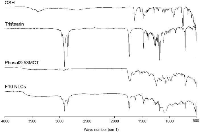
Fourier-Transform Infrared (FTIR) spectroscopy comparison chart displaying the molecular finger-printing of octasulfate heparin (OSH), tristearin (solid lipid), Phosal® 53MCT (liquid lipid), and the resulting F10 nanostructured lipid carrier (NLC) formulation. The spectra are plotted across a wavenumber range of 4000 to 500 cm-1. OSH shows a distinctive broad O-H stretching band near 3300 cm-1 and sharp fingerprint peaks below 1500 cm-1. Tristearin exhibits intense, sharp doublets near 2900 cm-1 (C-H stretching) and a prominent carbonyl peak (C=O) near 1740 cm-1. Phosal® 53MCT demonstrates similar but broader lipid features. The F10 NLCs spectrum represents a composite formulation; it retains the characteristic broad O-H feature of OSH and the sharp aliphatic and carbonyl peaks of the lipid matrix (tristearin and Phosal®), confirming the successful incorporation of the drug into the lipid carrier without significant chemical alteration. This diagnostic chart is used in pharmaceutical science to evaluate drug-excipient compatibility and the integrity of nanoparticle formulations.
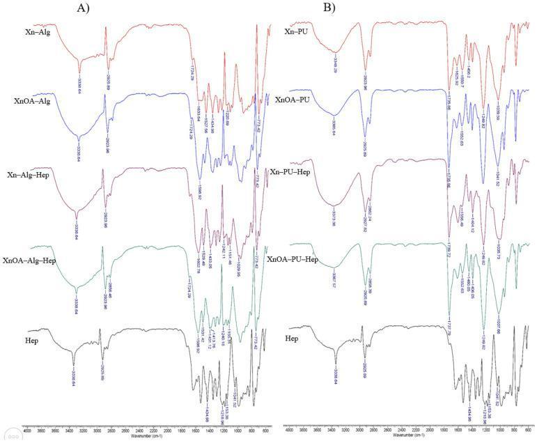
This diagnostic comparison chart displays Fourier-Transform Infrared (FTIR) spectra of biocompatible polymer matrices designed for tissue engineering and drug delivery. The image is divided into two panels: (A) xanthan-alginate (Xn-Alg) based materials and (B) xanthan-polyurethane (Xn-PU) based materials, both evaluated with and without chemical modification (XnOA) and heparin (Hep) loading. Key educational features include: - Broad absorption bands at approximately 3338–3373 cm⁻¹, signifying -OH and -NH- stretching vibrations essential for material hydration and structural stability. - Carbonyl group signals near 1737 cm⁻¹, used to verify the chemical modification of xanthan with oleic acid (XnOA). - Specific heparin markers, including sulphate group vibrations (S=O) at 1240–1249 cm⁻¹ and carboxylate (-COO-) stretching at 1433 cm⁻¹, which confirm successful incorporation of the anticoagulant into the scaffold. - Comparisons between matrices demonstrate the influence of polyurethane versus alginate on the hydrogen bonding and functional group distribution, critical for predicting material performance in human biomedical applications such as scaffold-based regeneration.
🩸 Coagulants & Anticoagulants - ADHD-Smart Notes
HOW TO MAKE NOTES (Strategy First)
Name → Target → What it does → Use it when → Watch out for → Antidote
STEP 1: Understand the CASCADE First (1 diagram, memorize it)
| Pathway | Triggered by | Key Factors |
|---|---|---|
| Intrinsic | Damaged vessel (collagen) | XII → XI → IX → VIII |
| Extrinsic | Tissue damage (Tissue Factor) | VII |
| Common | Both merge here | X → II (prothrombin → thrombin) → Fibrinogen → Fibrin clot |
STEP 2: Full Classification Tree
COAGULANTS & ANTICOAGULANTS
│
├── COAGULANTS (PROCOAGULANTS) - "Help make a clot"
│   ├── Systemic
│   │   ├── Vitamin K (phytonadione)
│   │   ├── Fresh Frozen Plasma (FFP)
│   │   └── Tranexamic acid (anti-fibrinolytic)
│   └── Local
│       ├── Thrombin (topical)
│       ├── Fibrin glue
│       └── Gelatin sponge (Gelfoam)
│
└── ANTICOAGULANTS - "Stop the clot from forming"
    ├── PARENTERAL (Injectable)
    │   ├── Unfractionated Heparin (UFH)
    │   ├── LMWHs: Enoxaparin, Dalteparin
    │   ├── Fondaparinux (selective Xa inhibitor)
    │   ├── Direct Thrombin Inhibitors (DTIs):
    │   │   ├── Bivalirudin
    │   │   ├── Argatroban
    │   │   └── Lepirudin, Desirudin
    └── ORAL
        ├── Vitamin K Antagonist: Warfarin
        └── DOACs (Direct Oral Anticoagulants)
            ├── Factor Xa inhibitors:
            │   ├── Rivaroxaban
            │   ├── Apixaban
            │   └── Edoxaban
            └── Thrombin (IIa) inhibitor:
                └── Dabigatran
STEP 3: Drug Cards (One-glance format)
🟠 HEPARIN (Unfractionated)
| Feature | Detail |
|---|---|
| Route | IV or SC injection |
| Target | Activates Antithrombin III → inhibits Thrombin (IIa) + Factor Xa |
| Onset | Immediate (minutes) |
| Monitor | aPTT |
| Uses | DVT, PE, ACS, cardiac surgery |
| Side effects | Bleeding, HIT (Heparin-Induced Thrombocytopenia), osteoporosis |
| Antidote | Protamine sulfate |
🟡 LMWHs (Enoxaparin, Dalteparin)
| Feature | Detail |
|---|---|
| Target | Mainly Factor Xa (less effect on thrombin) |
| Route | SC injection |
| Monitoring | Usually NOT needed (predictable kinetics) |
| Uses | DVT prophylaxis, ACS, pregnancy (safe in pregnancy!) |
| Side effects | Less HIT than UFH, less osteoporosis |
| Antidote | Partial reversal with protamine |
🔵 FONDAPARINUX
| Feature | Detail |
|---|---|
| Type | Synthetic pentasaccharide |
| Target | Only Factor Xa (via Antithrombin III) |
| Route | SC injection |
| Uses | DVT/PE treatment, orthopedic surgery prophylaxis |
| Key note | No HIT risk (no platelet binding). Eliminated renally - avoid in renal failure |
| Antidote | None |
🔴 WARFARIN (Vitamin K Antagonist)
| Feature | Detail |
|---|---|
| Target | Blocks Vitamin K epoxide reductase → reduces factors II, VII, IX, X (and protein C, S) |
| Route | Oral |
| Onset | Delayed - 3-5 days (depleting existing factors) |
| Monitor | PT/INR (target 2.0 - 3.0) |
| Uses | AF, mechanical heart valves, VTE |
| Side effects | Bleeding, skin necrosis (protein C depletion), teratogenic (crosses placenta) |
| Antidote | Vitamin K (slow), FFP / 4-factor PCC (fast) |
🟣 DOACs - The New Kids
| Drug | Target | Route | Monitor | Antidote |
|---|---|---|---|---|
| Rivaroxaban | Xa | Oral | Not needed | Andexanet alfa |
| Apixaban | Xa | Oral | Not needed | Andexanet alfa |
| Edoxaban | Xa | Oral | Not needed | Andexanet alfa |
| Dabigatran | IIa (thrombin) | Oral | Not needed | Idarucizumab |
- "-xaban" = Factor Xa inhibitor
- "Dabigatran" = Thrombin inhibitor
🟢 COAGULANTS (Pro-clotting drugs)
| Drug | Mechanism | Use |
|---|---|---|
| Vitamin K (phytonadione) | Restores synthesis of factors II, VII, IX, X | Warfarin reversal, bleeding |
| Tranexamic acid | Inhibits plasminogen → prevents fibrin breakdown (anti-fibrinolytic) | Surgical bleeding, trauma, heavy menstrual bleeding |
| Aminocaproic acid | Same as tranexamic acid | Bleeding post-surgery |
| FFP | Replaces all clotting factors | Acute reversal of coagulopathy |
| Protamine sulfate | Binds and neutralizes heparin | Heparin antidote |
STEP 4: ADHD-Friendly Memory System
The "WATCHDOG" rule for Warfarin interactions:
HIT (Heparin-Induced Thrombocytopenia) - Key Alert:
- Occurs 5-14 days after starting heparin
- PARADOX: Platelets drop BUT you get clots (not bleeding)
- Switch to Argatroban or Bivalirudin (DTIs, no cross-reactivity)
- Do NOT use LMWHs (cross-react with HIT antibodies)
STEP 5: ONE-PAGE COMPARISON TABLE (Stick this on your wall)
| Drug | Route | Target | Monitor | Antidote | Special note |
|---|---|---|---|---|---|
| Heparin (UFH) | IV/SC | IIa + Xa | aPTT | Protamine | HIT risk |
| Enoxaparin | SC | Xa >> IIa | None needed | Partial protamine | Safe in pregnancy |
| Fondaparinux | SC | Xa only | None needed | None | No HIT |
| Warfarin | Oral | Vit K epoxide reductase | INR | Vit K / FFP | Teratogenic |
| Rivaroxaban | Oral | Xa | None | Andexanet alfa | Once daily |
| Apixaban | Oral | Xa | None | Andexanet alfa | Twice daily |
| Dabigatran | Oral | IIa (thrombin) | None | Idarucizumab | Dialyzable |
| Tranexamic acid | Oral/IV | Plasminogen | None | None | Anti-fibrinolytic |
STEP 6: Note-Making Tips for ADHD
[ DRUG NAME ]
CLASS: ____________
TARGET: ___________
ROUTE: ____________
MONITOR: __________
ANTIDOTE: _________
USE: ______________
DANGER: ___________
- First pass (20 min): Just read the classification tree above - nothing else
- Second pass (30 min): Fill in blank drug cards for each drug from memory
- Third pass (15 min): Do the comparison table - focus only on columns you keep getting wrong
- Review (5 min daily): Quiz yourself on antidotes - that is where exams focus
- 🟠 Orange = parenteral anticoagulants (injectable)
- 🔴 Red = oral anticoagulants
- 🟢 Green = coagulants (pro-clotting)
- 🔵 Blue = antidotes
"Heparin is a fast bodyguard that grabs Antithrombin III and makes it 1000x stronger against thrombin - but if it goes rogue (HIT), you fire it and call protamine."
We are in the steps of clotting factor and coagulants and classification along mode of action
coagulation cascade clotting factors steps intrinsic extrinsic common pathway
Summary : This diagram illustrates the traditional coagulation cascade, detailing the sequence of events and factors involved in blood clotting via the intrinsic, extrinsic, and common pathways. flowchart: # Pathways : • Intrinsic pathway (green box): Initiated by factor XII, proceeds through XI, IX, and VIII, with activation steps indicated by "a" (e.g., XIIa, XIa, IXa). • Extrinsic pathway (red box): Initiated by tissue damage, which activates tissue factor (TF) and factor VIIa. • Common pathway (blue box): Both intrinsic and extrinsic pathways converge at factor X, leading to the activation of prothrombin to thrombin, which then converts fibrinogen to fibrin, forming a fibrin clot. # Nodes : • XII → XIIa (Intrinsic) • XI → XIa (Intrinsic) • IX → IXa (Intrinsic) • Ca²⁺, PT, V (Intrinsic, Common) • TF/VIIa (Extrinsic) • Tissue damage (Extrinsic) • X → Xa (Common) • Prothrombin → Thrombin (Common) • Fibrinogen → Fibrin (Common) • XIII (Common) • Fibrin clot (Common) # Connectors : • Arrows indicate the direction of activation or conversion between factors. • Intrinsic and extrinsic pathways both feed into the activation of factor X in the common pathway. • Ca²⁺ and PT, V are required cofactors at specific steps. # Layout : • Three main colored sections: green (intrinsic, top left), red (extrinsic, top right), blue (common, bottom). • Flow proceeds from top (initiation) to bottom (clot formation). • Arrows show sequential activation and convergence of pathways. # Analysis : • The diagram clearly separates the intrinsic and extrinsic initiation mechanisms, both converging on the common pathway at factor X. • The cascade is highly sequential, with each activated factor catalyzing the next step. • Calcium ions (Ca²⁺) and phospholipids (PT) are essential cofactors at multiple steps. • The final outcome is the formation of a stable fibrin clot, highlighting the coordinated nature of the coagulation process.
This medical schematic illustrates the physiological processes of hemostasis and fibrinolysis. Part A depicts the Coagulation Cascade, divided into the Intrinsic, Extrinsic, and Common pathways. The Intrinsic pathway (purple) is triggered by internal damaged surfaces, activating Factor XII to XIIa, followed by XI and IX. The Extrinsic pathway (green) begins with endothelial tissue damage and Tissue Factor (TF), activating Factor VII to VIIa. Both pathways converge at the Common pathway (blue) starting with the activation of Factor X to Xa. This leads to the conversion of prothrombin (II) to thrombin (IIa), and fibrinogen (I) to fibrin (Ia), culminating in a stable fibrin clot stabilized by Factor XIIIa. Activated factors are denoted by an 'a' suffix. Part B illustrates Fibrinolysis, where tissue-type (t-PA) and urokinase-type (u-PA) plasminogen activators convert plasminogen to plasmin, which then degrades the fibrin network of the blood clot. Inhibitory regulators including PAI-1, PAI-2, and ̡2-antiplasmin are also shown. The diagram represents the structural breakdown of a clot containing platelets and red blood cells into fibrin degradation products.
A comprehensive pathophysiology diagram illustrating the human coagulation cascade and the specific pharmacodynamic sites of action for various anticoagulants. The diagram is divided into three functional zones: the Intrinsic Pathway (blue), starting with Factor XII; the Extrinsic Pathway (red), initiated by Tissue Factor (TF) and Factor VII; and the Common Pathway (purple), where the pathways converge at Factor X. The cascade terminates in the conversion of Fibrinogen (I) to Fibrin (Ia), forming a fibrin clot. A secondary visual element depicts a platelet clot associated with the extrinsic pathway. Key clinical pharmacotherapeutic targets are marked with color-coded symbols: Warfarin inhibits Factors IX, VII, X, and II; Unfractionated Heparin (UHEP) + Antithrombin III (ATIII) inactivates Factors XIIa, XIa, IXa, Xa, and IIa; Low-Molecular-Weight Heparin (LMWHEP) and Fondaparinux target Factor Xa; Direct Factor Xa inhibitors act on Factor Xa; and Dabigatran acts as a direct thrombin inhibitor (Factor IIa). This schematic is designed for medical education regarding hemostasis and thromboprophylaxis.
coagulation factors table Roman numerals names functions
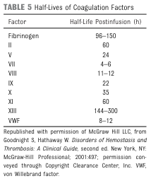
TABLE 5 Half-Lives of Coagulation Factors <table><thead><tr><th>Factor</th><th>Half-Life Postinfusion (h)</th></tr></thead><tbody><tr><td>Fibrinogen</td><td>96–150</td></tr><tr><td>II</td><td>60</td></tr><tr><td>V</td><td>24</td></tr><tr><td>VII</td><td>4–6</td></tr><tr><td>VIII</td><td>11–12</td></tr><tr><td>IX</td><td>22</td></tr><tr><td>X</td><td>35</td></tr><tr><td>XI</td><td>60</td></tr><tr><td>XIII</td><td>144–300</td></tr><tr><td>VWF</td><td>8–12</td></tr></tbody></table> Republished with permission of McGraw Hill LLC, from Goodnight S, Hathaway W. Disorders of Hemostasis and Thrombosis: A Clinical Guide, second ed. New York, NY: McGraw-Hill Professional; 2001:497; permission conveyed through Copyright Clearance Center, Inc. VWF, von Willebrand factor.
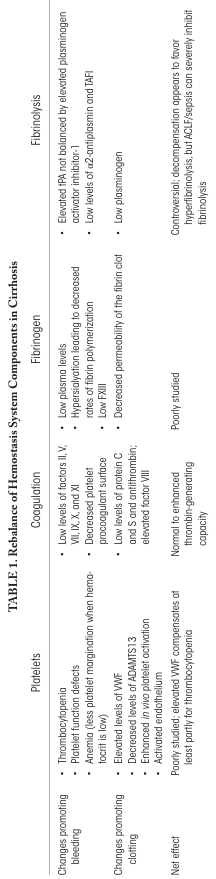
TABLE 1: Rebalance of Hemostasis System Components in Cirrhosis <table><tr><th rowspan="2">Platelets</th><td>Thrombocytopenia</td><td rowspan="2">Coagulation</td><td>• Low levels of factors II, V, VII, IX, X, and XI<br>• Decreased platelet procoagulant surface<br>• Low levels of protein C and S and antithrombin; elevated factor VIII</td><td rowspan="2">Fibrinogen</td><td>• Low plasma levels<br>• Hypersialylation leading to decreased rates of fibrin polymerization<br>• Low FXIII<br>• Decreased permeability of the fibrin clot</td><td rowspan="2">Fibrinolysis</td><td>• Elevated tPA not balanced by elevated plasminogen activator inhibitor-1<br>• Low levels of α2-antiplasmin and TAFI<br>• Low plasminogen</td></tr><tr><td>Platelet (less platelet) malfunction when hematocrit is low</td><td>Normal to enhanced thrombin-generating capacity</td><td>Poorly studied</td><td>Controversial: decompensation appears to favor hyperfibrinolysis, but ACLF/sepsis can severely inhibit fibrinolysis</td></tr><tr><th>Changes promoting bleeding</th><td>Anemia</td><td>Changes promoting bleeding</td><td>• Low levels of factors II, V, VII, IX, X, and XI<br>• Decreased platelet procoagulant surface<br>• Low levels of protein C and S and antithrombin; elevated factor VIII</td><td>Changes promoting bleeding</td><td>• Low plasma levels<br>• Hypersialylation leading to decreased rates of fibrin polymerization<br>• Low FXIII<br>• Decreased permeability of the fibrin clot</td><td>Changes promoting bleeding</td><td>• Elevated tPA not balanced by elevated plasminogen activator inhibitor-1<br>• Low levels of α2-antiplasmin and TAFI<br>• Low plasminogen</td></tr><tr><th>Changes promoting clotting</th><td>Elevated levels of VWF<br>Decreased levels of ADAMTS13<br>Enhanced in vivo platelet activation<br>Activated endothelium</td><td>Changes promoting clotting</td><td>Low levels of protein C and S and antithrombin; elevated factor VIII</td><td>Changes promoting clotting</td><td>Poorly studied</td><td>Changes promoting clotting</td><td>Controversial: decompensation appears to favor hyperfibrinolysis, but ACLF/sepsis can severely inhibit fibrinolysis</td></tr><tr><th>Net effect</th><td>Poorly studied; elevated VWF compensates at least partly for thrombocytopenia</td><td>Net effect</td><td>Normal to enhanced thrombin-generating capacity</td><td>Net effect</td><td>Poorly studied</td><td>Net effect</td><td>Controversial: decompensation appears to favor hyperfibrinolysis, but ACLF/sepsis can severely inhibit fibrinolysis</td></tr></table>
🩸 Clotting Factors + Coagulants - Deep Notes (ADHD Edition)
🔑 PART 1: THE 13 CLOTTING FACTORS - Know them cold
| Factor # | Common Name | Synonyms / Nickname | Remember it as... |
|---|---|---|---|
| I | Fibrinogen | - | "Fiber net" - makes the clot mesh |
| II | Prothrombin | - | "Pro" = before thrombin forms |
| III | Tissue Factor | Tissue Thromboplastin | Starts the extrinsic pathway |
| IV | Calcium (Ca²⁺) | - | "4th = Calcium" - cofactor everywhere |
| V | Proaccelerin | Labile factor, Ac-G | Accelerates the cascade |
| VII | Proconvertin | Stable factor, SPCA | Extrinsic pathway key player |
| VIII | Antihemophilic Factor A | AHF, AHG | Deficiency = Hemophilia A |
| IX | Christmas Factor | PTC, AHF-B | Deficiency = Hemophilia B |
| X | Stuart-Prower Factor | Stuart factor | Merges intrinsic + extrinsic |
| XI | PTA | Plasma Thromboplastin Antecedent | Deficiency = Hemophilia C |
| XII | Hageman Factor | Contact factor | Starts the intrinsic pathway |
| XIII | Fibrin-Stabilizing Factor | - | Locks the fibrin clot solid |
⚠️ There is NO Factor VI (it was found to be the same as Factor Va)
🔑 PART 2: THE CASCADE - Steps in Order
🟢 INTRINSIC PATHWAY (contact activation)
XII → XIIa
    ↓
XI → XIa
    ↓
IX → IXa  (+ VIII + Ca²⁺ + phospholipid)
    ↓
→ activates Factor X
🔴 EXTRINSIC PATHWAY (tissue factor pathway)
Tissue Factor (III) + VII → VIIa
    ↓
→ activates Factor X
🔵 COMMON PATHWAY (both merge here)
X → Xa  (+ V + Ca²⁺ + phospholipid = "Prothrombinase complex")
    ↓
Prothrombin (II) → Thrombin (IIa)
    ↓
Fibrinogen (I) → Fibrin (Ia) (loose)
    ↓
XIII → XIIIa
    ↓
Stable Fibrin Clot ✅
🔑 PART 3: WHERE DRUGS ACT ON THE CASCADE
| Drug | Hits which Factor? | Pathway blocked |
|---|---|---|
| Heparin (UFH) | XIIa, XIa, IXa, Xa, IIa (thrombin) | Intrinsic + Common |
| LMWH | Mainly Xa | Common |
| Fondaparinux | Only Xa | Common |
| Warfarin | II, VII, IX, X (synthesis blocked) | Extrinsic + Common |
| Rivaroxaban / Apixaban | Xa only | Common |
| Dabigatran | IIa (thrombin) only | Common |
🔑 PART 4: CLASSIFICATION OF COAGULANTS (Pro-clotting drugs)
COAGULANTS
│
├── SYSTEMIC (act throughout the body)
│   ├── Vitamin K (phytonadione/phylloquinone)
│   ├── Fresh Frozen Plasma (FFP)
│   ├── Protamine Sulfate (heparin antidote)
│   └── Factor Concentrates (e.g., Factor VIII, IX concentrates)
│
└── LOCAL (act at application site)
    ├── Absorbable
    │   ├── Gelatin sponge (Gelfoam)
    │   ├── Oxidized cellulose (Surgicel)
    │   └── Collagen sponge
    └── Non-absorbable
        ├── Topical thrombin
        ├── Fibrin glue (tissue sealant)
        └── Aluminum/ferric sulfate (astringents)
🔑 PART 5: COAGULANT DRUG CARDS - Mode of Action
🟢 VITAMIN K (Phytonadione)
CLASS: Systemic coagulant / Fat-soluble vitamin
TARGET: Vitamin K epoxide reductase enzyme
MOA: Vitamin K is required for the γ-carboxylation of
     glutamic acid residues on Factors II, VII, IX, X
     (also Protein C and S)
     → Without carboxylation, these factors can't bind Ca²⁺
     → Without Ca²⁺, they can't anchor to platelet membranes
     → Clotting cascade fails to assemble properly
USE: Warfarin reversal, newborn hemorrhage, dietary deficiency
ONSET: Slow (24 hours - needs new factor synthesis)
ROUTE: Oral or IV (never IM if INR elevated)
🟢 PROTAMINE SULFATE
CLASS: Systemic coagulant / Heparin antidote
MOA: Protamine is a strongly POSITIVELY charged protein
     Heparin is strongly NEGATIVELY charged
     → They bind ionically → form a stable inactive complex
     → Heparin is neutralized immediately
USE: Reversal of heparin overdose (post-cardiac surgery)
ROUTE: IV (slow infusion - rapid IV causes hypotension)
DOSE: 1 mg protamine neutralizes 100 units of heparin
WATCH: Anaphylaxis risk, especially in fish allergy patients
🟢 TRANEXAMIC ACID
CLASS: Anti-fibrinolytic (keeps clot from being broken down)
MOA: Blocks plasminogen from binding to fibrin
     → Plasmin cannot form
     → Fibrinolysis is inhibited
     → Existing clot is preserved
USE: Surgical bleeding, trauma, heavy periods (menorrhagia), haemophilia
ROUTE: Oral or IV
REMEMBER: It does NOT make new clots - it PROTECTS existing ones
🟢 AMINOCAPROIC ACID (epsilon-aminocaproic acid)
CLASS: Anti-fibrinolytic
MOA: Same as tranexamic acid - competitive inhibitor
     of plasminogen activators (blocks lysine-binding sites)
USE: Bleeding after cardiac surgery, hemophilia procedures
REMEMBER: Tranexamic acid is 6-10x more potent
🟢 FRESH FROZEN PLASMA (FFP)
CLASS: Blood product / Systemic coagulant
MOA: Contains ALL clotting factors (I, II, V, VII, VIII, IX, X, XI)
     + Protein C, Protein S, Antithrombin
     → Replaces multiple missing/consumed factors at once
USE: Acute reversal of warfarin, massive transfusion, liver failure
ONSET: Immediate
NOTE: Must ABO match. Risk of TRALI, volume overload
🔑 PART 6: ONE-LOOK HEMOSTASIS OVERVIEW (Stick this on your wall)
INJURY
  ↓
① Vascular Spasm (immediate, seconds)
  ↓
② Platelet Plug (primary hemostasis, minutes)
   [platelet adhesion → activation → aggregation]
  ↓
③ Coagulation Cascade (secondary hemostasis)
   Intrinsic (XII→) + Extrinsic (TF+VII→)
              ↓
        Common (X→II→I)
              ↓
      Fibrin Clot (stable)
  ↓
④ Fibrinolysis (clot breakdown, days later)
   Plasminogen → Plasmin → Fibrin degradation products
🔑 PART 7: ADHD STUDY FLOW for THIS CHAPTER
- Write out the 13 clotting factors from memory using the table above
- Draw the cascade: just boxes and arrows, no details yet
- Quiz: "Which factor starts intrinsic? Extrinsic? Common?"
- Add drugs to your cascade drawing - draw a red X where each drug acts
- Fill drug cards for Vitamin K and Protamine (these are coagulants = your current focus)
- Quiz: "What does Vitamin K do? What does tranexamic acid do?"
- Cover the MOA column and say each drug's mechanism out loud
- Key exam question: "Factor VII has the shortest half-life (4-6 hours) - so Warfarin's effect on PT/INR appears first!"
- Vitamin K-dependent factors = II, VII, IX, X (+ Protein C, S)
- Factor VIII deficiency = Hemophilia A
- Factor IX deficiency = Hemophilia B
- Protamine = specific antidote for heparin
- Tranexamic acid = anti-fibrinolytic (NOT an anticoagulant)
Not about drugs today it was classification of coagulants
classification of coagulants pharmacology local systemic mode of action KD Tripathi
https://dl.icdst.org/pdfs/files3/f87428a2f2a044977b3c26eff96…
coagulants classification local systemic hemostatic agents pharmacology
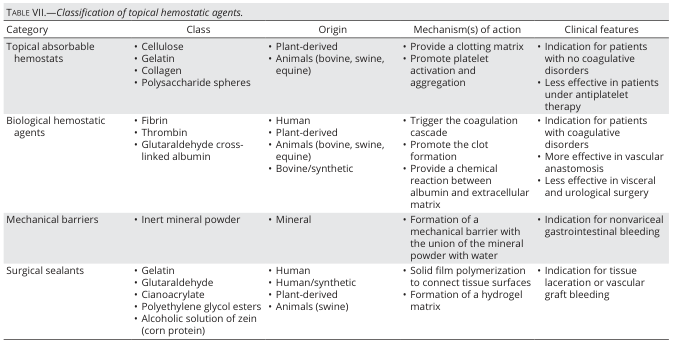
TABLE VII.—Classification of topical hemostatic agents. <table><thead><tr><th>Category</th><th>Class</th><th>Origin</th><th>Mechanism(s) of action</th><th>Clinical features</th></tr></thead><tbody><tr><td>Topical absorbable hemostats</td><td>• Cellulose<br>• Gelatin<br>• Collagen<br>• Polysaccharide spheres</td><td>• Plant-derived<br>• Animals (bovine, swine, equine)</td><td>• Provide a clotting matrix<br>• Promote platelet activation and aggregation</td><td>• Indication for patients with no coagulative disorders<br>• Less effective in patients under antiplatelet therapy</td></tr><tr><td>Biological hemostatic agents</td><td>• Fibrin<br>• Thrombin<br>• Glutaraldehyde cross-linked albumin</td><td>• Human<br>• Plant-derived<br>• Animals (bovine, swine, equine)<br>• Bovine/synthetic</td><td>• Trigger the coagulation cascade<br>• Promote the clot formation<br>• Provide a chemical reaction between albumin and extracellular matrix</td><td>• Indication for patients with coagulative disorders<br>• More effective in vascular anastomosis<br>• Less effective in visceral and urological surgery</td></tr><tr><td>Mechanical barriers</td><td>• Inert mineral powder</td><td>• Mineral</td><td>• Formation of a mechanical barrier with the union of the mineral powder with water</td><td>• Indication for nonvariceal gastrointestinal bleeding</td></tr><tr><td>Surgical sealants</td><td>• Gelatin<br>• Glutaraldehyde<br>• Cyanoacrylate<br>• Polyethylene glycol esters<br>• Alcoholic solution of zein (corn protein)</td><td>• Human<br>• Human/synthetic<br>• Plant-derived<br>• Animals (swine)</td><td>• Solid film polymerization to connect tissue surfaces<br>• Formation of a hydrogel matrix</td><td>• Indication for tissue laceration or vascular graft bleeding</td></tr></tbody></table>
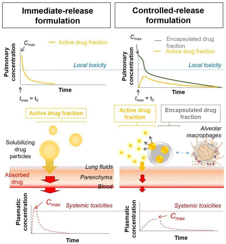
A comparison chart and pathophysiology diagram illustrating the local and systemic pharmacokinetic profiles of immediate-release versus controlled-release inhalation formulations. The top row features line graphs of pulmonary drug concentration over time. The immediate-release graph shows a sharp, high peak (Cmax) at tmax = t0, significantly exceeding the local toxicity threshold. The controlled-release graph depicts two profiles: a high initial encapsulated drug fraction and a lower, sustained active drug fraction that remains below the local toxicity limit. The middle section shows the mechanism of action: immediate-release involves rapid solubilizing drug particles penetrating lung fluids and parenchyma, while controlled-release shows nanocarriers releasing active drug fractions and interacting with alveolar macrophages. The bottom row presents plasmatic concentration graphs; the immediate-release formulation exhibits a rapid systemic Cmax correlated with higher systemic toxicities, whereas the controlled-release formulation demonstrates a blunted, delayed peak, indicating improved systemic safety. This educational graphic is intended for pharmacology and oncology instruction regarding inhaled drug delivery systems.
🩸 COAGULANTS - Classification + Mode of Action
(ADHD-Smart Notes | No drug dosing, just classification & MOA)
🔑 WHAT IS A COAGULANT?
Coagulants = Substances that PROMOTE clot formation Used when: bleeding won't stop on its own (haemorrhagic states, surgery, wounds)
🗂️ MASTER CLASSIFICATION TREE
COAGULANTS
│
├── A. LOCAL COAGULANTS
│   (applied directly at bleeding site)
│   │
│   ├── 1. Absorbable Mechanical Agents
│   │       • Gelatin sponge (Gelfoam)
│   │       • Oxidized cellulose (Surgicel)
│   │       • Microfibrillar collagen (Avitene)
│   │
│   ├── 2. Biological / Active Agents
│   │       • Topical Thrombin
│   │       • Fibrin sealant (fibrin glue)
│   │       • Platelet sealant (collagen + thrombin + fibrinogen)
│   │
│   ├── 3. Astringents (precipitate proteins)
│   │       • Ferric chloride / Alum
│   │       • Silver nitrate
│   │
│   └── 4. Vasoconstrictors (local)
│           • Adrenaline (epinephrine) - applied locally
│
└── B. SYSTEMIC COAGULANTS
    (act throughout the body via bloodstream)
    │
    ├── 1. Vitamin K (phytonadione / phylloquinone)
    │
    ├── 2. Anti-fibrinolytics (prevent clot breakdown)
    │       • Tranexamic acid
    │       • Epsilon-aminocaproic acid (EACA)
    │
    ├── 3. Blood / Plasma Products
    │       • Fresh Frozen Plasma (FFP)
    │       • Cryoprecipitate
    │       • Factor concentrates (VIII, IX, VWF)
    │
    └── 4. Protamine Sulfate
            (specific antidote to heparin)
🔬 MODE OF ACTION - Each Group (Simple Cards)
🟧 A1 - ABSORBABLE MECHANICAL AGENTS
| What they do | How |
|---|---|
| Provide a physical scaffold / matrix | Blood fills the sponge/mesh → clotting cascade is triggered naturally |
| Promote platelet adhesion | Collagen activates platelets → platelet plug forms |
| Absorb and disappear | Body digests them over days (no removal needed) |
⚠️ Work best when the patient's own clotting system is intact (not useful in haemophilia alone)
🟥 A2 - BIOLOGICAL / ACTIVE AGENTS
| Agent | MOA |
|---|---|
| Topical Thrombin | Directly converts Fibrinogen → Fibrin (skips the whole cascade above) |
| Fibrin sealant (glue) | Contains thrombin + fibrinogen together → instantly forms fibrin clot on contact |
| Platelet sealant | Provides collagen + thrombin + patient's own fibrinogen + platelets → immediate complete seal |
⚠️ Thrombin must stay LOCAL - if it enters a large vessel it can cause DIC (systemic clotting)
🟫 A3 - ASTRINGENTS
| What they do | How |
|---|---|
| Precipitate surface proteins | Chemically denature proteins at wound surface → forms a tough protein film |
| Physically seal small vessels | Protein coagulum acts like a plug |
🟩 B1 - VITAMIN K (The most exam-important systemic coagulant)
MOA in 4 steps:

Liver needs Vitamin K
    ↓
Vitamin K activates an enzyme (γ-carboxylase)
    ↓
This enzyme adds COOH groups to Factors: II, VII, IX, X
(also Protein C and S)
    ↓
These factors can now bind Ca²⁺
    ↓
Clotting cascade can proceed normally ✅
| Vitamin K type | Source | Notes |
|---|---|---|
| K1 (Phylloquinone) | Green leafy vegetables | Main therapeutic form |
| K2 (Menaquinone) | Gut bacteria, fermented food | Natural |
| K3 (Menadione) | Synthetic | More toxic, not preferred |
🟦 B2 - ANTI-FIBRINOLYTICS
Normal fibrinolysis:
Plasminogen → (activated by tPA) → Plasmin → breaks down Fibrin → clot dissolves

Anti-fibrinolytics BLOCK this:
Tranexamic acid blocks plasminogen from binding to fibrin
    ↓
Plasmin cannot form
    ↓
Existing fibrin clot is PROTECTED from breakdown
    ↓
Bleeding stops / clot lasts longer ✅
- They do NOT make new clots
- They PRESERVE clots that have already formed
- This is why they're used in surgical bleeding, trauma, heavy periods
🟪 B3 - BLOOD / PLASMA PRODUCTS
| Product | What it contains | MOA |
|---|---|---|
| FFP (Fresh Frozen Plasma) | ALL clotting factors (I, II, V, VII, VIII, IX, X, XI) | Replaces multiple missing factors at once |
| Cryoprecipitate | Factor VIII, XIII, fibrinogen, vWF | Targeted - used in haemophilia A, vWD, DIC |
| Factor VIII concentrate | Purified Factor VIII | Used in Haemophilia A specifically |
| Factor IX concentrate | Purified Factor IX | Used in Haemophilia B specifically |
🟨 B4 - PROTAMINE SULFATE
MOA:
Protamine = STRONGLY POSITIVE charge (+)
Heparin   = STRONGLY NEGATIVE charge (-)
    ↓
They bind to each other ionically
    ↓
Form a stable, inactive complex
    ↓
Heparin is neutralized immediately ✅
🧠 ADHD QUICK RECALL TABLE (Stick this somewhere visible)
| Group | Example | MOA in ONE line |
|---|---|---|
| Absorbable mechanical | Gelfoam, Surgicel | Provides scaffold → platelets stick → natural clot |
| Biological active | Thrombin, Fibrin glue | Directly makes fibrin - bypasses cascade |
| Astringents | Ferric Cl, Alum | Denatures proteins → physical protein plug |
| Vitamin K | Phytonadione (K1) | Activates γ-carboxylase → working clotting factors II, VII, IX, X |
| Anti-fibrinolytics | Tranexamic acid, EACA | Blocks plasminogen → preserves existing fibrin clot |
| Plasma products | FFP, Cryo, Factor VIII | Replaces missing clotting factors |
| Protamine | Protamine sulfate | (+) charge binds heparin's (-) charge → neutralizes it |
📌 EXAM HOTSPOTS FROM THIS CHAPTER
- Vitamin K-dependent factors = II, VII, IX, X (and Protein C, S)
- Tranexamic acid is NOT a coagulant - it is an anti-fibrinolytic (indirect coagulant)
- Topical thrombin - stays local only! Systemic entry = DIC
- Cryoprecipitate contains fibrinogen + Factor VIII + vWF (used in DIC, haemophilia A)
- Protamine is not a coagulant per se - it's a heparin antidote that restores coagulation
About only vitamin K
vitamin K mechanism action coagulation factors gamma carboxylation warfarin
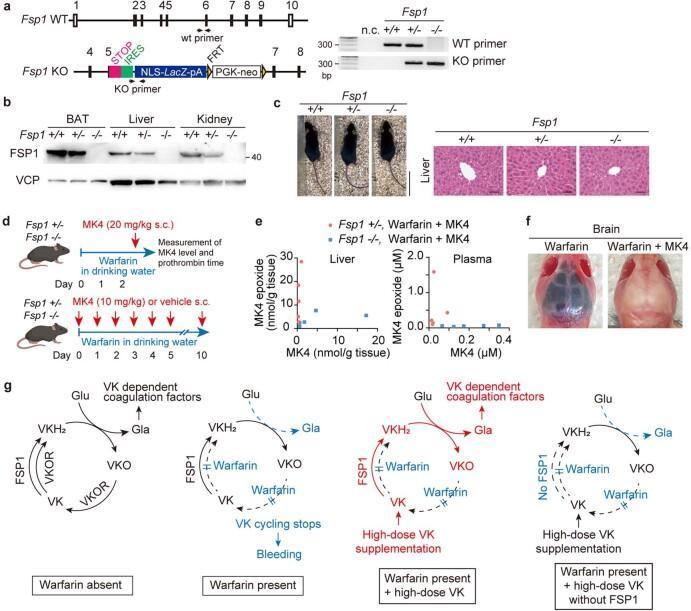
This composite educational graphic illustrates the role of Ferroptosis Suppressor Protein 1 (FSP1) as a warfarin-resistant vitamin K (VK) reductase. Panel (a) shows genomic schematic maps of Fsp1 wild-type and knockout alleles alongside genotyping PCR results. Panel (b) provides an immunoblot for FSP1 expression in adipose tissue, liver, and kidney across genotypes. Panel (c) displays gross morphological and H&E stained liver histology of Fsp1 mice, demonstrating normal architecture. Panel (d) outlines a clinical study design evaluating MK4 (vitamin K2) levels and prothrombin time under high-dose warfarin treatment. Panel (e) uses scatter plots to show the concentrations of MK4 and MK4 epoxide in liver and plasma. Panel (f) presents a clinical photograph comparing mouse brains: the warfarin-only specimen exhibits massive cerebral hemorrhage (dark discoloration), while the warfarin plus MK4 specimen appears normal, illustrating the antidotal effect of MK4. Finally, panel (g) provides pathophysiology flowcharts of the vitamin K cycle, detailing the enzymatic pathways involving VKOR and FSP1, and the mechanism by which high-dose vitamin K bypasses warfarin inhibition to enable carboxylation of coagulation factors.
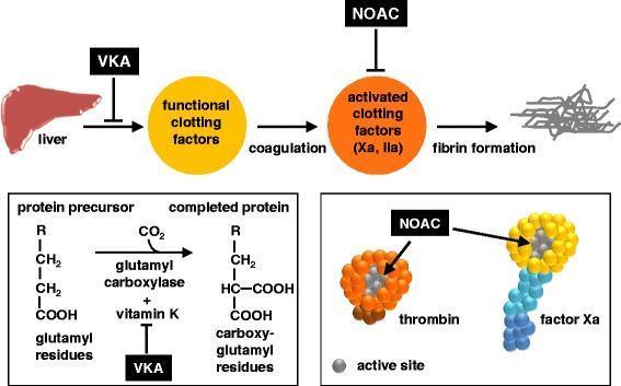
A multi-panel pathophysiology diagram illustrating the mechanism of action of oral anticoagulants on the coagulation cascade. The top panel shows a linear pathway: the liver produces functional clotting factors, which undergo coagulation to become activated clotting factors (Xa and IIa/thrombin), ultimately leading to fibrin formation. Vitamin K antagonists (VKA) are shown inhibiting the liver's production of functional factors, while non-vitamin K antagonist oral anticoagulants (NOACs) inhibit the activated factors. The bottom-left panel details the biochemical mechanism of VKAs, showing they inhibit the glutamyl carboxylase and vitamin K-dependent conversion of glutamyl residues on protein precursors into carboxy-glutamyl residues on completed clotting proteins. The bottom-right panel provides a molecular-level view of NOAC action, depicting thrombin (orange sphere) and factor Xa (yellow head with blue tail) being inhibited at their respective active sites (indicated by grey coloring). This educational graphic highlights the distinction between indirect synthesis inhibition by VKAs and direct enzymatic inhibition by NOACs/DOACs.
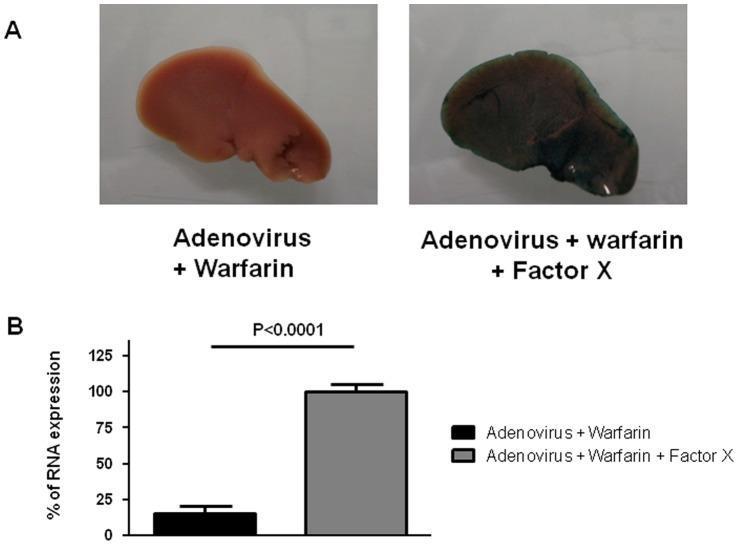
This composite educational image, categorized as Clinical Imaging/Specimen, illustrates the role of blood coagulation Factor X in adenovirus transduction using a mouse model. Panel A presents two gross liver specimens stained for β-galactosidase activity (Lac-Z reporter gene). The specimen labeled 'Adenovirus + Warfarin' appears pale pink/tan, indicating minimal viral transduction due to warfarin-induced depletion of vitamin K-dependent factors. In contrast, the 'Adenovirus + Warfarin + Factor X' specimen shows intense dark blue-green staining across the hepatic surface, demonstrating that exogenous Factor X supplementation restores adenoviral infection. Panel B contains a corresponding bar graph quantifying the percentage of RNA expression in adrenal glands. The 'Adenovirus + Warfarin' group (black bar) shows significantly lower expression compared to the 'Adenovirus + Warfarin + Factor X' group (gray bar), which is set as the 100% reference point. Statistical significance is indicated by a p-value < 0.0001. This figure demonstrates that adenovirus type 5 infection in both liver and adrenal tissues is dependent on Factor X, bypassing traditional CAR receptor-mediated pathways.
🌿 VITAMIN K - Complete Notes
(ADHD-Smart | Classification + MOA + Everything exam-worthy)
📦 WHAT IS VITAMIN K?
Vitamin K = Fat-soluble vitamin that acts as a cofactor for making working clotting factors in the liver.
🗂️ CLASSIFICATION OF VITAMIN K (3 Types)
VITAMIN K
│
├── K1 - Phylloquinone / Phytonadione
│         Source: Green leafy vegetables (spinach, kale, broccoli)
│         Main NATURAL + THERAPEUTIC form
│         Used clinically for warfarin reversal, deficiency
│
├── K2 - Menaquinone
│         Source: Gut bacteria (synthesized in colon) + Fermented foods
│         Also plays role in bone metabolism (activates osteocalcin)
│         NOT used clinically for bleeding
│
└── K3 - Menadione
          Source: SYNTHETIC (not found naturally)
          Most POTENT (converted to K2 in body)
          TOXIC at high doses → causes haemolytic anaemia, jaundice
          CONTRAINDICATED in neonates and pregnant women
          Rarely used now
⚙️ MODE OF ACTION - The Vitamin K Cycle
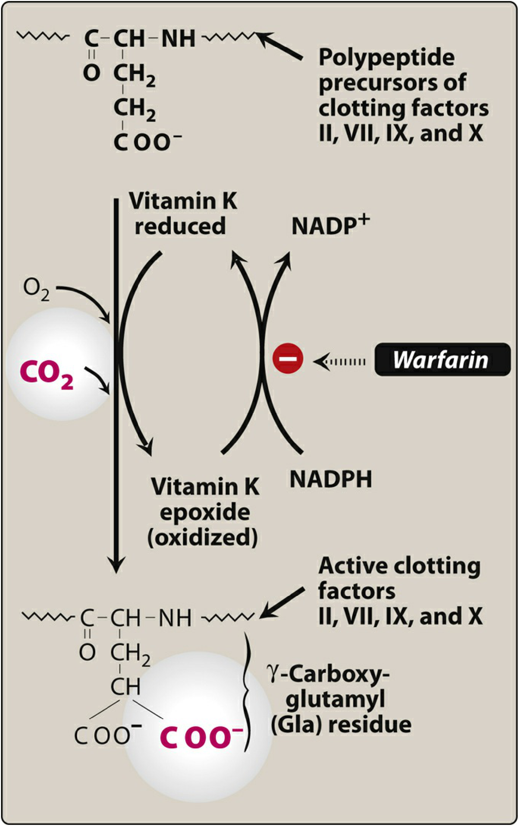
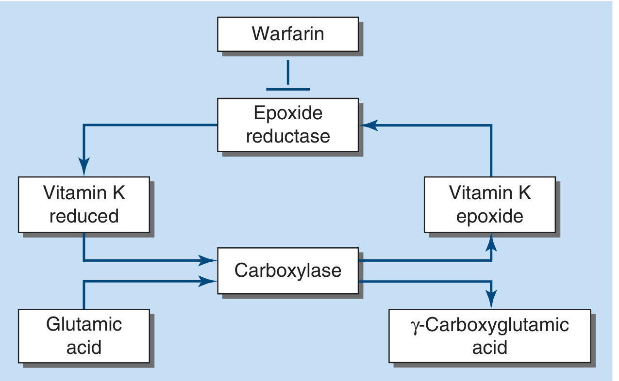
Step-by-step (write this as a flowchart in your notes):
STEP 1:
Vitamin K (quinone form) enters hepatocyte
         ↓
STEP 2:
Reduced to → Vitamin K HYDROQUINONE (KH₂) = ACTIVE FORM
[enzyme: quinone reductase]
         ↓
STEP 3:
KH₂ acts as cofactor for → γ-Glutamylcarboxylase enzyme
This enzyme adds a COOH group to Glutamic acid residues
on clotting factors II, VII, IX, X
→ Forms γ-Carboxyglutamic acid (Gla) residues
         ↓
STEP 4:
These Gla residues can now BIND CALCIUM (Ca²⁺)
→ Clotting factors anchor to platelet membranes
→ Cascade proceeds normally ✅
         ↓
STEP 5 (Recycling):
In the process, Vitamin KH₂ → oxidized → Vitamin K EPOXIDE
This epoxide is "used up" Vitamin K
         ↓
STEP 6 (Recovery):
Vitamin K epoxide reductase (VKOR) converts it back
→ Vitamin K → KH₂ (active again)
→ Cycle continues ♻️
WHERE WARFARIN ACTS:
Warfarin BLOCKS Vitamin K epoxide reductase (VKOR) at Step 6 → Vitamin K epoxide cannot be recycled back to active form → No KH₂ → No carboxylation → Factors II, VII, IX, X are inactive → Anticoagulation
🎯 VITAMIN K-DEPENDENT FACTORS (Exam Gold)
| Procoagulant | Anticoagulant |
|---|---|
| Factor II (Prothrombin) | Protein C |
| Factor VII | Protein S |
| Factor IX | Protein Z |
| Factor X |
⚠️ Protein C has the shortest half-life of all Vitamin K-dependent proteins → When Warfarin is started, Protein C drops FIRST → brief procoagulant/thrombotic state → That's why Warfarin needs bridging with heparin at initiation
💊 VITAMIN K AS A DRUG (Phytonadione / K1)
CLASS:     Systemic coagulant / Fat-soluble vitamin

ROUTE:     Oral (preferred) | IV (use slow infusion only!)
           IM is AVOIDED when INR is elevated (risk of haematoma)

ONSET:     Oral → 6-12 hours
           IV   → 1-2 hours (faster but anaphylaxis risk)
           Peak effect: 24 hours (time needed to synthesize new factors)

USES:
  1. Warfarin overdose / reversal
  2. Newborn haemorrhagic disease (VKDB)
  3. Obstructive jaundice / malabsorption (fat-soluble vitamin not absorbed)
  4. Dietary deficiency (rare in adults)

DOSE:      1-10 mg oral (for warfarin reversal)
           5-20 mg for severe deficiency

ANTIDOTE:  It IS the antidote for warfarin!
🔴 VITAMIN K DEFICIENCY - Causes + What Happens
CAUSES:
  ├── Newborns → gut not colonized yet, no K2 from bacteria
  │             → breast milk has LOW K1
  │             → VKDB (Vitamin K Deficiency Bleeding) = classic exam scenario
  │
  ├── Malabsorption → obstructive jaundice, celiac, Crohn's
  │                 → fat not digested → fat-soluble vitamins not absorbed
  │
  ├── Prolonged antibiotics → kill gut bacteria → less K2 made
  │
  └── Warfarin therapy → pharmacological Vitamin K antagonism

RESULT OF DEFICIENCY:
  Factors II, VII, IX, X + Protein C, S → cannot be carboxylated
         ↓
  Clotting cascade FAILS to assemble properly
         ↓
  BLEEDING (especially surgical, GI, intracranial in newborns)

TESTS:
  PT/INR elevated (Factor VII drops first - shortest half-life ~4-6 hrs)
  aPTT also prolonged (Factors IX, X affected)
📊 QUICK COMPARISON: K1 vs K2 vs K3
| Feature | K1 (Phylloquinone) | K2 (Menaquinone) | K3 (Menadione) |
|---|---|---|---|
| Source | Green vegetables | Gut bacteria | Synthetic |
| Water/fat soluble | Fat | Fat | Water soluble |
| Clinical use | Yes (warfarin reversal) | Bone health | Rarely (toxic) |
| Safe in neonates? | Yes | Yes | NO (haemolytic) |
| Safe in pregnancy? | Yes | Yes | NO |
🧠 ADHD STUDY CARD FOR VITAMIN K
┌──────────────────────────────────────────┐
│             VITAMIN K                    │
│                                          │
│  Types: K1 (plant) K2 (bacteria) K3(lab)│
│                                          │
│  JOB: Activates γ-carboxylase           │
│  → carboxylates Factors 2, 7, 9, 10     │
│  → they bind Ca²⁺ → clotting works      │
│                                          │
│  CYCLE: KH₂ → Epoxide → VKOR → KH₂     │
│  WARFARIN blocks: VKOR (step 6)          │
│                                          │
│  USE AS DRUG:                            │
│   ✅ Warfarin reversal                   │
│   ✅ Newborn VKDB prevention             │
│   ✅ Malabsorption bleeding              │
│                                          │
│  ONSET: 6-12h oral, 1-2h IV             │
│  Peak: 24 hours                         │
│  Route: Oral preferred, NO IM if INR↑   │
└──────────────────────────────────────────┘
📌 3 EXAM TRAPS TO REMEMBER
- Warfarin reversal takes 24 hours even with Vitamin K - because existing broken factors must degrade first before new working ones are made
- Protein C drops first when warfarin starts → momentary procoagulant state → bridge with heparin
- K3 (Menadione) is toxic to neonates - causes haemolytic anaemia + jaundice - never use in newborns