
Hiw to oresent pulse after examination
When presenting a pulse after clinical examination, you describe it in a structured way covering rate, rhythm, volume, character, and any special features. Here's the standard format:
Searching sources...
Reading File
Finding Sources
Finding Sources
Reading File
Reading File
Reading File
Loading Image
Reading File
Here is a complete guide on how to present the pulse after clinical examination:
How to Present the Pulse After Examination
When presenting the pulse in a clinical setting (OSCE, bedside, case presentation), you describe it systematically across 6 parameters:
The 6 Parameters of Pulse Presentation
1. Rate
- Count beats per minute (over 60 seconds at the radial pulse, or 15 seconds x4)
- Normal: 60-100 bpm
- Tachycardia >100 bpm (fever, shock, thyrotoxicosis, anemia, sepsis)
- Bradycardia <60 bpm (heart block, hypothyroidism, athletes, beta-blockers)
"The pulse rate is 88 beats per minute"
2. Rhythm
- Regular or irregular
- If irregular: is it "regularly irregular" (patterned, e.g. 2nd degree heart block, bigeminy) or "irregularly irregular" (no pattern, e.g. atrial fibrillation)
"The rhythm is regular" or "The rhythm is irregularly irregular, consistent with atrial fibrillation"
3. Volume (Pulse Pressure)
- The perceived amplitude of the pulse
- Normal volume
- High volume / bounding (wide pulse pressure): aortic regurgitation, thyrotoxicosis, CO2 retention, PDA, fever
- Low volume / weak / thready (narrow pulse pressure): aortic stenosis, shock, cardiac tamponade, severe LV dysfunction
"The volume is normal" or "The volume is increased (bounding)"
4. Character (Waveform / Contour)
This is the most clinically informative part. Assessed best at the carotid or brachial artery:
| Character | Description | Condition |
|---|---|---|
| Normal | Smooth, single peak | Normal |
| Parvus et tardus | Small amplitude + slow rise | Severe aortic stenosis |
| Water-hammer (Corrigan's) | Bounding, collapsing pulse | Severe aortic regurgitation |
| Bisferiens | Two palpable systolic peaks | Mixed aortic valve disease (AR > AS) |
| Spike-and-dome | Rapid rise then mid-systolic obstruction | Hypertrophic obstructive cardiomyopathy (HOCM) |
| Dicrotic | Exaggerated dicrotic notch, two beats felt | Septic shock, severe peripheral vasoconstriction |
| Pulsus alternans | Alternating strong and weak beats | Severe LV systolic dysfunction |
| Pulsus paradoxus | Amplitude falls >10 mmHg on inspiration | Cardiac tamponade, severe asthma |
"The character is normal" or "There is a collapsing, water-hammer character"
5. Condition of the Vessel Wall
- Normally the vessel wall is not palpable between beats
- Thickened / tortuous / pipe-stem wall: atherosclerosis (especially in elderly)
- Assessed by rolling the vessel under the fingers
"The vessel wall is soft and compressible" or "The vessel wall is thickened and tortuous, suggesting atherosclerosis"
6. Radio-Femoral Delay (Special Feature)
- Palpate radial and femoral simultaneously
- Normally felt at the same time
- Delay (femoral pulse felt later): coarctation of the aorta
"There is no radio-femoral delay"
Example of a Complete Pulse Presentation
"On examination of the pulse at the right radial artery, the rate is 78 beats per minute, the rhythm is regular, the volume is increased with a collapsing quality, and the character is consistent with a water-hammer pulse. The vessel wall is soft. There is no radio-femoral delay."
This presentation would point towards aortic regurgitation.
Pulse Waveform Patterns (Visual Guide)
The diagram below from Fuster and Hurst's The Heart shows normal and abnormal arterial pulse waveforms, illustrating what you would expect to feel on palpation:
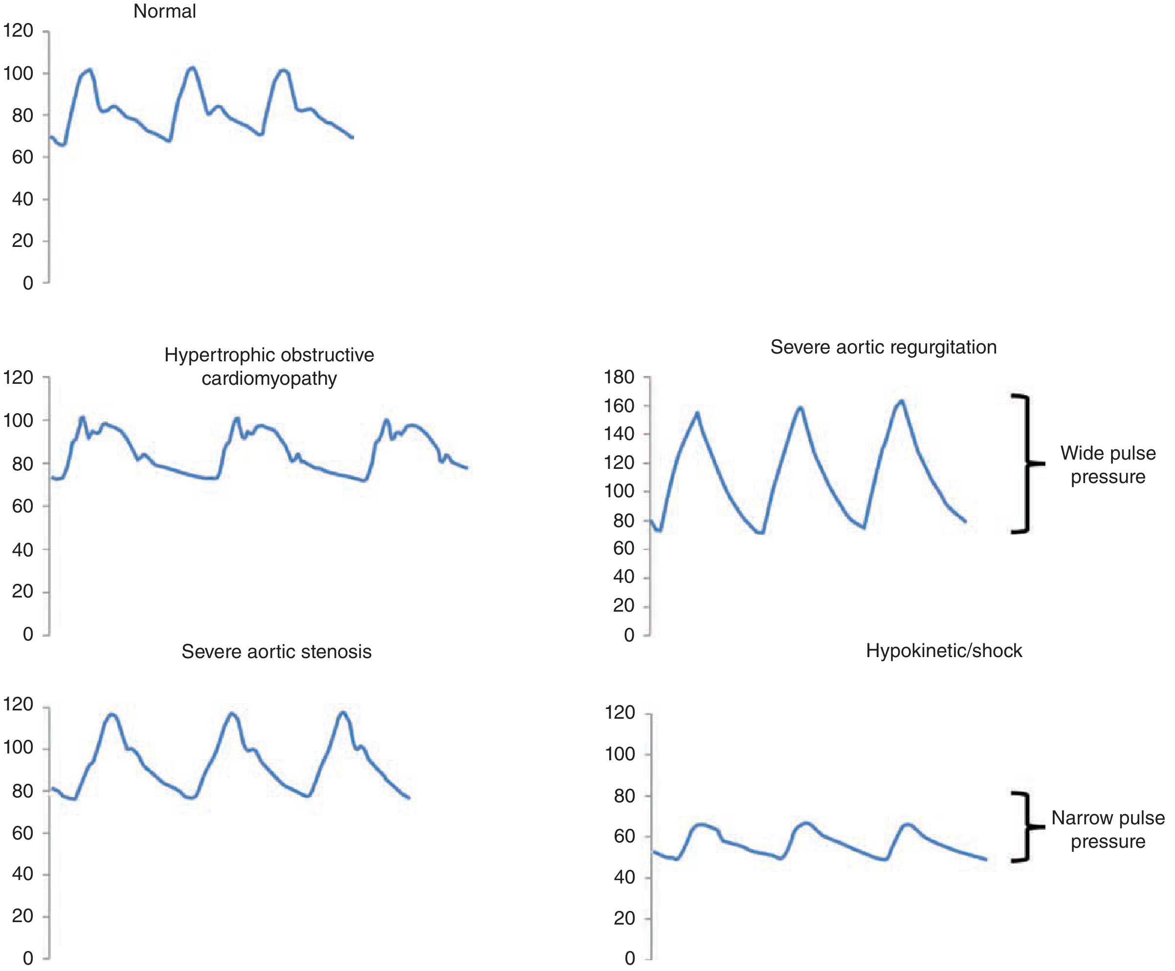
Normal pulse has a smooth upstroke; Severe aortic regurgitation shows wide pulse pressure (bounding); Hypokinetic/shock shows narrow pulse pressure (weak); Severe aortic stenosis (parvus et tardus) shows a dampened, delayed peak - Fuster and Hurst's The Heart, 15th Ed.
Practical Tips
- Site: Radial is routine; carotid/brachial is best for assessing character
- Simultaneous bilateral palpation: Check for symmetry and radio-femoral delay
- Always compare both sides: Absent/diminished on one side suggests arterial occlusion or dissection
- Pulsus paradoxus: Requires a sphygmomanometer - not purely a palpation finding
Sources: S Das - A Manual on Clinical Surgery, 13th Ed. | Fuster and Hurst's The Heart, 15th Ed.
In examination pf cvs. How will i present it?
A full CVS examination presentation follows a structured format: General inspection → Hands/peripheries → Pulse → Blood pressure → JVP → Precordium (inspection, palpation, auscultation) → Peripheral findings. Let me get the detailed textbook content for you.
Searching sources...
Finding Sources
Finding Sources
Reading File
Reading File
Reading File
Reading File
Reading File
Finding Sources
Reading File
I now have comprehensive content. Let me compile the full CVS examination presentation guide.
How to Present the CVS Examination
The cardiovascular examination is presented in a fixed sequence. Each section flows into the next.
Standard Presentation Sequence
"On examination of the cardiovascular system..."
1. General Appearance (from the end of the bed)
Before touching the patient, comment on:
- Conscious level - alert, comfortable at rest, or distressed
- Dyspnoea at rest - labored breathing, orthopnoea (sitting up)
- Pallor / cyanosis / jaundice
- Nutritional status - cachexia (cardiac failure)
- Dysmorphic features - Marfan syndrome (tall, arachnodactyly - aortic root disease), Down syndrome, Turner syndrome
- Scars - midline sternotomy, lateral thoracotomy (previous cardiac surgery), pacemaker device bulge
"The patient is a middle-aged male, sitting comfortably at rest with no obvious dyspnoea, pallor, or cyanosis. No dysmorphic features are noted."
2. Hands and Peripheries
| Finding | Significance |
|---|---|
| Clubbing | Cyanotic congenital heart disease, infective endocarditis |
| Splinter haemorrhages | Infective endocarditis |
| Janeway lesions | Infective endocarditis (painless, palms/soles) |
| Osler's nodes | Infective endocarditis (painful, finger pads) |
| Koilonychia | Iron-deficiency anaemia (high-output failure) |
| Capillary refill >2 sec | Peripheral vasoconstriction, low output |
| Peripheral cyanosis | Reduced peripheral perfusion |
| Xanthomata (tendons) | Hypercholesterolaemia |
"Hands are warm and well-perfused. Capillary refill time is less than 2 seconds. No clubbing, splinter haemorrhages, or peripheral stigmata of infective endocarditis."
3. Pulse
(As covered in detail previously - present rate, rhythm, volume, character, vessel wall, radio-femoral delay)
"Pulse is 78 bpm, regular, normal volume and character. No radio-femoral delay."
4. Blood Pressure
- Measured in both arms (difference >10 mmHg = subclavian disease, aortic dissection, coarctation)
- State: systolic / diastolic, and which arm
- Pulse pressure = systolic - diastolic (normal ~40 mmHg)
- Wide pulse pressure (>60): aortic regurgitation, thyrotoxicosis
- Narrow pulse pressure (<25): cardiac tamponade, severe AS, cardiogenic shock
- Pulsus paradoxus (>10 mmHg drop in systolic on inspiration): cardiac tamponade, massive PE, severe asthma
"Blood pressure is 128/78 mmHg in the right arm. No significant inter-arm difference. Pulse pressure is normal."
5. Face and Eyes
- Malar flush (mitral facies) - mitral stenosis
- Xanthelasma (around eyelids) - hypercholesterolaemia
- Corneal arcus - hypercholesterolaemia (significant if <50 years)
- Pallor of conjunctivae - anaemia
- Central cyanosis - inspect tongue and lips
- Fundoscopy (if asked): hypertensive/diabetic retinopathy, Roth spots (endocarditis)
"No malar flush, xanthelasma, or corneal arcus. Conjunctivae are pink. No central cyanosis."
6. Jugular Venous Pressure (JVP)
- Patient at 45°, head turned slightly left
- Locate the internal jugular vein pulsation (medial to sternocleidomastoid)
- Measure vertical height above the sternal angle (angle of Louis) - normal ≤4.5 cm at 45°
- JVP >5 cm above sternal angle = elevated CVP
- Character of the waveform:
| Feature | Significance |
|---|---|
| Absent 'a' wave | Atrial fibrillation |
| Giant 'a' wave | Pulmonary hypertension, tricuspid stenosis, RVOTO |
| Cannon 'a' waves | Complete heart block, VT (AV dissociation) |
| Giant 'v' wave | Tricuspid regurgitation |
| Prominent 'y' descent | Constrictive pericarditis, tricuspid regurgitation |
| Non-pulsatile, fixed elevation | SVC obstruction |
- Hepatojugular reflux - press over liver for 15 sec; JVP rises >3 cm = right heart failure
"JVP is not elevated. It is 3 cm above the sternal angle at 45°. Normal 'a' and 'v' waveforms visible. No hepatojugular reflux."
7. Precordium - Inspection
Look at the chest with tangential lighting:
- Shape - pectus excavatum/carinatum, kyphoscoliosis (affect cardiac position)
- Visible pulsations:
- Apex beat (5th ICS, midclavicular line) - normal
- Left parasternal heave - right ventricular enlargement
- Visible pulsation elsewhere - abnormal
- Scars - midline sternotomy, valve surgery, pacemaker pocket, CABG drain scars
- Deformities - precordial bulge in children = cardiomegaly from infancy
"The chest is normal in shape. Apex beat visible at the 5th intercostal space, midclavicular line. No abnormal pulsations or surgical scars."
8. Precordium - Palpation
Apex beat:
- Normal position: 5th ICS, midclavicular line
- Displaced laterally/inferiorly = LV enlargement
- Character:
| Apex Character | Cause |
|---|---|
| Tapping | Palpable S1 - mitral stenosis |
| Heaving (sustained, forceful) | Pressure overload - AS, hypertension |
| Thrusting (hyperdynamic, non-sustained) | Volume overload - AR, MR, VSD |
| Diffuse, dyskinetic | Dilated cardiomyopathy, large anterior MI |
| Impalpable | COPD, obesity, pericardial effusion, dextrocardia |
| Double impulse | HOCM |
Parasternal heave:
- Place heel of right hand on lower left sternal border
- Sustained lift = right ventricular hypertrophy (pulmonary hypertension, pulmonary stenosis)
Thrills:
- Palpable murmurs (grade 4+)
- Systolic thrill at base = AS or pulmonary stenosis
- Systolic thrill at apex = MR or VSD
"Apex beat is palpable in the 5th intercostal space, midclavicular line. It has a normal character - not sustained, not displaced. No parasternal heave. No thrills palpable."
9. Precordium - Auscultation
Positions of auscultation:
| Area | Location | Best Hears |
|---|---|---|
| Aortic area | 2nd ICS, right sternal border | Aortic valve sounds |
| Pulmonary area | 2nd ICS, left sternal border | Pulmonary valve sounds |
| Tricuspid area | Lower left sternal border (4th ICS) | Tricuspid valve |
| Mitral area (Apex) | 5th ICS, midclavicular line | Mitral valve |
Also auscultate with patient:
- In left lateral decubitus position with bell (low frequency sounds): mitral stenosis rumble, S3, S4
- Sitting forward, breath held in expiration with diaphragm: aortic regurgitation (early diastolic murmur)
Present in this order:
- Heart sounds S1 and S2 - present, normal intensity, single or split
- Added sounds - S3 (ventricular gallop, heart failure/volume overload), S4 (atrial gallop, stiff ventricle)
- Murmurs - describe: timing (systolic/diastolic), location, radiation, grade (1-6), character, variation with posture/respiration
Murmur grading (Levine scale):
| Grade | Description |
|---|---|
| 1/6 | Barely audible, only with concentration |
| 2/6 | Soft but immediately heard |
| 3/6 | Moderately loud, no thrill |
| 4/6 | Loud + thrill |
| 5/6 | Very loud, audible with stethoscope partly off chest |
| 6/6 | Audible without stethoscope |
"Heart sounds S1 and S2 are heard, normal in intensity. No added sounds. A grade 3/6 ejection systolic murmur is audible at the aortic area, radiating to the carotids, loudest with the patient sitting forward in expiration."
10. Lung Bases
Always examine lung bases after the precordium:
- Bibasal crepitations (crackles) - pulmonary oedema (left heart failure)
- Pleural effusion (stony dull + absent breath sounds): heart failure, constrictive pericarditis
"Air entry is equal bilaterally. No basal crepitations or signs of pleural effusion."
11. Peripheral Oedema and Abdomen
- Pedal/ankle oedema - right heart failure, hypoalbuminaemia
- Check up to sacrum in bed-bound patients
- Hepatomegaly - pulsatile in tricuspid regurgitation; tender + smooth in right heart failure
- Ascites - advanced right heart failure, constrictive pericarditis
- Splenomegaly - infective endocarditis
"No peripheral oedema. Abdomen - no hepatomegaly or ascites."
Full Example Summary Presentation
"On examination of the cardiovascular system, the patient is comfortable at rest with no dyspnoea or cyanosis. The hands are warm, with a capillary refill time less than 2 seconds. There is no clubbing or peripheral stigmata of endocarditis. The pulse is 76 bpm, regular, normal volume and character. Blood pressure is 130/80 mmHg in the right arm. There is no central cyanosis. The JVP is not elevated. On examination of the precordium, the apex beat is palpable in the 5th intercostal space at the midclavicular line and has a heaving character. There is no parasternal heave and no thrills. On auscultation, heart sounds S1 and S2 are heard. There is a grade 3/6 ejection systolic murmur at the aortic area radiating to the carotids. No added sounds. Lung bases are clear. There is no peripheral oedema, hepatomegaly, or ascites."
Impression: "These findings are consistent with aortic stenosis."
Sources: Harrison's Principles of Internal Medicine, 22nd Ed. | Goldman-Cecil Medicine | Fuster and Hurst's The Heart, 15th Ed.
This is a shared conversation. Sign in to Orris to start your own chat.