

Help to understand and also which surgery is planned
intertrochanteric fracture proximal femur nailing PFN X-ray
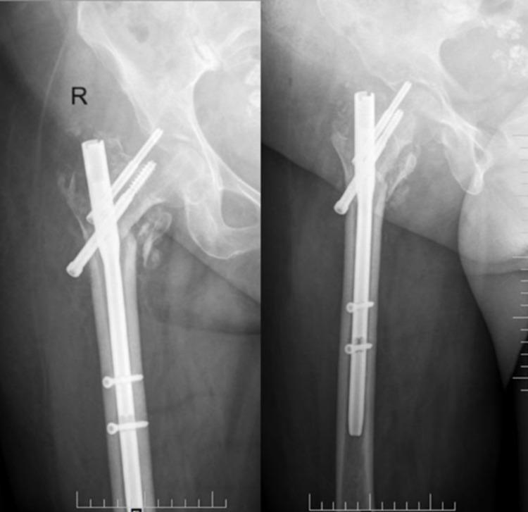
This diagnostic X-ray image demonstrates an anteroposterior (AP) and lateral view of a right proximal femur following internal fixation of an intertrochanteric fracture with a proximal femoral nail (PFN). The orthopedic hardware consists of a metallic intramedullary nail with distal locking screws and two cephalocervical screws extending through the femoral neck into the head. The AP view clearly illustrates a mechanical complication known as the 'Z-effect.' This phenomenon is characterized by the differential migration of the two proximal screws in opposite directions: the superior (stabilizing) screw has migrated medially and penetrated the hip joint space, while the inferior (lag) screw has migrated laterally through the nail. The lateral view confirms the intramedullary position of the main nail and the abnormal orientation of the cephalic screws. This visual is a classic educational example of hardware failure and implant instability in geriatric fracture management, typically resulting from poor bone quality or inadequate fracture reduction.
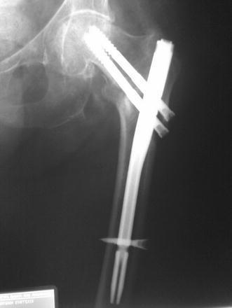
This anteroposterior (AP) diagnostic X-ray of the proximal femur illustrates a post-surgical state following the treatment of an intertrochanteric fracture with a cephalomedullary nail. The orthopedic construct consists of a radiopaque intramedullary nail positioned within the femoral canal. Two cephalic lag screws are inserted through the proximal portion of the nail, extending into the femoral head and neck to provide rotational stability and compression. A notable technical finding is visualized distally: the distal locking screws appear to have missed the nail's locking aperture, as they are seen passing adjacent to the nail rather than through it. Despite this malpositioning, the overall alignment of the femur appears maintained. The image serves as a clinical teaching example for identifying technical complications during intramedullary nailing, specifically the 'missed' distal locking screw, and assessing the relationship between orthopedic implants and native bone anatomy in the context of fracture fixation.
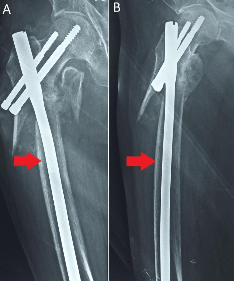
Diagnostic radiographic images (X-rays) of the proximal femur demonstrating surgical stabilization of an intertrochanteric fracture using a Proximal Femoral Nail (PFN). Panels A and B show an anteroposterior view of the hip and proximal femur. The hardware consists of a metallic intramedullary nail (indicated by red arrows) inserted into the femoral shaft canal, through which cephalocervical screws are passed into the femoral neck and head to provide stability and compression. In image A, a distinct intertrochanteric fracture line with fragmentation is visible, showing signs of internal fixation and alignment maintenance. The bone demonstrates varying radiolucency, which may suggest underlying osteopenia or osteoporosis. This imaging is characteristic of orthopedic trauma management, specifically the internal fixation of hip fractures to facilitate healing and early mobilization. The specialty focus is Orthopedic Surgery and Traumatology.
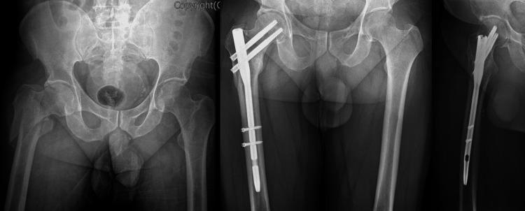
This composite diagnostic image consists of three X-ray radiographs illustrating the surgical management of a proximal femur fracture. The leftmost image is a preoperative AP pelvis radiograph showing a reverse oblique intertrochanteric fracture of the right femur with significant lateral displacement of the distal fragment. The central and rightmost images are postoperative AP and lateral views showing fracture reduction and internal fixation with a Proximal Femoral Nail (PFN). The intramedullary device is secured proximally by two parallel cephalomedullary lag screws extending into the femoral head and neck to provide rotational stability. Distally, the nail is anchored by two transverse interlocking cortical screws in the femoral shaft. This educational content demonstrates orthopedic trauma management for unstable hip fractures using intramedullary nail systems. It is intended for intermediate to advanced medical learners focusing on radiology and orthopedic surgery.
Clinical Note - Full Explanation
Patient Details
- Date: 5/8/26
- Doctor: Dr. Hiten Kayasth, Orthopedic Surgeon, Ved Hospital
Left Side of the Note (History & Examination)
| Abbreviation | Full Form | Meaning |
|---|---|---|
| C/O | Complaining of | The main symptom the patient presents with |
| Lt (circled) | Left | Refers to the left side of the body |
| H/O | History of | How the injury happened |
| O/E | On Examination | Clinical findings when the doctor examined the patient |
| DP/DM | Distal Pulse / Distal Motor | Checking blood flow and nerve function below the injury - noted as (++) meaning intact/normal |
| X-ray PBH | X-ray Pelvis with Both Hips | X-ray ordered to look at both hips and the pelvis |
| AP / Lat | Anteroposterior / Lateral | Two standard X-ray views (front-to-back and side view) |
| IT # | Intertrochanteric Fracture | The diagnosis - a fracture between the greater and lesser trochanters of the left femur (thigh bone) |
- Patient presented with left hip pain that started today
- Fell at home around 2 PM today
- On examination: swelling and tenderness over the left hip
- Distal pulse and motor function are intact (no vascular or nerve damage below the fracture)
- X-ray of pelvis with both hips (AP and lateral views) confirmed a Left Intertrochanteric Fracture (#17 - likely fracture number or age 17 is unlikely; more likely refers to fracture classification or patient age noted separately)
Right Side of the Note (Plan/Advice)
| Abbreviation | Full Form | Meaning |
|---|---|---|
| Adv. | Advice / Plan | Treatment plan |
| Lt Proximal Femur Nailing | Left Proximal Femoral Nail (PFN) | The planned surgery |
| Admission | Hospital Admission | Patient to be admitted |
| Preop major | Pre-operative workup for major surgery | Blood tests, fitness assessment before operation |
| CXR | Chest X-ray | Pre-operative chest X-ray to assess cardiac/lung fitness |
| ECG | Electrocardiogram | Pre-operative heart trace to assess cardiac fitness for anaesthesia |
The Planned Surgery: Left Proximal Femur Nailing (PFN)
- The patient is placed on a fracture table, and traction is applied to the injured leg to reduce (realign) the fracture, often without a large incision.
- A metal nail (rod) is inserted down the hollow canal inside the femur (thigh bone) through a small incision near the hip.
- A large lag screw is then passed through the nail, through the femoral neck, and into the femoral head - this anchors the fracture fragments together.
- Distal locking screws are placed at the bottom of the nail to prevent rotation.
- The two proximal screws allow the fracture to compress dynamically as the patient walks, promoting bone healing.
Pre-operative Workup Ordered
- Admission to hospital
- Pre-op workup (blood tests: CBC, coagulation, renal function, blood group/cross-match, etc.)
- CXR - chest X-ray to check heart and lung health before anaesthesia
- ECG - to check heart rhythm and rule out cardiac issues before general/spinal anaesthesia