
Explain malignant melanoma surgery mbbs for 10 marks from Bailey and love and srb textbook surgery
malignant melanoma surgery wide local excision sentinel node biopsy
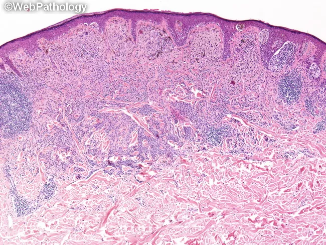
A histopathologic skin biopsy evaluated by light microscopy following routine hematoxylin and eosin (H&E) staining. The specimen is an excisional biopsy of cutaneous tissue. The image shows malignant melanocytic proliferation with invasion through the epidermal-dermal junction into the dermis. Tumor nests and single-cell invasion are evident at the dermoepidermal junction with dermal penetration by pigmented melanocytes. Breslow thickness, the most important prognostic parameter in melanoma, is measured from the most superficial granular cell layer of the epidermis to the deepest point of tumor invasion; in this case the depth is 1.0 millimeter. If an ulceration is present, depth is measured from the base of the ulcer; ulceration is not described here. Some tumors may extend along adnexal structures, and if present this finding is documented as part of depth assessment. Polypoid tumor components, if any, are measured through their thickest region. Although invasion depth is continuous, AJCC guidelines categorize 1 mm, 2 mm, and 4 mm for staging (T1, T2, T3). Clinically, Breslow depth informs prognosis, sentinel lymph node biopsy decisions, and surgical margins for wide local excision, as well as consideration for adjuvant therapy depending on ulceration, mitotic rate, and nodal status.
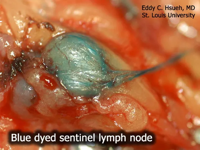
Imaging modality: Intraoperative surgical photography of a sentinel lymph node biopsy for malignant melanoma. Technique: blue dye mapping using Lymphazurin blue (isosulfan blue) injected near the primary lesion to trace afferent lymphatics to the sentinel node, which is visually stained a blue-green hue. Anatomical context: axillary regional lymphatics draining the tumor bed are highlighted by dye transport, with the sentinel lymph node identified by conspicuous color uptake. Visual features: a single lymph node is enlarged and engorged, with surrounding adipose tissue and operating field in clear view; the node exhibits uniform blue coloration within the cortex and hilum, with surrounding soft tissues appearing erythematous from operative exposure. Notable findings: blue-stained sentinel node ready for targeted excision; no gross metastatic nodal disease is discernible on this view, though microscopic metastasis may be present. Clinical significance: sentinel lymph node biopsy is a standard staging procedure in melanoma, enabling nodal assessment while minimizing extensive lymphadenectomy. Differential considerations: accidental intradermal staining, dye leakage, or allergic reaction to isosulfan blue. Clinical correlation: histopathologic examination of the excised node is required to determine nodal metastasis status, guiding adjuvant therapy and prognosis. This image serves educational and surgical planning purposes for clinicians, trainees, and researchers studying lymphatic mapping in cutaneous melanoma.
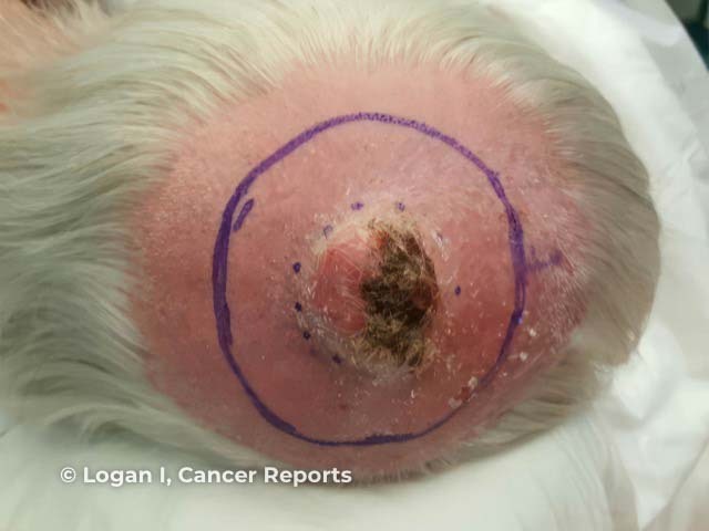
Clinical photography of a solitary scalp lesion prior to wide local excision. Modality: clinical photography with high‑resolution digital color imaging. Anatomical location: scalp skin over the cranial vault; exact laterality not specified. Imaging perspective: frontal/occlusal view bias, close-up of the lesion. The lesion appears as a raised, dome‑shaped to indurated nodule with an erythematous to pink surface, focal crusting, and a central necrotic/ulcerated area. Surrounding perilesional erythema and mild edema are evident; there may be subtle superficial induration at the lesion margins. The circumscribed lesion is outlined in purple ink to aid surgical planning. This morphology raises concern for a high‑grade cutaneous neoplasm, most consistent with pleomorphic dermal sarcoma (cutaneous undifferentiated pleomorphic sarcoma, UPS), a malignant dermal/subcutaneous spindle cell tumor. Differential diagnoses include basal cell carcinoma, squamous cell carcinoma, amelanotic melanoma, keratoacanthoma, spindle cell carcinoma, and other cutaneous sarcomas. The image provides essential preoperative context to guide biopsy strategy, margin planning, and definitive wide excision. Diagnostic significance lies in distinguishing aggressive skin cancers from benign mimics, informing staging, sentinel lymph node assessment considerations, and adjuvant therapy decisions when histology confirms UPS. This preoperative image supports risk stratification and informs multidisciplinary discussion with dermatopathology and surgical oncology.
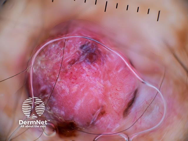
In this dermoscopy examination, a solitary cutaneous lesion is imaged in vivo with dermoscopic close-up (polarized light, approximately 10x magnification). The lesion presents as pink to light red, showing atypical vascular patterns and peripheral speckles of pigment. Morphology is asymmetric with irregular, poorly defined borders, and a raised, enface appearance typical of an active melanocytic neoplasm. The primary diagnosis is malignant melanoma; Breslow thickness is reported as 8.3 mm (thick melanoma), compatible with substantial risk of subcutaneous invasion and metastatic potential. Dermoscopic features include irregular, dilated vessels and subtle pigment peppering at the lesion periphery, which correlate with invasive growth and angiogenesis. Differential considerations include pigmented nevi with vascular irregularity, Spitz nevi, blue nevus, or amelanotic melanoma; however, the combination of pink coloration, atypical vasculature, and peripheral pigment supports melanoma in this context. Clinically, these findings are relevant for diagnostic decision-making, prognostication, and treatment planning. Potential clinical applications encompass education for dermoscopy interpretation, imaging-based risk stratification, and guiding biopsy/surgical planning. Management implications include wide local excision with margins defined by thickness and consideration of sentinel lymph node biopsy, lymphatic assessment, and comprehensive staging per guidelines. This image demonstrates high-risk melanoma characteristics suitable for educational and research datasets. Resources.
Malignant Melanoma - Surgery (10 Marks)
Definition and Introduction
Epidemiology and Risk Factors
- Rising incidence worldwide
- More common in Caucasians; extremely rare in dark-skinned people
- Age: majority between 20-40 years; rare before puberty
- Sex: girls > boys (ratio 2-3:1 per S. Das); male gender associated with thicker tumours at diagnosis
- Risk factors:
- Genetic syndromes (FAMMM - familial atypical multiple mole melanoma)
- Personal or first-degree family history of melanoma
-
30 sun-acquired naevi, or ≥5 significant sunburns before age 16
- Fair skin / red hair / living near the equator
- Excessive UV radiation (environmental or tanning salons)
- Immunosuppression (increases incidence 20-30 fold)
Macroscopic Types (Variants)
| Type | Frequency | Features |
|---|---|---|
| Superficial Spreading Melanoma (SSM) | 70% (most common) | Arises in pre-existing naevus; slow radial then rapid vertical growth |
| Nodular Melanoma (NM) | 15% | Aggressive; arises de novo; blue/black papule; head/neck/trunk; up to 5% amelanotic |
| Lentigo Maligna Melanoma (LMM) | ~10% | Arises from Hutchinson's freckle; face in elderly; treated with wide excision + radiotherapy |
| Acral Lentiginous Melanoma (ALM) | ~5% | Palms, soles, subungual; most common in Asians/Africans; poor prognosis |
- Sudden increase in size
- Change in colour (patchy darkening; rarely amelanotic)
- Itching / bleeding when rubbed
- Pigmented halo around tumour (indicates local spread)
- Satellite nodules in intradermal lymphatics
- Regional lymph node enlargement or distant metastases
Microscopic Pathology
- Malignant change occurs in melanocytes in the basal epidermis
- Horizontal (radial) growth phase: cells spread along the dermoepidermal junction; may breach dermis but migration is predominantly radial - lower metastatic risk
- Vertical growth phase: dermis is invaded - greater depth = greater metastatic potential
- Clark's levels (I-V): depth of invasion through skin layers
- Breslow thickness: measured in mm from the granular layer to the base of the tumour - the single most important prognostic indicator
Staging - AJCC 8th Edition (Bailey & Love, Table 45.2)
| T Stage | Breslow Thickness |
|---|---|
| T1a | <0.8 mm, no ulceration |
| T1b | 0.8-1.0 mm, or <1.0 mm with ulceration |
| T2 | 1.0-2.0 mm |
| T3 | 2.0-4.0 mm |
| T4 | >4.0 mm |
- N staging: based on number of nodes and presence of macro vs micrometastasis
- M staging: based on site of distant metastasis and LDH level
Investigations
- Excision biopsy with 2-3 mm margin and a cuff of subdermal fat (primary diagnostic step)
- Incision biopsy may be used in large facial lesions to avoid disfigurement
- Histopathology for Breslow thickness, Clark level, ulceration, mitotic rate
- Sentinel Node Biopsy (SNB): offered to T2a disease and greater (Breslow >1 mm); maps lymphatic drainage to the first "sentinel" node in the regional basin
- CT/PET scan for T3a and above to detect distant metastases
- Dermoscopy (with serial photography when observation is chosen over biopsy)
SURGICAL MANAGEMENT
1. Local Treatment - Wide Local Excision (WLE)
| Breslow Thickness | Recommended Excision Margin |
|---|---|
| In situ (Lentigo maligna) | 5 mm |
| <1 mm | 1 cm margin |
| >1 mm (deeper lesions) | 2 cm margin |
No evidence supports wider margins beyond 2 cm.
2. Regional Lymph Node Management
- Elective lymph node dissection at the time of WLE (now abandoned - no survival benefit)
- Therapeutic lymphadenectomy when clinical metastases become evident
- Sentinel Node Biopsy (SNB) - the current standard
- Based on the principle that lymphatic metastases proceed in an orderly fashion
- The primary tumour drains to the "sentinel node" first before spreading further
- Blue dye (isosulfan blue/Patent V blue) and/or radioisotope (Tc-99m) is injected around the primary lesion
- The sentinel node(s) are identified and excised
- Histopathological and immunohistochemical analysis follows
- Positive SNB = microscopic metastasis present
- Prospective controlled studies show no survival benefit from completion lymphadenectomy after positive SNB with micrometastasis <0.2 mm in a single node
- However, completion lymphadenectomy after positive SNB remains the optimum method for regional control if the patient accepts associated morbidity
3. Spread and Metastasis (S. Das)
- Lymphatic spread: regional lymph nodes are involved early; satellite nodules may appear between primary and regional nodes (embolism in lymphatics)
- Haematogenous spread: to liver, lungs, bones, brain
- In-transit metastases: satellite nodules along lymphatics between primary tumour and regional nodes
- Subungual melanoma: occurs under nail (thumb / great toe); nail lifted and eventually lost
Adjuvant and Systemic Therapy (Bailey & Love)
- BRAF mutations (V600) present in ~50% of melanomas - locks BRAF protein signalling "on", promoting MAPK pathway and tumour progression
- BRAF inhibitors: Dabrafenib, Vemurafenib (for BRAF V600-mutant Stage IV disease)
- MEK inhibitor: Trametinib (acts on MAPK pathway; combined with dabrafenib to counter acquired resistance)
- Immune checkpoint inhibitors: Anti-PD1 (pembrolizumab/nivolumab), anti-CTLA4 (ipilimumab) - show significant response in advanced disease
- Radiotherapy: used as adjuvant for LMM and for palliation in metastatic disease
- Chemotherapy: limited role (dacarbazine historically)
Prognosis
- Breslow thickness is the single most important prognostic factor in the absence of nodal metastases
- Ulceration, high mitotic rate, and nodal positivity worsen prognosis
- 5-year survival: ~98% for localised thin melanoma; ~23% for metastatic disease
Summary (Bailey & Love Summary Box 45.4)
- Rising incidence
- Genetic and acquired risk factors
- Superficial spreading form is the most common type
- Breslow thickness is the most important prognostic indicator
- Sentinel node biopsy (SNB) is useful for staging
- The only cure for malignant melanoma is appropriate surgery
- Bailey and Love's Short Practice of Surgery, 28th Ed., Chapter 45 (Skin and Subcutaneous Tissue), pp. 675-681
- S. Das, Manual on Clinical Surgery, 13th Ed., Chapter 3 (Swellings), pp. 54-60
Note: SRB's Concise Textbook of Surgery by Dr. Sriram Bhat is not currently in this textbook library. The content above is drawn from Bailey & Love (the primary reference requested) supplemented by S. Das's Manual on Clinical Surgery, which is the other widely used Indian MBBS surgery reference available. The surgical principles align with SRB's coverage of this topic.