
What is secular microscope, principal why it's used and interpitesion
The term "secular microscope" doesn't match a standard instrument - you likely mean the ocular microscope (the eyepiece) or perhaps the specular microscope. Let me look this up properly.
Searching sources...
Reading File
Searching the Web
specular microscope principle uses interpretation corneal endothelium
Finding Sources
Reading Web Page
https://pmc.ncbi.nlm.nih.gov/articles/PMC7942069
Reading File
Loading Image
You almost certainly mean the Specular Microscope - a key ophthalmic diagnostic instrument. Here is a complete explanation.
Specular Microscopy
What Is It?
The specular microscope (also called a specular photomicroscope) is a non-invasive ophthalmic instrument used to image and analyze the corneal endothelium - the innermost layer of the cornea - at very high magnification. It provides about 100 times greater magnification than standard slit lamp biomicroscopy.
- Kanski's Clinical Ophthalmology, 10th Ed., p. 225
Principle
The instrument works on the physics of specular reflection - the same principle as a mirror:
- A focused beam of light is projected through the cornea
- As the light travels through the cornea, it hits a series of optical interfaces between tissues with different refractive indices
- At the endothelium-aqueous humor interface, the endothelial cells have a refractive index greater than 1.336 (the refractive index of aqueous humor), so they reflect approximately 0.022% of the projected light back toward the microscope
- This reflected (specular) light forms a magnified image that is captured, photographed, and analyzed
- The angle of reflected light equals the angle of incident light - this is the defining principle of specular reflection
Why It Is Used (Indications)
| Indication | Details |
|---|---|
| Pre-surgical evaluation | Most common use - assesses the functional reserve of the corneal endothelium before intraocular surgery (e.g., cataract surgery) |
| Donor cornea evaluation | Checks endothelial health of corneas intended for transplant |
| Diagnosis of pathology | Detects cornea guttata, Descemet membrane irregularities, posterior polymorphic dystrophy, Fuchs' endothelial dystrophy |
| Contact lens monitoring | Evaluates chronic endothelial changes in long-term contact lens wearers |
| Post-operative follow-up | Monitors endothelial health after corneal surgery (e.g., DSAEK, DMEK) |
| ICE syndrome | Detects iridocorneal endothelial syndrome |
Key point: A cornea can appear clear with normal thickness on pachymetry and still have abnormal endothelial morphology or reduced cell density. Specular microscopy reveals what slit lamp alone cannot.
Interpretation
The image produced is called a specular micrograph, and three key parameters are analyzed:
1. Endothelial Cell Density (ECD)
- Normal in a young adult: ~3000 cells/mm²
- Decreases with age (normal loss ~0.6%/year)
- Below 1000 cells/mm² - unlikely to develop corneal oedema
- Below 700 cells/mm² - corneal oedema is considerably more likely
- The risk threshold for surgery is generally considered 500-700 cells/mm²
2. Pleomorphism (Change in Shape)
- Normal endothelial cells are regular hexagons - the most energy-efficient polygon
- Pleomorphism = deviation from hexagonality (cells become irregular in shape)
- Normal hexagonality: ~60-70% hexagonal cells in young adults
- Increased pleomorphism is a sign of endothelial stress or disease
3. Polymegathism (Change in Size)
- Polymegathism = increased variability in cell size (coefficient of variation of cell area)
- When cells are lost, neighboring cells migrate and enlarge to fill the gap (cells do NOT regenerate or divide in vivo)
- High polymegathism = sign of chronic endothelial damage
Specular Micrograph - Normal vs. Diseased
Here is a direct comparison from Kanski's Clinical Ophthalmology:
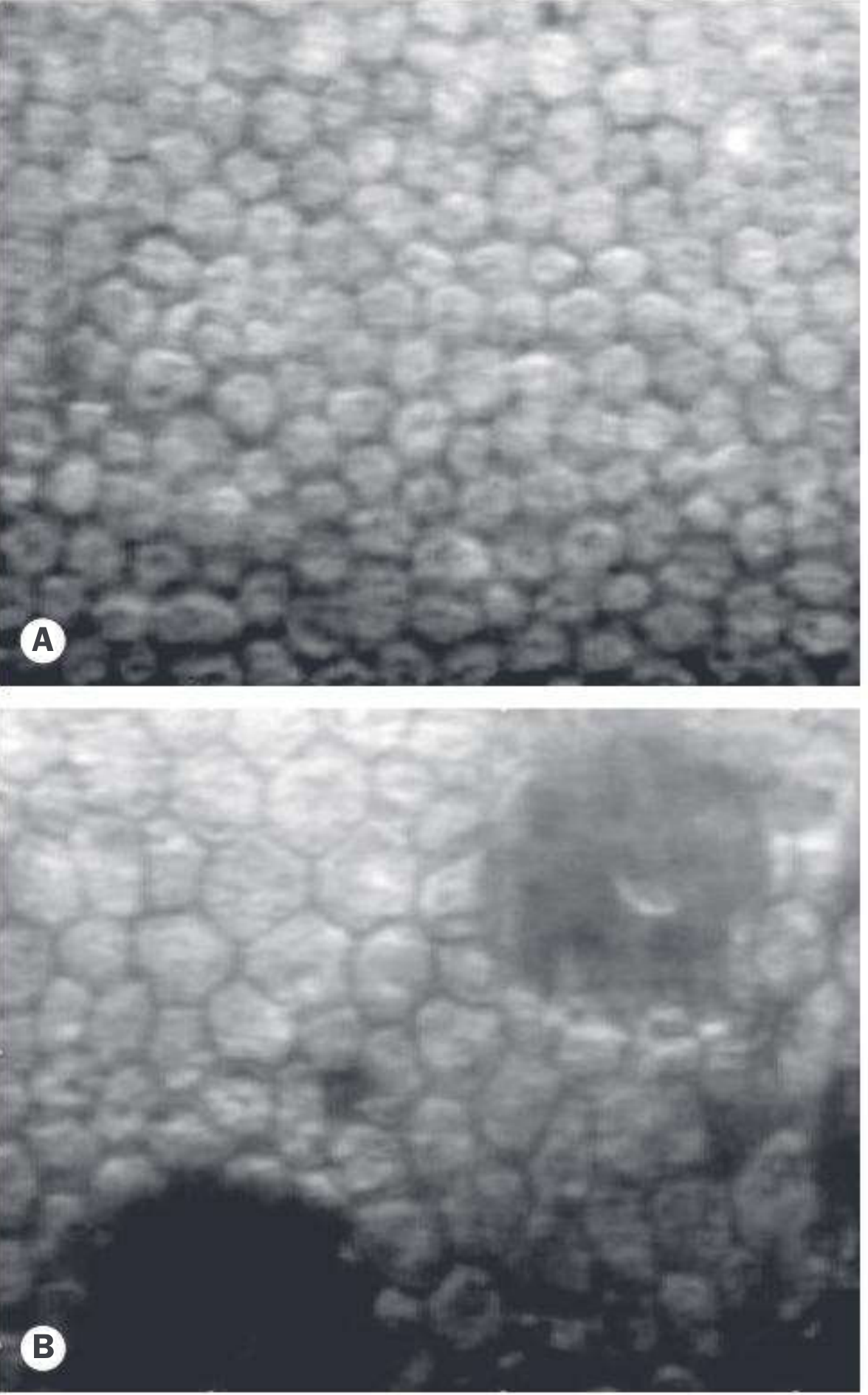
- Image A (top): Normal endothelium - tightly packed, uniform, regular hexagonal cells (~3000/mm²)
- Image B (bottom): Cornea guttata - markedly fewer cells, larger and irregular cells, dark areas indicating lost cells or guttae deposits
Types of Specular Microscopes
| Type | Features |
|---|---|
| Contact | Probe touches the cornea; older design; provides wide-field views |
| Non-contact | No corneal contact; patient-friendly; more widely used today |
Summary
| Feature | Detail |
|---|---|
| Tissue visualized | Corneal endothelium |
| Principle | Specular (mirror-like) reflection at the endothelium-aqueous interface |
| Key parameters | Cell density (ECD), pleomorphism, polymegathism |
| Main use | Pre-op assessment before intraocular surgery |
| Normal ECD | ~3000 cells/mm² (young adult) |
| Critical threshold | <700 cells/mm² = high oedema risk |
Sources: [Kanski's Clinical Ophthalmology, 10th Ed.] | Chaurasia S, Specular microscopy in clinical practice, PMC7942069, 2021
This is a shared conversation. Sign in to Orris to start your own chat.