
Autoimmune diseases with a rash
Reading File
Now let me search the medical library for autoimmune diseases with rashes in parallel.
Finding Sources
Finding Sources
Finding Sources
Good results. Let me now read the key sections - the Goldman-Cecil cutaneous manifestations section and search for specific diseases.
Reading File
Finding Sources
Finding Sources
Excellent! Let me read the continuation of the cutaneous manifestations section and search for more specific diseases.
Reading File
Finding Sources
Finding Sources
Let me get detailed info on specific diseases - SLE rash, dermatomyositis, and others.
Reading File
Finding Sources
Finding Sources
Let me now fetch the dermatomyositis image and read the malar rash section for SLE, plus get more detail on specific rashes.
Reading File
Reading File
Loading Image
Excellent content gathered. Let me get a couple more images and the psoriasis/vasculitis info.
Loading Image
Finding Sources
Finding Sources
I now have comprehensive information from the medical textbooks. Let me compile a thorough response.
Here is a detailed overview of autoimmune diseases that classically present with a rash, drawn from Firestein & Kelley's Textbook of Rheumatology, Goldman-Cecil Medicine, and Dermatology 5e.
Autoimmune Diseases with a Rash
1. Systemic Lupus Erythematosus (SLE)
SLE has the richest variety of cutaneous manifestations, classified as lupus-specific and lupus-nonspecific.
Acute Cutaneous Lupus Erythematosus (ACLE) - the "butterfly rash"
- The hallmark is the malar (butterfly) rash: confluent, macular or papular erythema appearing symmetrically on the cheeks and bridge of the nose, lasting days to weeks.
- A key distinguishing feature: spares the nasolabial folds (a sun-protected area). This separates it from rosacea and dermatomyositis, both of which involve the nasolabial folds.
- Triggered and worsened by UV light. Over 90% of SLE patients show an abnormal skin reaction to UV provocation testing.
- Can generalize to any photosensitive area of the body. On the hands, erythema typically spares the MCP joints and sits between the interphalangeal joints - the opposite of dermatomyositis.
- Heals without scarring (unlike discoid lupus).
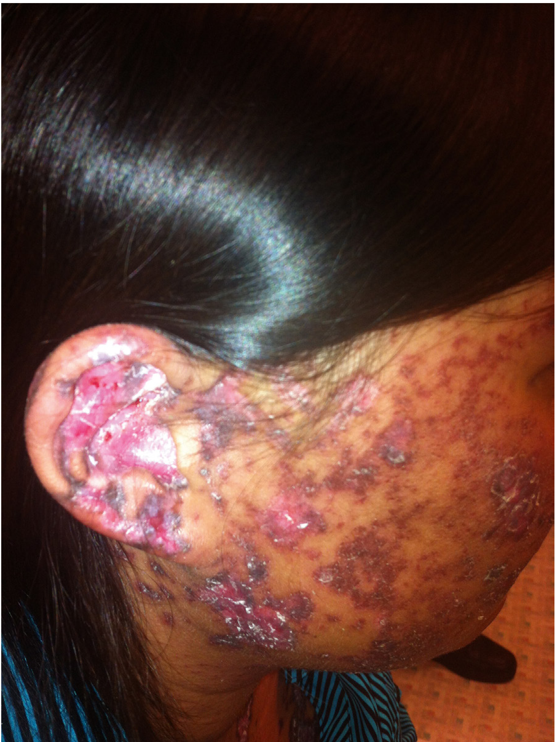
Discoid Lupus Erythematosus (DLE)
- Chronic, scarring plaques - active lesions shown here on the lateral face and ear:
Other SLE skin manifestations (lupus-nonspecific):
- Palpable purpura (lower extremities) - cutaneous leukocytoclastic vasculitis
- Livedo reticularis / livedo racemosa - violaceous, net-like pattern; strongly associated with antiphospholipid syndrome (APS)
- Bullous lupus - rare subepidermal blistering from anti-type VII collagen antibodies
- Periungual erythema - dilated capillaries at the nail base (also seen in scleroderma and MCTD)
- Lupus tumidus - edematous, erythematous smooth plaques; highly photosensitive; favors the zygomatic region
Source: Firestein & Kelley's Textbook of Rheumatology, 2-Volume Set
2. Dermatomyositis
Dermatomyositis has pathognomonic skin findings that are unique among autoimmune diseases.
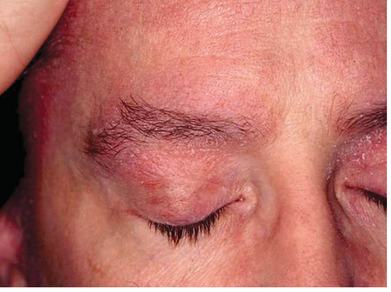
Heliotrope sign
- Pink-violet discoloration of the eyelids and periorbital skin, often with edema. Can be subtle. Shown here:
Gottron papules / Gottron sign
- Flat-topped, erythematous-to-violaceous papules on the knuckles (MCPs and interphalangeal joints) - the exact opposite distribution from SLE on the hands.
- Gottron sign = involvement of the elbows and/or knees.
Photodistributed poikiloderma
- V-neck sign (upper chest), shawl sign (upper back), holster sign (lateral thighs).
- Pink-violet hue; can be misdiagnosed as psoriasis when on extensor surfaces.
Additional clues:
- Nail-fold telangiectasias with capillary dropout ("ragged cuticles")
- Intensely pruritic - pruritus is uncommon in lupus and helps distinguish the two
- Calcinosis cutis - especially in juvenile dermatomyositis (25-70% of pediatric cases)
Source: Dermatology 2-Volume Set 5e
3. Psoriatic Arthritis / Psoriasis
Psoriasis is almost always present in psoriatic arthritis (a spondyloarthropathy variant). The rash may be subtle and easily missed in intertriginous regions (ear, umbilicus, buttock creases, scalp) or may involve only the nails.
- Classic appearance: sharply demarcated, thick, erythematous plaques covered by dry silvery-white scales, predominantly on extensor surfaces (elbows, knees).
- Inverse psoriasis lacks silvery scaling and instead presents as erythematous plaques in skin folds (inguinal, inframammary, axillary).
- Nail pitting and onycholysis are common associated features.
Source: Harrison's Principles of Internal Medicine 22E; Goldman-Cecil Medicine
4. Systemic Sclerosis (Scleroderma)
Scleroderma's skin findings are distinct from a typical "rash":
- Skin tightening beginning with swollen, puffy digits, then progressive tightening of the hands, face, and (in diffuse SSc) trunk and legs
- Telangiectasias on the face, hands
- Digital ulcers at fingertips
- Periungual erythema with capillary dropout (more prominent than in SLE)
- Raynaud phenomenon often precedes cutaneous involvement by years
Source: Goldman-Cecil Medicine
5. Adult-Onset Still Disease (AOSD)
A distinctive, often underrecognized rash pattern:
- Evanescent, salmon-colored blanching rash that appears during daily spiking fevers (peaking late in the day)
- Lasts only 1-2 hours, coinciding with fever spikes
- Associated with high ferritin, arthritis, and lymphadenopathy
Source: Goldman-Cecil Medicine
6. ANCA-Associated Vasculitis
- Palpable purpura - non-blanching, purpuric rash from necrotizing small-vessel vasculitis
- Present for days, often palpable; biopsy is essential for diagnosis
- Associated with conditions like granulomatosis with polyangiitis (GPA) and microscopic polyangiitis
Source: Goldman-Cecil Medicine
Quick Diagnostic Summary
| Disease | Rash Type | Key Location | Distinguishing Feature |
|---|---|---|---|
| SLE (ACLE) | Macular/papular erythema | Cheeks + nasal bridge | Spares nasolabial folds; UV-triggered |
| Discoid Lupus | Scarring plaques | Face, ears, scalp | Scarring; follicular plugging |
| Dermatomyositis | Heliotrope; Gottron papules | Eyelids; knuckles | Pathognomonic; pruritic; nail-fold changes |
| Psoriatic Arthritis | Silvery scaly plaques | Extensor surfaces, scalp | Nail pitting; well-demarcated |
| Scleroderma | Skin tightening + telangiectasias | Hands, face | Progressive fibrosis; Raynaud |
| Still Disease | Salmon-colored evanescent rash | Trunk | Lasts 1-2h with fever spikes |
| ANCA Vasculitis | Palpable purpura | Lower extremities | Non-blanching; requires biopsy |
A key clinical pearl: rashes in dermatomyositis and SLE are both photosensitive, but the nasolabial fold and knuckle distribution are the key differentiators. SLE spares the nasolabial folds and the MCP/IP joint surfaces; dermatomyositis involves both. - Firestein & Kelley's Textbook of Rheumatology, 2-Volume Set
Rash on whole body
Asking for Preferences
What context are you asking about?
Autoimmune diseases that cause a generalized (whole-body) rash - narrowing down from our previous discussion. Let me search the textbooks for this specifically.
Searching sources...
Finding Sources
Finding Sources
Finding Sources
Reading File
Finding Sources
Finding Sources
Finding Sources
Finding Sources
Finding Sources
Reading File
Finding Sources
I have solid textbook content now. Here is the focused answer on autoimmune diseases causing a whole-body (generalized) rash:
Autoimmune Diseases with a Whole-Body (Generalized) Rash
1. Generalized ACLE (Acute Cutaneous Lupus Erythematosus) - SLE
The generalized form of ACLE is one of the most characteristic whole-body autoimmune rashes:
- Widespread macular or maculopapular erythema in a photosensitive distribution across any area of the body - arms, trunk, legs, hands
- Palmar surfaces, dorsa of hands, and extensor surfaces of fingers commonly involved
- On the hands, erythema sits between the MCP and IP joints (sparing the joint surfaces themselves - opposite of dermatomyositis)
- Severe cases can progress to a TEN-like bullous eruption when interface inflammation at the dermo-epidermal junction causes epidermal detachment
- Heals without scarring; may leave post-inflammatory hyperpigmentation
Source: Firestein & Kelley's Textbook of Rheumatology
2. Subacute Cutaneous Lupus Erythematosus (SCLE)
SCLE produces a widespread nonscarring, photosensitive rash with a predilection for the back, neck, shoulders, and extensor arms, usually sparing the central face:
- Two forms: (1) papulosquamous (resembling psoriasis) or (2) annular-polycyclic with peripheral scale and central clearing - both forms can coexist in the same patient
- Strongly associated with anti-SSA/Ro antibody and HLA-DR3
- Drug-induced SCLE is common: ACE inhibitors, hydrochlorothiazide, terbinafine, calcium channel blockers are key culprits. Onset can be delayed up to 6 years after starting the drug.
- Lesions last weeks to months and heal without scarring
Source: Firestein & Kelley's Textbook of Rheumatology
3. Dermatomyositis - Photodistributed Poikiloderma
In dermatomyositis, the skin disease is often extensive and generalized beyond just the eyelids and knuckles:
- Photodistributed poikiloderma: involves the upper chest (V-neck sign), upper back (shawl sign), and lateral thighs (holster sign)
- Can cover most of the body surface in a photosensitive distribution
- Pink-violet coloration with telangiectasias, atrophy, and mottled pigmentation
- Often intensely pruritic (which helps distinguish it from SLE)
- Can mimic psoriasis when plaques occur on extensor surfaces, or mimic cutaneous T-cell lymphoma when poikiloderma is widespread
Source: Dermatology 2-Volume Set 5e
4. Pemphigus Vulgaris
An autoimmune blistering disease causing generalized whole-body erosions:
- Anti-desmoglein antibodies (IgG) attack epidermal cell adhesion, producing flaccid, thin-walled blisters that rupture almost immediately into painful erosions
- Erosions can become extensive over the entire body surface - oozing, bleeding, crusting with no tendency to heal spontaneously
- Nikolsky sign: gentle lateral pressure on skin slides off the upper epidermis (sign of active disease)
- Mucous membranes always involved (oral erosions in essentially 100% of cases); can also affect throat, esophagus, genitalia, conjunctivae
- Without treatment, erosions become generalized and life-threatening
Source: Dermatology 2-Volume Set 5e
5. Erythrodermic / Generalized Pustular Psoriasis
Psoriasis (an immune-mediated disease) has two severe forms that become generalized:
Erythrodermic psoriasis:
- >90% of body surface covered in red, scaling skin
- Typical psoriatic features are often lost; skin becomes uniformly red and exfoliating
- Can be precipitated by abrupt withdrawal of systemic steroids, or by UVB phototherapy
Generalized Pustular Psoriasis (von Zumbusch):
- Waves of sterile subcorneal pustules on an erythematous base, spreading over the entire body
- Systemic features: fever, malaise, leukocytosis
- Can be life-threatening due to fluid/electrolyte loss
Source: Firestein & Kelley's Textbook of Rheumatology; Fitzpatrick's Dermatology
6. Diffuse Cutaneous Systemic Sclerosis (dcSSc)
While not a "rash" in the classic sense, diffuse scleroderma produces widespread skin induration covering the entire body:
- Begins with sclerodactyly (finger tightening), then ascends from distal to proximal limbs, and eventually spreads to the trunk - distinguishing it from limited SSc which stays below the elbows/knees
- Associated with anti-Scl-70 (topoisomerase I) antibodies
- Raynaud phenomenon, telangiectasias, and digital ulcers accompany the skin changes
Source: Harrison's Principles of Internal Medicine 22E
Quick Summary Table
| Disease | Rash Character | Distribution | Key Clue |
|---|---|---|---|
| Generalized ACLE (SLE) | Macular/maculopapular erythema | Photosensitive - anywhere | Spares nasolabial folds; UV-triggered |
| SCLE | Annular/papulosquamous | Back, neck, shoulders | Anti-Ro antibody; drug-inducible |
| Dermatomyositis | Poikiloderma (pink-violet) | V-neck, shawl, holster signs | Intensely pruritic; nail-fold changes |
| Pemphigus Vulgaris | Flaccid blisters → erosions | Whole body + mucosae | Nikolsky sign; oral involvement first |
| Erythrodermic Psoriasis | Diffuse erythema + scaling | >90% body surface | Steroid withdrawal trigger |
| Generalized Pustular Psoriasis | Sterile pustules on red base | Whole body | Fever; waves of pustulation |
| Diffuse SSc | Skin induration/tightening | Fingers → trunk | Anti-Scl-70; Raynaud |
Key differentiating principle: the photosensitive pattern (UV-exposed areas sparing covered skin) in SLE and dermatomyositis, versus the trauma-independent spread in pemphigus or the inflammatory wave pattern of generalized pustular psoriasis, and the ascending fibrosis of diffuse SSc all point in different diagnostic directions even when the disease appears "total-body" in extent.
This is a shared conversation. Sign in to Orris to start your own chat.