
Explain me prostate cancer. I need to study this for my urology class. Our proffoser asks questions like we are doctors in urology speciality for 10yrs. I hope you understand how deep he asks questions. So explain everything about prostate cancer to me. Some of the things that you can't miss are Classification, Etiology, pathogenesis, symptoms and signs (where, how,severity etc etc), diagnosis, clinical manifestations, prognosis, treatment. These are some must give details you have to give me more information than this. Understood?
Reading File
Finding Sources
Finding Sources
Finding Sources
The key sources are Campbell Walsh Wein Urology (the gold standard urology textbook), Robbins Pathology, and Harrison's. Let me now read the most important blocks in parallel.
Reading File
Reading File
Reading File
Excellent content from Harrison's and Robbins. Let me read more from the prostate cancer sections, particularly from Harrison's (block10.md continues) and Robbins, plus Campbell Walsh for staging/treatment.
Reading File
Reading File
Finding Sources
Excellent detail from Harrison's and Robbins. Now let me continue reading Harrison's prostate cancer section and get more content on staging, treatment, and prognosis, plus get images.
Reading File
Reading File
Searching Images
prostate cancer Gleason grading histology adenocarcinoma
Excellent. Now let me read more of Harrison's treatment section and Robbins' grading/staging section to get the full clinical picture, plus search for more images.
Reading File
Reading File
Searching Images
prostate cancer TNM staging bone metastases osteoblastic
Excellent data. Let me continue reading the Harrison's treatment section and get the full Robbins staging/treatment content.
Reading File
Reading File
I have the full TNM staging table and Gleason groups from Robbins. Let me now read the Harrison's treatment section further and get the treatment of metastatic disease content.
Reading File
Reading File
Searching PubMed
prostate cancer treatment guidelines 2024
I now have a comprehensive picture from Harrison's, Robbins, and current EAU guidelines. Let me mark the research tasks done and now synthesize the complete answer.Here is a comprehensive, specialist-level deep dive on prostate cancer. This is compiled from Campbell Walsh Wein Urology, Harrison's Principles of Internal Medicine 22e, Robbins & Cotran Pathologic Basis of Disease, and the 2024 EAU Guidelines (PMID 38614820, 38688773).
Prostate Cancer — A Complete Reference
1. EPIDEMIOLOGY
Prostate cancer is the most common non-skin malignancy in men and the second leading cause of cancer death in males in the United States. The American Cancer Society estimated 299,010 new cases and 35,250 deaths in the US in 2024 alone. The incidence/mortality ratio is extremely high — most men diagnosed will not die from their disease.
- Prevalence by autopsy: ~70% of men in their 8th decade show histologic evidence of prostate cancer
- Age: Peak incidence after 65; rare before 40
- Race: African-American men have significantly higher incidence, present at more advanced stage, with higher-grade, more aggressive disease — attributed to genetic, environmental, and societal factors
- Geography: Autopsy-detected prevalence is similar worldwide; clinical incidence varies enormously due to PSA screening availability — Asian men who migrate to Western environments show increased risk
— Harrison's Principles of Internal Medicine 22e, p. 739
2. ETIOLOGY & RISK FACTORS
Non-modifiable
| Factor | Detail |
|---|---|
| Age | Most important single risk factor; incidence rises steeply after 50 |
| Family history | 2.5× higher risk if one first-degree relative affected; risk amplifies with more relatives and if diagnosed <65 |
| Hereditary | 40% of early-onset and 5–10% of all prostate cancers are hereditary |
| Germline mutations | BRCA2 (most significant), BRCA1, HOXB13, ATM, PALB2 — these substantially increase risk |
| Ethnicity | Black > White > Asian |
| GWAS risk loci | >100 susceptibility loci identified; explain up to 25% of prostate cancer risk |
Modifiable / Environmental
- High dietary fat (α-linolenic acid, polycyclic aromatic hydrocarbons from charred red meat) — increases risk
- Obesity — associated with more aggressive disease
- Smoking — increases risk of disease progression
- Protective: lycopene (tomatoes), statins (cholesterol inhibition), possible protective effect of selenium and vitamin E (though large trials negative)
— Harrison's 22e, p. 740; Robbins, p. 909
3. ANATOMY & ZONES OF ORIGIN
The prostate has four zones:
- Peripheral zone (70% of glandular tissue): ~70% of prostate cancers arise here — posterior location, often palpable on DRE
- Transition zone: Site of BPH; ~25% of prostate cancers arise here (usually incidentally found at TURP)
- Central zone: ~5% of cancers
- Periurethral (anterior fibromuscular stroma): Rarely site of cancer
Critical distinction for exams: BPH originates in the transition zone; carcinoma predominantly in the peripheral zone.
The prostate's anatomical neighbors are critical for understanding spread: the rectum (posterior), bladder neck (superior), external sphincter (inferior), neurovascular bundles (posterolateral — responsible for erection), and ureters.
4. PATHOGENESIS & MOLECULAR BIOLOGY
Androgen Dependence
Both normal prostate epithelium and prostate cancer cells are androgen dependent. Testosterone is converted to dihydrotestosterone (DHT) by 5α-reductase type 2 within prostate cells. DHT binds androgen receptors (ARs) with higher affinity than testosterone, translocates to the nucleus, and activates androgen-dependent gene transcription driving proliferation and survival.
Key Molecular Events
- ETS gene fusions (most common driver): The TMPRSS2:ERG fusion occurs in ~50% of prostate cancers. TMPRSS2 is androgen-regulated; this places ERG (an oncogenic transcription factor) under androgen control — creating a self-amplifying oncogenic loop. Fusions with ETV1, ETV4 also occur.
- PTEN loss: PTEN is a tumor suppressor that negatively regulates the PI3K/AKT/mTOR pathway. Loss of PTEN (found in ~40% of cancers) drives proliferation and survival. Inversely correlated with ERG fusion.
- p53 mutations: More common in advanced/castration-resistant disease
- RB1 deletion: Promotes cell cycle entry
- AR amplification/mutation: Central to castration-resistant prostate cancer (CRPC) — AR is re-activated despite castrate testosterone levels
- CDK12 mutations: Associated with genomic instability and AR-independent signaling
- BRCA2/ATM/CDK12: DNA repair pathway mutations — important for PARP inhibitor sensitivity
Precursor Lesion
High-grade Prostatic Intraepithelial Neoplasia (HGPIN): architecturally benign large acini lined by cytologically atypical cells (enlarged nuclei, prominent nucleoli). HGPIN retains a basal cell layer (unlike cancer). Found in ~80% of prostatic specimens harboring carcinoma. Considered the main precursor.
— Robbins, p. 908; Harrison's 22e
5. PATHOLOGY & CLASSIFICATION
Histological Type
- Acinar adenocarcinoma: >95% of cases — this is "prostate cancer" by default
- Ductal adenocarcinoma
- Mucinous adenocarcinoma
- Signet ring cell carcinoma
- Squamous cell carcinoma
- Transitional cell carcinoma (urothelial)
- Neuroendocrine (small cell): Rare but highly aggressive; androgen-independent; associated with treatment-induced neuroendocrine differentiation
Gross Pathology
- Arises in peripheral zone → gritty, firm, gray-white on cross-section (contrast to spongy BPH nodules)
- May be difficult to visualize by eye in early disease
Microscopic Features of Adenocarcinoma
| Feature | Malignant Glands | Benign Glands |
|---|---|---|
| Gland architecture | Small, crowded, back-to-back | Larger, branching, papillary |
| Basal cell layer | Absent (hallmark) | Present |
| Nuclei | Enlarged, prominent nucleoli | Small, inconspicuous |
| Cytoplasm | Amphophilic (pale-clear to dark) | Pale |
| Branching | Absent | Present |
Key diagnostic markers:
- AMACR (α-methylacyl-CoA racemase / P504S): Positive in 82–100% of prostate cancers — marks the malignant glands
- p63 / CK5/6 / CK903: Basal cell markers — positive in benign glands, negative in cancer
- PSA: Positive in both benign and malignant prostatic epithelium (not cancer-specific)
- Perineural invasion: Highly specific for malignancy when present
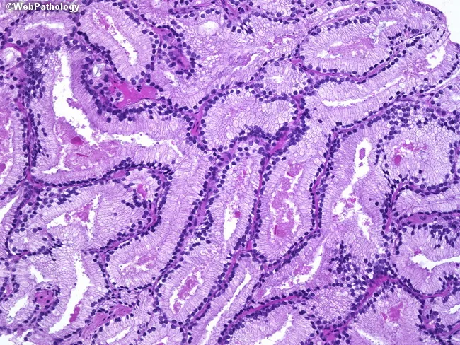
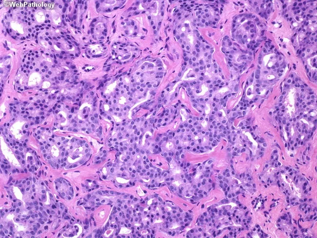
6. GRADING — GLEASON SYSTEM & GRADE GROUPS
The Gleason system grades the two most prevalent architectural patterns (primary + secondary) from 1–5, and sums them for a score from 2–10. In modern practice, Gleason score 2–5 is no longer assigned; minimum assigned is 6.
Five Grade Groups (ISUP 2014 / WHO 2016)
| Grade Group | Gleason Score | Description |
|---|---|---|
| 1 | ≤6 (3+3) | Only individual, discrete, well-formed glands |
| 2 | 3+4=7 | Predominantly well-formed glands + lesser component poorly formed/fused/cribriform |
| 3 | 4+3=7 | Predominantly poorly formed/fused/cribriform + lesser well-formed |
| 4 | 4+4=8; 3+5=8; 5+3=8 | Only poorly formed/fused/cribriform glands OR mix of well-formed and lack of glands |
| 5 | 4+5=9; 5+4=9; 5+5=10 | Lack of gland formation (or necrosis) ± poorly formed/cribriform glands |
Clinical correlation: Grade Group 1 = favorable, often managed by active surveillance. Grade Groups 4–5 = high-risk, typically require aggressive treatment.
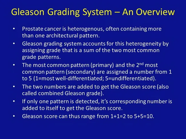
Important nuance: When only one pattern is present, it is doubled (e.g., 3+3=6). The highest-grade pattern (even if tertiary/minor) must be reported if it is grade 4 or 5, because it significantly impacts prognosis.
— Robbins, p. 909; Harrison's 22e, p. 741
7. STAGING — TNM SYSTEM (AJCC 8th Edition)
Clinical T Stage
| Stage | Definition |
|---|---|
| T1 | Clinically inapparent — not palpable, not visible on imaging |
| T1a | Incidental histologic finding in ≤5% of resected tissue (TURP) |
| T1b | Incidental in >5% resected tissue |
| T1c | Found by needle biopsy (elevated PSA only) |
| T2 | Tumor confined within prostate |
| T2a | ≤ half of one lobe |
| T2b | > half of one lobe, not both |
| T2c | Both lobes |
| T3 | Extracapsular extension |
| T3a | Extracapsular extension (unilateral or bilateral) |
| T3b | Seminal vesicle invasion |
| T4 | Fixed, or invades adjacent structures (external sphincter, rectum, bladder, levator muscles, pelvic wall) |
Pathological T Stage (pTNM)
Note: pT1 does not exist (can't assess T1 on a radical prostatectomy specimen). pT2 = organ confined; pT3a = extraprostatic extension ± bladder neck; pT3b = seminal vesicle invasion; pT4 = other adjacent structures.
N Stage
| N0 | No regional nodal metastasis |
| N1 | Regional lymph node metastasis (obturator → iliac → paraaortic) |
M Stage
| M0 | No distant metastasis |
| M1a | Non-regional lymph nodes |
| M1b | Bone metastases (most common) |
| M1c | Other distant sites (visceral) |
Risk Stratification (D'Amico / AUA / EAU)
| Risk Group | PSA | Grade Group | cT Stage |
|---|---|---|---|
| Very Low | <10 ng/mL | 1 | T1c; <3 biopsy cores; <50% any core |
| Low | <10 ng/mL | 1 | T1–T2a |
| Intermediate Favorable | 10–20 ng/mL | 2 | T2b–c |
| Intermediate Unfavorable | 10–20 ng/mL | 3 | or multiple intermediate factors |
| High | >20 ng/mL | 4–5 | T3a |
| Very High | Any | 5 | T3b–T4, >4 cores GG4+, primary Gleason 5 |
| Metastatic (M1) | Any | Any | N1 or M1 |
8. CLINICAL MANIFESTATIONS & SYMPTOMS
Localized Disease (T1–T2)
Often completely asymptomatic — most cases today are detected via PSA screening.
Locally Advanced Disease (T3–T4)
- Lower urinary tract symptoms (LUTS): Hesitancy, weak stream, nocturia, frequency, urgency — due to urethral compression (overlap with BPH; symptom profile alone cannot distinguish)
- Hematuria / hemospermia: With invasion into urethra or seminal vesicles
- Ureteral obstruction: Hydronephrosis, renal failure when tumor extends to bladder base
- Erectile dysfunction: From neurovascular bundle involvement
- Rectal symptoms: From posterior invasion — rare
DRE Findings
- Hard, nodular, irregular nodule — typically in the posterior peripheral zone
- Asymmetric or fixed gland in advanced disease
- 20–25% of patients with an abnormal DRE have prostate cancer; DRE sensitivity for early disease is poor
Metastatic Disease
The pattern of metastasis follows anatomical lymphatics and vascular routes:
Lymphatic spread (in order): Obturator nodes → Internal/external iliac → Common iliac → Para-aortic nodes
Hematogenous spread: Preferentially to axial skeleton via the Batson's vertebral venous plexus (valve-less, bidirectional):
- Lumbar spine (most common)
- Proximal femur
- Pelvis
- Thoracic spine
- Ribs
Bone metastases are characteristically OSTEOBLASTIC (sclerotic) — this is pathognomonic of prostate cancer origin in adult males. The mechanism is tumor-derived growth factors stimulating osteoblast activity while simultaneously stimulating osteoclasts (net osteoblastic effect).
Symptoms of metastatic disease:
- Bone pain: Deep, constant, progressive back/hip pain — initially nocturnal, later continuous
- Pathological fractures
- Spinal cord compression: Epidural extension → acute lower limb weakness, saddle anesthesia, bowel/bladder dysfunction — oncologic emergency
- Hypercalcemia: Less common than osteolytic tumors but can occur
- Anemia: Marrow infiltration → leukoerythroblastic picture
- Lymphedema: Lower limb edema from lymph node obstruction
- Ureteral obstruction / hydronephrosis
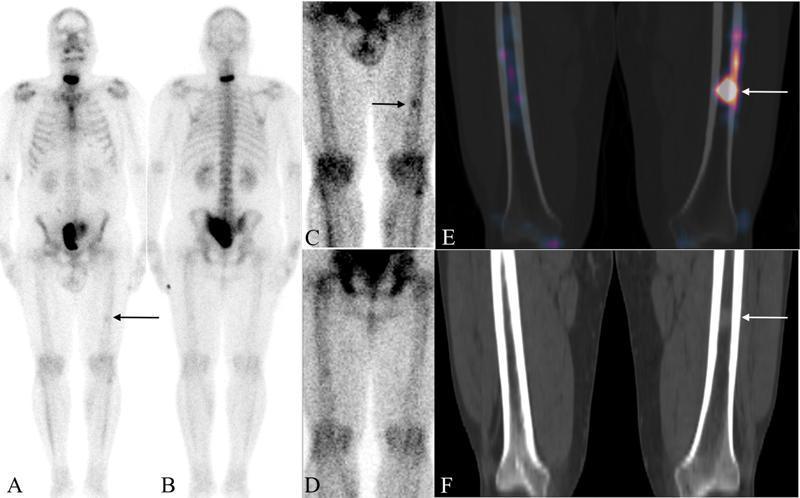
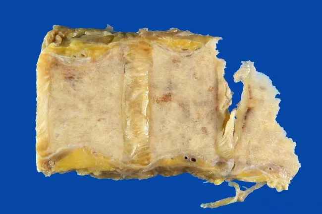
— Robbins, p. 908–910; Harrison's 22e
9. DIAGNOSIS
PSA (Prostate-Specific Antigen)
- A kallikrein-related serine protease (KLK3) — produced by both benign and malignant prostatic epithelium
- Organ-specific, not cancer-specific: Also elevated in BPH, prostatitis, prostate infarction, ejaculation, urethral instrumentation
- In blood: exists as free PSA and complexed (mainly with α₁-antichymotrypsin)
- Lower free:total PSA ratio → higher probability of cancer
- Standard cut-off: ≥4 ng/mL for biopsy; however there is no zero-risk PSA level
- After radical prostatectomy: should be undetectable by 6 weeks; any detectable PSA = biochemical recurrence/residual disease
PSA derivative tests to improve specificity:
| Test | Description |
|---|---|
| PSA density | PSA / prostate volume (TRUS); >0.15 suspicious |
| PSA velocity | Rate of PSA rise >0.75 ng/mL/year |
| Free PSA ratio | Free/total PSA <10% → higher cancer risk |
| PSA doubling time | Rapid doubling (<3 months) = aggressive biology |
| 4K Score | Combines total PSA, free PSA, intact PSA, hK2 + clinical factors → % risk of high-grade cancer |
| PHI (Prostate Health Index) | Combines [-2]proPSA, free PSA, total PSA → better predictor than PSA alone |
| SelectMDx / ExoDx | Urine-based mRNA/exosome markers |
Digital Rectal Examination (DRE)
- Mandatory component of evaluation
- Detects posterior peripheral zone tumors
- Sensitivity/specificity insufficient for standalone screening
- ~20–25% of abnormal DREs have cancer on biopsy
Multiparametric MRI (mpMRI) — Now Standard of Care
Before biopsy in most guidelines. Combines:
- T2-weighted (anatomy, peripheral zone, capsular integrity)
- Diffusion-weighted imaging (DWI) — restricted diffusion in high-cellularity tumors (low ADC)
- Dynamic contrast-enhanced (DCE) — abnormal vascularity
- MR spectroscopy (optional) — elevated choline:citrate ratio
Findings scored using PI-RADS v2.1 (1–5):
- PI-RADS 1–2: Low probability of clinically significant cancer
- PI-RADS 3: Intermediate; biopsy based on clinical context
- PI-RADS 4–5: High probability; biopsy recommended
Prostate Biopsy
- TRUS-guided systematic biopsy: Standard; 10–12 core systematic sampling
- MRI-targeted biopsy (fusion biopsy): MRI-visible lesion + systematic cores — significantly improves detection of clinically significant cancers (GG ≥2) and reduces overdetection of GG1
- Transperineal approach: Lower risk of sepsis vs. transrectal; increasingly preferred post-antibiotic resistance concerns
Imaging for Staging
| Modality | Use |
|---|---|
| Bone scan (Tc-99m MDP) | Standard for bone metastasis detection; indicated when PSA >10–20 or high-risk features |
| CT abdomen/pelvis | Lymph node staging (limited sensitivity for <8mm nodes) |
| PSMA PET/CT | Highly sensitive/specific for nodal and distant metastasis; FDA-approved (⁶⁸Ga-PSMA-11, ¹⁸F-DCFPyL); superior to bone scan + CT; increasingly replacing conventional imaging |
| Whole-body MRI | Alternative for metastasis detection, especially bone marrow |
| ¹¹C-choline PET | Earlier agent; useful for biochemical recurrence detection |
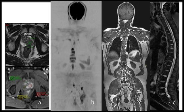
Laboratory Investigations
- Serum PSA (baseline; serial measurements for monitoring)
- Alkaline phosphatase (ALP): Elevated in osteoblastic bone metastases (bone ALP isoform)
- LDH: Tumor burden marker in metastatic disease
- Testosterone: Baseline pre-ADT; castrate level = <50 ng/dL (<1.7 nmol/L)
- CBC: For anemia (marrow infiltration); pre-treatment baseline
- BMP/creatinine: For obstruction assessment
- Germline testing: Recommended for high-risk, metastatic, or family history (BRCA1/2, ATM, PALB2, MLH1, MSH2, MSH6, PMS2, HOXB13)
- Somatic tumor testing: For BRCA2/ATM (PARP inhibitor eligibility), MSI-H/dMMR (pembrolizumab eligibility), CDK12 mutation
10. CLINICAL STATES MODEL (Harrison's Framework)
Prostate cancer management is organized around clinical states:
- No cancer diagnosis (screening/prevention)
- Clinically localized disease (organ-confined)
- Biochemical recurrence after local therapy (rising PSA, no imaging evidence of metastasis)
- Metastatic disease — further subdivided:
- mHSPC: Metastatic hormone-sensitive (castration-naive)
- nmCRPC: Non-metastatic castration-resistant (rising PSA on ADT, no imaging metastasis)
- mCRPC: Metastatic castration-resistant
This model is critical because treatment decisions and goals differ at each state.
11. TREATMENT
11a. Localized Disease
Very Low / Low Risk
- Active Surveillance (AS): First-line for GG1 (Gleason 6) and selected GG2 patients. Serial PSA, DRE, mpMRI, and repeat biopsies. Deferred treatment until evidence of progression. Avoids overtreatment. >90% of GG1 patients will not need immediate intervention.
- Watchful Waiting: Different from AS — no curative intent; palliative intervention only if symptomatic. Used for older men with limited life expectancy.
Localized Intermediate/High Risk — Definitive (Curative) Treatment
Radical Prostatectomy (RP):
- Open, laparoscopic, or robotic-assisted (RALP) — robotic now predominates
- Standard: nerve-sparing RP (if oncologically safe) to preserve potency + continence
- Extended pelvic lymph node dissection (ePLND): Required for intermediate/high-risk
- Specimen assessment: pT stage, Gleason grade, surgical margins (positive margin = higher recurrence risk), lymph node status
- Oncologic outcomes: >90% 15-year survival for high-risk disease treated definitively
- Complications: Urinary incontinence (stress incontinence; 5–15% long-term), erectile dysfunction (30–80% depending on nerve-sparing and patient age)
Radiation Therapy:
- External Beam Radiation Therapy (EBRT): Intensity-modulated RT (IMRT) is standard; daily fractions; total ~70–80 Gy; MRI-guided RT emerging
- Stereotactic Body Radiation Therapy (SBRT): Hypofractionated; 5 sessions; equivalent oncologic outcomes; growing evidence
- Low-dose rate brachytherapy (LDR-BT): Permanent seed implantation (¹²⁵I, ¹⁰³Pd); for low/selected intermediate risk; excellent long-term outcomes
- High-dose rate brachytherapy (HDR-BT): Temporary implant; used as boost with EBRT for high-risk
- Combined EBRT + ADT for 2–3 years: Standard of care for high-risk localized disease (Gleason 8–10, T3–T4)
- Complications: Radiation proctitis, urinary frequency/urgency, erectile dysfunction (delayed onset)
Other Local Therapies:
- Focal therapy (for carefully selected intermediate risk): HIFU (High-Intensity Focused Ultrasound), focal cryoablation, irreversible electroporation — ablate only MRI-visible index lesion. Still investigational for most guidelines but EAU considers it in selected patients.
11b. Biochemical Recurrence (BCR) After Primary Treatment
- PSA rising post-prostatectomy (detectable/rising PSA) or post-radiation (Phoenix definition: PSA rise ≥2 ng/mL above nadir)
- Salvage RT to prostate bed: First-line after RP if PSA is rising; most effective when PSA <0.5 ng/mL
- Salvage RP: After failed radiation; technically demanding; increased morbidity
- ADT: For those with short PSA doubling time (<3 months), high Gleason grade, or nodal recurrence on PSMA PET
- PSMA PET/CT: Critical to identify site of recurrence; guides salvage treatment
11c. Metastatic Hormone-Sensitive Prostate Cancer (mHSPC)
The backbone of treatment is Androgen Deprivation Therapy (ADT) — surgical or medical castration:
Surgical: Bilateral orchiectomy — permanent, immediate, cost-effective; castrate testosterone within hours
Medical (LHRH agonists): Leuprolide, goserelin, triptorelin — chronic administration causes receptor downregulation → pituitary desensitization → ↓ LH → ↓ testicular testosterone. Testosterone flare in first 1–2 weeks (requires short-term antiandrogen coverage with bicalutamide/flutamide).
LHRH antagonists: Degarelix, relugolix — direct receptor blockade, no flare, faster castration; relugolix is oral
Androgen receptor pathway inhibitors (ARPIs): Second-generation; crucial now
| Agent | Class | Mechanism |
|---|---|---|
| Enzalutamide | ARPI | AR antagonist; inhibits nuclear translocation and DNA binding |
| Apalutamide | ARPI | AR antagonist |
| Darolutamide | ARPI | AR antagonist; minimal CNS penetration |
| Abiraterone + prednisone | CYP17A1 inhibitor | Blocks androgen synthesis in adrenal glands AND tumor |
Current standard of care for mHSPC: ADT + intensification
- ADT + docetaxel (CHAARTED, STAMPEDE trials): Benefit mostly in high-volume disease
- ADT + ARPI (enzalutamide, apalutamide, or darolutamide + docetaxel): Demonstrated significant overall survival benefit; now standard
- ADT + abiraterone + prednisone (LATITUDE trial): Improved OS in high-risk mHSPC
- Triplet therapy (ADT + docetaxel + abiraterone, or ADT + docetaxel + darolutamide) — ARASENS, PEACE-1 trials support triplet in high-volume/high-risk
11d. Castration-Resistant Prostate Cancer (CRPC)
Defined as disease progression (PSA or radiographic) despite castrate testosterone levels (<50 ng/dL). AR re-activation is the dominant mechanism:
- AR amplification/overexpression
- AR ligand-binding domain mutations (e.g., F877L from enzalutamide selection; W742C from bicalutamide)
- AR splice variants (AR-V7): Constitutively active; lack ligand-binding domain → resistance to ARPIs
- Intratumoral androgen synthesis
- Lineage plasticity — neuroendocrine differentiation
Treatment of mCRPC
| Agent | Mechanism | Indication |
|---|---|---|
| Enzalutamide, apalutamide | ARPI | mCRPC; pre/post-docetaxel |
| Abiraterone + prednisone | CYP17A1 inhibitor | mCRPC |
| Docetaxel (75 mg/m², q3wk + prednisone) | Taxane | First chemo line |
| Cabazitaxel (25 mg/m², q3wk) | Taxane | Post-docetaxel |
| Olaparib | PARP inhibitor | mCRPC with BRCA1/2 or other HRR mutations (PROfound) |
| Rucaparib | PARP inhibitor | Post-docetaxel + ARPI with BRCA mutations |
| Pembrolizumab | Anti-PD-1 | MSI-H/dMMR or TMB-high tumors |
| ¹⁷⁷Lu-PSMA-617 (Lutetium PSMA) | Targeted radioligand therapy | mCRPC post-ARPI + taxane (TheraP, VISION trials); FDA approved 2022 |
| Radium-223 (Ra-223) | Alpha emitter | Symptomatic bone-only mCRPC; improves OS and delays SREs |
| Sipuleucel-T | Autologous dendritic cell vaccine | Asymptomatic/minimally symptomatic mCRPC |
| Denosumab / Zoledronic acid | Anti-RANKL / Bisphosphonate | Bone-targeted agents; prevent skeletal-related events (SREs); used throughout metastatic disease |
Non-metastatic CRPC (nmCRPC): Rising PSA on ADT, no imaging metastasis, PSA doubling time ≤10 months → Enzalutamide, apalutamide, or darolutamide delay metastasis-free survival significantly (SPARTAN, PROSPER, ARAMIS trials).
11e. Symptomatic Management
- Bone pain: NSAIDs → opioids; bone-targeting agents (denosumab, zoledronic acid); Ra-223 for multiple symptomatic bone metastases
- Spinal cord compression: Dexamethasone immediately → emergency RT or surgical decompression
- Ureteral obstruction: Ureteral stents or percutaneous nephrostomy
- Urinary obstruction/retention: TURP (palliative) or catheterization
- ADT side effects management: Hot flashes (venlafaxine, medroxyprogesterone), osteoporosis (denosumab/bisphosphonates + calcium/D3), cardiovascular surveillance, metabolic syndrome monitoring
12. PROGNOSIS
Localized Disease
-
90% of patients with high-risk localized disease treated with definitive therapy survive 15 years
- Gleason grade group is the single strongest histopathologic prognostic factor
- pT stage + margin status + PSA level = key post-operative prognostic variables
Nomograms
- Partin Tables: Pre-operative — predict pathologic stage based on PSA, Gleason score, clinical T stage
- Kattan nomogram / Memorial Sloan Kettering tools: Predict biochemical recurrence-free survival after RP
- D'Amico risk classification: Standard risk stratification for localized disease
Metastatic Disease
- mHSPC → mCRPC conversion: Median time ~18–24 months on ADT; progression accelerated by high-volume disease, high Gleason grade
- mCRPC: Median OS was historically ~14–18 months with docetaxel; modern combination approaches (ARPI + ADT, PARP inhibitors, lutetium-PSMA) have extended median OS
- BRCA2-mutated mCRPC: More aggressive; however PARP inhibitors (olaparib) provide meaningful benefit
- Biochemical recurrence after RP: Median time to metastasis ~8 years; time to cancer-specific death ~5 years after metastases (PSA doubling time <3 months strongly predicts lethal outcome)
Key Prognostic Factors Summary
| Factor | Favorable | Unfavorable |
|---|---|---|
| Gleason Grade Group | 1–2 | 4–5 |
| PSA at diagnosis | <10 | >20 |
| Clinical stage | T1–T2 | T3–T4, N1, M1 |
| PSA doubling time | >12 months | <3 months |
| Metastasis volume | Low | High |
| AR-V7 status | Negative | Positive (ARPI resistance) |
| BRCA2 mutation | — | Worse prognosis (but PARP targetable) |
| Visceral metastases | Absent | Present (worst prognosis) |
13. GENOMIC TESTING — CLINICALLY USED TODAY
| Test | Type | Clinical Use |
|---|---|---|
| Oncotype DX GPS | 17-gene expression | Active surveillance vs. definitive treatment in low-risk |
| Prolaris | 46-gene cell-cycle panel | Risk of biochemical recurrence; treatment intensification |
| Decipher | 22-gene expression | Predict metastasis after RP; guide adjuvant RT |
| ProMark | 8-protein IHC | Risk stratification at biopsy |
| Germline BRCA1/2 | Germline DNA | PARP inhibitor eligibility; family counseling |
| Somatic BRCA2/ATM | Tumor DNA | PARP inhibitor eligibility |
| MSI/dMMR | Tumor DNA | Pembrolizumab eligibility |
14. SCREENING RECOMMENDATIONS (2024 Guidelines)
| Organization | Recommendation |
|---|---|
| AUA 2023 | Shared decision-making age 50–69; start at 40–45 for Black men, BRCA2 carriers, strong family history; every 2–4 years |
| USPSTF | Individual decision age 55–69; against screening ≥70 |
| EAU 2024 | Risk-adapted early detection; baseline PSA at 45–50; individualized intervals based on PSA level |
PSA <1 ng/mL at age 60 → can cease screening; PSA >1 ng/mL at 60 → continue screening.
Quick-Reference Key Concepts for Your Exam
| Point | Core Fact |
|---|---|
| Most common site of origin | Peripheral zone (~70%) |
| Most common histology | Acinar adenocarcinoma (>95%) |
| Key hallmark on histology | Absent basal cell layer in malignant glands |
| Immunohistochemical marker of cancer | AMACR/P504S positive; p63/CK5/CK903 negative |
| Precursor lesion | HGPIN (retains basal cells) |
| Key molecular alteration | TMPRSS2:ERG fusion (~50%); PTEN loss (~40%) |
| Bone metastasis character | Osteoblastic (sclerotic) |
| Order of bone metastasis | Lumbar spine > femur > pelvis > thoracic spine > ribs |
| Oncologic emergency | Spinal cord compression → dexamethasone + emergency decompression |
| PSA nature | Organ-specific, not cancer-specific; produced by normal AND malignant epithelium |
| ADT mechanism (LHRH agonist) | Receptor downregulation → ↓LH → ↓testosterone |
| CRPC definition | Progression despite castrate testosterone (<50 ng/dL) |
| AR-V7 significance | Constitutively active AR; predicts ARPI resistance |
| BRCA2 clinical relevance | More aggressive; predicts PARP inhibitor response |
| New standard mHSPC | ADT + ARPI (enzalutamide/apalutamide/darolutamide) |
| Lutetium-PSMA | Targeted alpha therapy for mCRPC post-ARPI + taxane |
| 5-year survival (localized) | ~99% |
| 5-year survival (distant metastasis) | ~34% (SEER) |
Sources: Campbell Walsh Wein Urology (9780323546423); Harrison's Principles of Internal Medicine 22e (9781265977061), Ch. 92; Robbins & Cotran Pathologic Basis of Disease (9780443264528), Ch. 21; EAU Guidelines on Prostate Cancer 2024 Part I (PMID 38614820) and Part II (PMID 38688773).That covers everything your professor is likely to ask at a 10-year urology specialist level. Here's a quick map of what was covered so you can navigate:
What you just got — section by section:
- Epidemiology — incidence, mortality, racial/ethnic disparities, autopsy prevalence
- Etiology & Risk Factors — family history, germline mutations (BRCA2, HOXB13, ATM), GWAS loci, diet, obesity, protective factors
- Anatomy & Zones — why peripheral zone matters; contrast with BPH in transition zone
- Pathogenesis — androgen/DHT/AR axis, TMPRSS2:ERG fusion, PTEN loss, AR mutations in CRPC, HGPIN as precursor
- Pathology & Classification — histologic subtypes, gross/microscopic features, IHC markers (AMACR, p63, CK5), perineural invasion
- Gleason Grading — patterns 1–5, grade groups 1–5 with descriptions, tertiary pattern rule
- TNM Staging — full AJCC 8th edition clinical + pathological, risk stratification table
- Clinical Manifestations — asymptomatic localized → LUTS → bone pain → spinal cord compression; lymphatic then hematogenous spread order; osteoblastic nature of bone mets
- Diagnosis — PSA and all derivatives (PHI, 4K Score, free PSA), DRE, mpMRI/PI-RADS, biopsy techniques, PSMA PET, labs, germline/somatic testing
- Clinical States Model — the modern framework (localized → BCR → nmCRPC → mHSPC → mCRPC)
- Treatment — active surveillance, radical prostatectomy, radiation (EBRT/brachytherapy/SBRT), ADT mechanisms and agents, treatment intensification for mHSPC (ARPI, docetaxel, triplet), mCRPC therapies (taxanes, PARP inhibitors, lutetium-PSMA, Ra-223), nmCRPC
- Prognosis — nomograms, Partin tables, PSA doubling time, 5-year survival by stage
- Genomic Testing — Oncotype GPS, Decipher, Prolaris, MSI/BRCA testing
- Screening Guidelines — AUA 2023, USPSTF, EAU 2024
The two most frequently tested "exam traps" at this level: the distinction between clinical vs. pathological T1 (pT1 does not exist in the surgical specimen), and the fact that Gleason score 6 is the minimum assigned today — patients mistakenly think it's "middle of the scale."
This is a shared conversation. Sign in to Orris to start your own chat.