
Right shoulder bankart lesion Right shoulder arthroscopic bankart repair
Reading File
A Bankart lesion is a tear/avulsion of the anterior inferior glenoid labrum (with or without attached periosteum) caused by anterior shoulder dislocation. The arthroscopic Bankart repair reattaches this labrum to the glenoid rim using suture anchors. Let me pull the detailed textbook content on this.
Searching sources...
Finding Sources
Finding Sources
Excellent results from both Campbell's and Rockwood & Green's. Let me read all the key sections in parallel.
Reading File
Reading File
Reading File
Excellent content found. Let me read more of the arthroscopic technique and rehabilitation sections, plus get the Rockwood & Green content on outcomes and indications.
Reading File
Reading File
Reading File
Good, I have extensive technique and rehab data. Let me fetch the key surgical images and read the Rockwood outcomes section plus the full rehab protocol.
Reading File
Reading File
Loading Image
Loading Image
I now have comprehensive information from Campbell's Operative Orthopaedics 2026 and Rockwood & Green's 2025. Let me fetch one more key image showing the arthroscopic repair step, and also read the Bankart lesion pathology section.
Finding Sources
Loading Image
I now have all the information needed to construct a comprehensive answer. Let me compile the full response.
Right Shoulder Bankart Lesion & Arthroscopic Bankart Repair
1. The Bankart Lesion — Pathology
A Bankart lesion is an avulsion of the anteroinferior glenoid labrum from the glenoid rim, occurring as a result of anterior shoulder dislocation. It is considered the "essential lesion" of recurrent anterior shoulder instability.
Types:
| Type | Description |
|---|---|
| Soft tissue Bankart | Avulsion of the labrum and anterior band of the inferior glenohumeral ligament (IGHL) from the glenoid rim, with intact periosteum |
| Bony Bankart | Labral avulsion with a fragment of the anterior glenoid rim bone |
| ALPSA lesion | Anterior Labroligamentous Periosteal Sleeve Avulsion - labrum displaced medially with intact periosteum |
Associated lesions commonly seen at arthroscopy:
- Hill-Sachs lesion - posterolateral humeral head impaction fracture (occurs at the time of anterior dislocation)
- HAGL lesion - Humeral Avulsion of the Glenohumeral Ligament
- SLAP tear - Superior labrum anterior to posterior tear
- Rotator cuff tears (especially in older patients)
A soft tissue Bankart lesion is specifically associated with a tear of the anterior band of the inferior glenohumeral ligament and anterior instability. - Firestein & Kelley's Textbook of Rheumatology
2. Preoperative Workup
Imaging
- Plain radiographs: AP, lateral, axillary views to screen for bony lesions
- MRI or MRA: MRA is more sensitive and specific than MRI for anteroinferior labral tears
- CT scan with 3D reconstruction: Mandatory to quantify glenoid bone loss before surgery
Bone Loss Assessment - the Critical Decision
The amount of glenoid bone loss determines whether arthroscopic Bankart repair (ABR) is appropriate:
"The critical bone loss that changes the indication from ABR to a bone procedure is between 13.5% and 17.3% according to the literature. In high-demand/contact sports patients, a bone-based procedure should be considered at 13.5%; in low-demand patients, 17.3% is the critical threshold." - Rockwood and Green's Fractures in Adults, 10th ed. 2025
- Glenoid bone loss <13.5%: ABR is appropriate
- 13.5-17.3%: Surgeon discretion based on patient demands (contact athlete vs. sedentary)
- >17.3%: Open bony reconstruction (Latarjet procedure) should be considered
A glenoid defect that creates an "inverted pear" shape on arthroscopic evaluation is a sign of significant bone loss requiring a bone procedure.
3. Indications for Arthroscopic Bankart Repair
Arthroscopic Bankart repair is the current treatment of choice for:
- Recurrent traumatic anterior shoulder instability
- First-time dislocation in young, active patients or athletes in certain scenarios
- No significant humeral or glenoid bone loss (below the critical threshold)
- No capsular deficiency
Contraindications / Factors Favoring Open or Bony Procedure:
- Glenoid bone loss >13.5-17.3%
- Engaging Hill-Sachs lesion (may require remplissage procedure concurrently)
- Young male contact-sport athlete (higher recurrence risk)
- Multiple prior dislocations
- Capsular hyperlaxity / generalized ligamentous laxity
4. Surgical Technique - Arthroscopic Bankart Repair
(Based on Campbell's Operative Orthopaedics, Technique 57.10, 15th ed. 2026)
Patient Positioning
Two options:
- Lateral decubitus (preferred by many surgeons): Patient on a beanbag; arm placed in 45-60° abduction, 20° forward flexion with 12-14 lb of traction
- Beach chair: Head holder with patient sat at 70-80° flexion; arm in spider arm holder
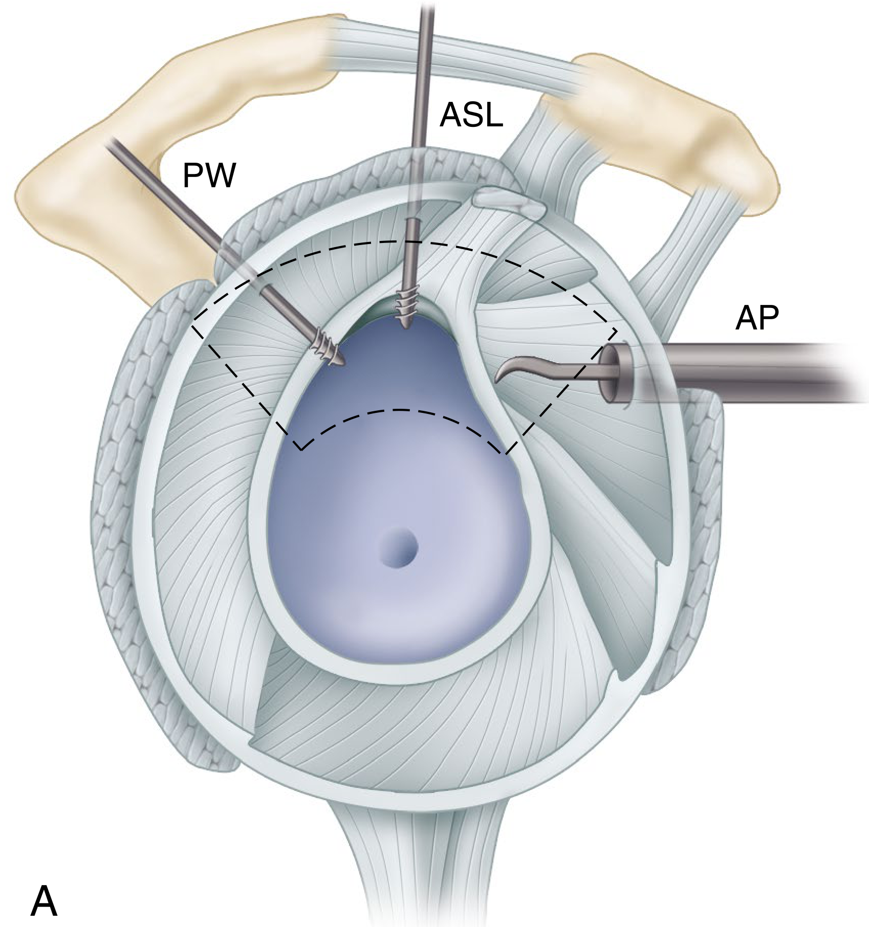
Portals
- Posterior portal: ~2 cm below and 1 cm lateral to the posterolateral edge of the acromion (viewing portal)
- Anterolateral portal: ~1 cm from the anterolateral edge of the acromion (working portal)
- Anteroinferior / 5:30 portal: Just above the subscapularis tendon, slightly above the glenoid fossa (drilling and suture passing)
The diagram below shows portal placement for the four-quadrant approach to labral repair:
Step-by-Step Surgical Steps
1. Diagnostic Arthroscopy
Through the posterior portal with a 30° scope - complete evaluation of labrum, cartilage, rotator cuff, humeral head, and all intra-articular structures.
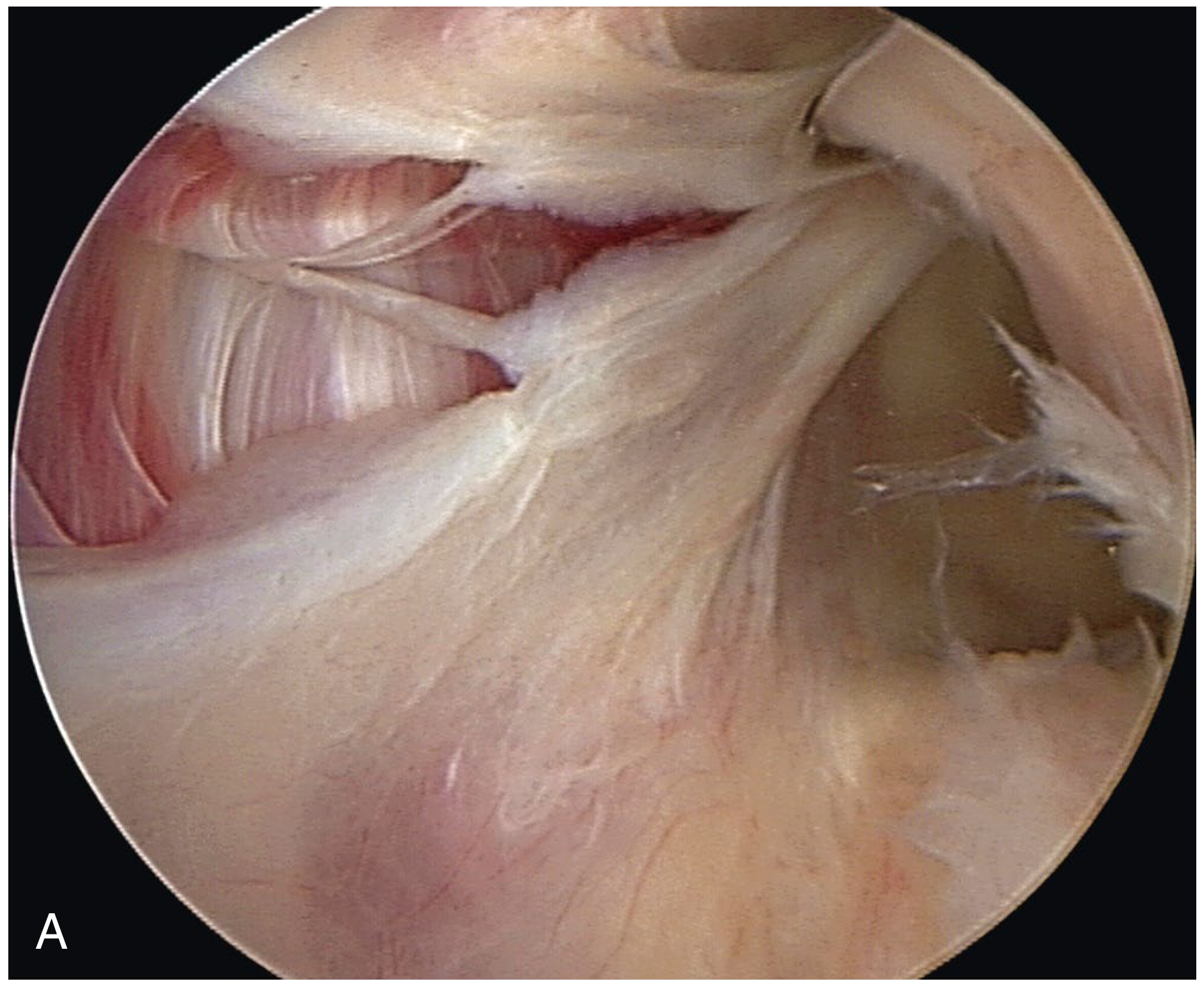
2. Assessment of Pathology
The arthroscopic image below shows a classic Bankart tear with labral avulsion from the anterior glenoid rim:
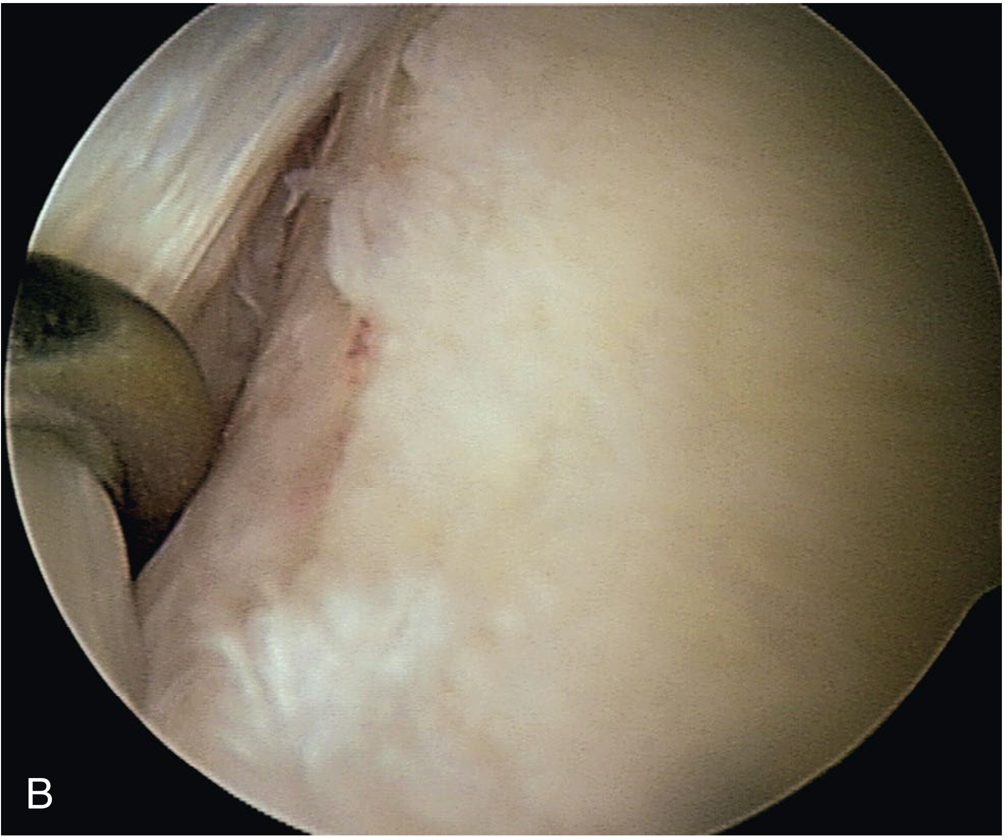
The image below shows the glenoid after labral release and preparation with a rasp - a curved elevator is used to mobilize the capsulolabral tissue down to the 6 o'clock position:
3. Release of the Capsulolabral Complex
Using a curved elevator through the anterior portal, mobilize the capsulolabral tissue from approximately 2 o'clock down to the 6 o'clock position, so the underlying subscapularis muscle can be clearly seen. Complete circumferential release is essential to allow superior advancement.
4. Preparation of the Glenoid Neck
Abrade the glenoid neck with a rasp or motorized burr to create a vascularized bleeding bed for optimal capsulolabral healing.
5. Suture Anchor Placement
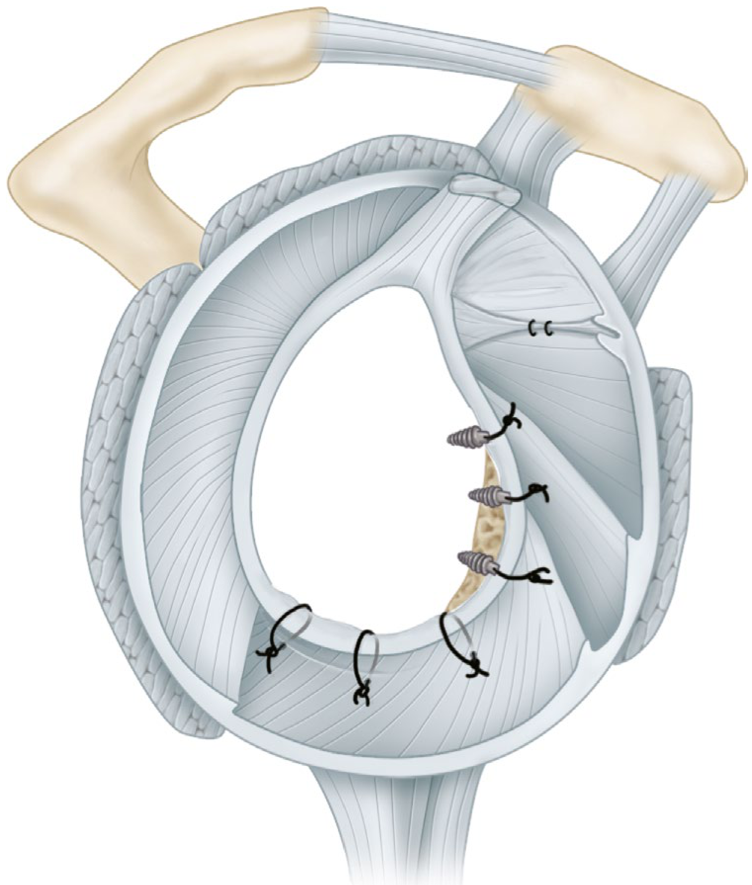
- Place anchors 1-2 mm over the articular surface (at the glenoid rim, not medially on the neck)
- Minimum of 3 suture anchors are required; start inferiorly (6 o'clock position) and work superiorly
- Either knotted or knotless anchors can be used - no reported difference in clinical outcomes
- If knotted anchors are used, ensure knots do not cause articular impingement
6. Suture Passage and Labral Reattachment
Using a suture-passing device or self-retrieving suture passer:
- Start at the 6 o'clock position
- Take a bite of approximately 1 cm of capsule
- Needle should come out through the capsule and pass up under the labrum in correct anatomic position
- Superiorly advance the capsulolabral complex as each suture is tied - this restores physiologic tension and eliminates the "drive-through sign"
7. Address Concomitant Pathology
- Posterior capsular laxity: Posterior capsular plication if PIGHL injury is present
- Hill-Sachs lesion (engaging): Remplissage procedure (posterior capsulodesis) - except in throwing athletes' dominant shoulder
- SLAP tear: Repair through anterosuperior portals
- Rotator interval: Close if significant defect exists
Key Technical Points for Success (Campbell's)
- Careful preoperative assessment of bone defects (3D CT, MRI)
- Arthroscopic examination through anterosuperior portal to evaluate "inverted pear" glenoid defect
- Complete mobilization of capsulolabral complex to 6 o'clock
- Abrasion of glenoid neck for vascularized healing bed
- Superior advancement of the glenohumeral complex to restore physiologic tension
- Minimum 3 suture anchors with secure fixation 1-2 mm over articular surface
- Anchor/knot placement to avoid impingement
- Goal-oriented supervised rehabilitation
5. Postoperative Rehabilitation Protocol
(Campbell's Bankart Repair Rehabilitation Protocol, Table 57.3)
Preoperative Goals
- Independent with postoperative exercise program
- Preoperative strengthening with isometrics in pain-free range
Phase I - Protection & Early Motion
| Timeframe | Goals & Exercises |
|---|---|
| Weeks 1-2 | Sling at all times; pendulum exercises; elbow/forearm/wrist AROM; grip exercises |
| Weeks 3-4 | PT initiated ~day 15; PROM: FL <160°, scaption <150°, ER 30-40°; gentle AAROM; scapular mobility |
| Weeks 5-6 | PROM: FL <170°, ER 45°; AAROM with cane/pulley/wall walks; 25% submaximal isometrics for IR/ER/ABD |
Precautions Phase I:
- Sling at all times except PT
- No true ABD PROM
- No ER with arm abducted from body
Goals by end of Week 4:
- PROM: 150° FL, 150° scaption, 40° ER, 60° IR in 45° scaption
Phase II - Graded AROM and Strengthening
| Timeframe | Goals & Exercises |
|---|---|
| Weeks 7-8 | PROM ER to 70° (week 7) then 90° (week 8); AROM FL/scaption to 90°; isotonics 1-2 lb; UBE; proprioceptive training |
| Weeks 9-10 | PROM ER 90°; progress all AROM to WFL; Jobe rotator cuff program; progressive proprioceptive work |
Goals by end of Week 8:
- Discontinue sling
- PROM FL to WNL, scaption to WNL, ER 70° at 90° scaption
- AROM FL and scaption to at least 90° with proper scapular mechanics
- Independent ADLs (grooming, dressing, car door)
Phase III - Return to Activity
- Progressive strength and endurance
- Sport-specific training from ~months 4-5
- Return to contact sports: typically 6-9 months
6. Outcomes
From Rockwood and Green's Fractures in Adults, 10th ed. 2025:
Recurrence rates after ABR:
- Overall recurrence of shoulder instability: ~10.7-13.1% at follow-up
Risk factors for recurrence:
- Young age
- Higher number of preoperative dislocations
- Significant bone loss (humeral head or glenoid)
- Inferior capsule hyperlaxity
- Male sex / contact sport athlete
Functional scores (long-term, 13 years follow-up, n=180 patients):
- VAS pain: 0.0 ± 1.7 (minimal)
- Rowe score: 90.0 ± 20.5
- ASES score: 92.0 ± 17.0
- Patient satisfaction: 92.3%
ROM changes after ABR:
- Loss of external rotation at side: 3-9°
- Loss of ER at 90° abduction: 3.5-6°
- Loss of forward flexion: 1-3° (minimal)
- ROM after arthroscopic repair is superior to open repair - an important consideration for overhead throwing athletes
Arthroscopic vs. Open Bankart:
| Parameter | Arthroscopic | Open |
|---|---|---|
| Recurrence | ~10-13% | ~2-5% |
| Postoperative ROM | Better | Worse |
| Best for | Most anterior instability, athletes | High bone loss, failed ABR |
7. Special Considerations
Remplissage (for engaging Hill-Sachs):
- Posterior capsulodesis + infraspinatus tenodesis into the Hill-Sachs defect
- Performed alongside ABR for engaging lesions
- Avoid in the dominant shoulder of overhead throwing athletes
Bony Bankart:
If a bony fragment is present but small (non-critical), it can be repaired arthroscopically. If glenoid bone loss is in the critical range, Latarjet procedure (coracoid bone block transfer) is preferred.
Latarjet Procedure (when ABR fails or is contraindicated):
- Transfers the coracoid process to the anterior glenoid rim
- Provides both a bony augment and a sling effect via the conjoint tendon
- Preferred for glenoid bone loss >13.5-17.3%, contact athletes, and revision cases
Sources: Campbell's Operative Orthopaedics, 15th Ed. 2026; Rockwood and Green's Fractures in Adults, 10th ed. 2025; Firestein & Kelley's Textbook of Rheumatology 2022
Complications of this procedure
Searching sources...
Finding Sources
Finding Sources
Finding Sources
Finding Sources
Finding Sources
Finding Sources
FTS searches are returning empty - let me search the Rockwood and Campbell's blocks directly where the Bankart content was found previously.
Reading File
Finding Sources
Good - the Rockwood & Green's block has complications data. Let me search that block more and also check the Campbell's orthopaedics block for complications.
Reading File
Finding Sources
Excellent - the Rockwood block already has detailed complications content starting at line 2109. Let me read more of that section and also check the Campbell's block 29 for complications near the Bankart technique.
Reading File
Reading File
I have the completed repair image. Let me fetch it and also read the full complications section from Rockwood block 12 more carefully.
Reading File
Loading Image
I now have comprehensive, well-sourced data on complications. Here is the full response:
Complications of Arthroscopic Bankart Repair
The overall complication rate of arthroscopic Bankart repair (ABR) is low compared with open procedures. Data from the American Board of Orthopaedic Surgery shows perioperative morbidity is exceedingly rare - infection 0.2%, neurologic injury 0.3%. For comparison: open Bankart has a complication rate of ~4.3%, and Latarjet 10.6-15%. - Rockwood and Green's Fractures in Adults, 10th ed. 2025
Below is the completed repair diagram for reference - three suture anchors reattaching the anteroinferior labrum to the glenoid rim:
Complications Summary
| Complication | Incidence | Notes |
|---|---|---|
| Recurrent instability | 10.7-13.1% | Most common adverse outcome |
| Loss of external rotation | 3-9° (side), 3.5-6° (90° abduction) | Expected, usually tolerated |
| Nerve injury | 0.3% | Axillary, musculocutaneous |
| Infection | 0.2% | Rare |
| Stiffness requiring reoperation | 0.5% | Rare |
| Anchor pullout / hardware issues | Uncommon | Related to technique |
| Glenohumeral arthrosis | Late complication | Related to recurrence & cartilage damage |
1. Recurrent Anterior Instability (Most Common)
The most common adverse outcome after ABR. Overall recurrence rate is approximately 10.7-13.1% across literature reviews.
Risk factors for recurrence:
- Young age (especially males <25 years)
- Contact sport athlete (collision sport, overhead throwing)
- Higher number of preoperative dislocations before surgery
- Significant glenoid bone loss (>13.5-17.3%)
- Engaging Hill-Sachs lesion not addressed at primary surgery
- Inferior capsule hyperlaxity / generalized ligamentous laxity
- Failure to mobilize capsulolabral complex sufficiently to 6 o'clock
- Placement of anchors medially on the glenoid neck rather than at the rim
Comparative recurrence rates by procedure:
| Procedure | Redislocation (%) | Overall Instability (%) |
|---|---|---|
| Arthroscopic Bankart | 15.1% | 20.2% |
| Open Bankart | 7.7% | 20.8% |
| Latarjet | 2.7% | 14.8% |
(From Rollick et al. systematic review - Rockwood and Green's 2025)
Management of recurrence:
- For recurrence without critical bone loss: revision open or arthroscopic Bankart can still be successful. Neviaser et al. reported on 30 patients who failed prior ABR and underwent open repair - at 10.2 years, no patient had apprehension, pain, or instability; ASES 89.44, Rowe scores excellent.
- For recurrence with bone loss: Latarjet coracoid transfer or allograft bone block reconstruction.
2. Loss of Range of Motion / Stiffness
Expected and acceptable loss of ROM after ABR:
- External rotation at side: 3-9° loss
- External rotation at 90° abduction: 3.5-6° loss
- Forward flexion: 1-3° loss (minimal)
Postoperative ROM after arthroscopic repair is superior to open repair - an important factor for overhead throwing athletes.
Pathologic stiffness requiring intervention:
- Incidence requiring secondary surgical release is very low at 0.5%
- Managed first with physiotherapy and glenohumeral corticosteroid injections
- If refractory: arthroscopic lysis of adhesions
3. Neurologic Injury
Incidence: ~0.3%
Nerves at risk during arthroscopic Bankart repair:
| Nerve | Mechanism | Clinical Result |
|---|---|---|
| Axillary nerve | Injury during inferior capsular mobilization, suture passage at 5-6 o'clock position, portal placement | Deltoid weakness, axillary patch hypoesthesia |
| Musculocutaneous nerve | Traction injury, medial retraction with conjoined tendon | Biceps/brachialis weakness, lateral forearm numbness |
| Suprascapular nerve | Posterior portal placement too medial | Supraspinatus/infraspinatus weakness |
| Brachial plexus | Excessive traction in lateral decubitus, improper positioning | Variable motor/sensory deficit |
Prevention:
- Careful suture passage with awareness of axillary nerve location (passes along inferior capsule at 5-6 o'clock)
- Limited traction weight (12-14 lb) in lateral decubitus position
- Proper padding and positioning
4. Anchor-Related Complications
- Anchor pullout: Usually due to osteoporotic bone, anchor placed medially on glenoid neck (not at rim), or inadequate number of anchors (<3)
- Knot impingement / chondral damage: Knotted anchors placed too laterally can abrade the humeral articular cartilage; knotless anchors avoid this
- Anchor migration: Loose anchors can become intra-articular loose bodies
- Suture failure: Suture breakage or cut-through if tissue is thin or repair is overtightened
Prevention:
- Minimum 3 anchors placed 1-2 mm over the articular surface
- Use knotless anchors or carefully bury knots to avoid impingement
- Adequate capsulolabral tissue bite (~1 cm)
5. Infection
Incidence: ~0.2% (exceedingly rare with arthroscopic technique)
- Superficial wound infection: managed with oral antibiotics
- Deep septic arthritis: requires arthroscopic washout, IV antibiotics, possible hardware removal
- Risk factors: diabetes, immunosuppression, prolonged operative time, steroid injections within 3 months
6. Overtightening / Iatrogenic Stiffness
An overcorrected or overtightened repair restricts external rotation excessively. This is more common with open procedures but can occur arthroscopically if:
- Capsular plication is too aggressive
- Rotator interval is closed excessively
- Capsular tissue is advanced too superiorly
Result: loss of ER > 20-30°, functional limitation, and risk of obligate posterior humeral head translation ("Putti-Platt" effect - posterior subluxation from over-constraining the anterior structures)
7. Glenohumeral Arthrosis (Late)
A recognized long-term complication, particularly in patients with:
- Recurrent dislocations before or after surgery
- Intraoperative chondral damage (anchor placement, aggressive glenoid abrasion)
- Prior metallic hardware (historical - less relevant with modern bioabsorbable/all-suture anchors)
- Missed or undertreated bone loss leading to recurrent instability and progressive joint damage
Rates of significant arthritis after ABR are low at long-term follow-up when the repair is successful and instability does not recur.
8. Positioning-Related Complications
| Complication | Position | Mechanism |
|---|---|---|
| Brachial plexus neuropraxia | Lateral decubitus | Excess traction; poor head/neck positioning |
| Common peroneal nerve palsy | Lateral decubitus | Pressure over fibular head |
| Cerebral hypoperfusion | Beach chair | Hypotensive anesthesia + upright position |
| Air embolism | Beach chair | Rare; venous injury near acromion |
9. Fluid Extravasation
Arthroscopic fluid irrigation can extravasate into surrounding soft tissues:
- Anterior chest/mediastinum: rare but potentially serious
- Soft tissue swelling: common, self-resolving
- Risk increases with prolonged operative time and high pump pressures
Key Takeaways
- Recurrence (~10-13%) is the dominant concern, not perioperative complications
- Recurrence risk is heavily driven by patient factors (age, sport, bone loss) more than surgical technique
- Nerve injury is rare (0.3%) but the axillary nerve is the most at-risk structure during inferior capsular mobilization
- Stiffness requiring reoperation is very rare (0.5%) - ABR preserves ROM better than open repair
- Anchor complications are minimized by correct placement at the glenoid rim (not neck) with ≥3 anchors
- For high-risk patients (young male contact athlete, bone loss), primary Latarjet may be preferable to ABR despite its higher complication rate, given far lower recurrence
Sources: Rockwood and Green's Fractures in Adults, 10th ed. 2025; Campbell's Operative Orthopaedics, 15th ed. 2026
This is a shared conversation. Sign in to Orris to start your own chat.