
Pain in sacral region,at back,only on hard touch
Reading File
Pain in the sacral region that occurs only on hard/firm touch (deep pressure or percussion tenderness) is a clinically important finding. The key possibilities include sacral insufficiency fractures, sacroiliitis, coccydynia, sacral bone lesions, or referred pain. Let me search the medical library for a thorough answer.
Searching sources...
Finding Sources
Finding Sources
Searching the Web
sacral region pain only on deep palpation hard touch differential diagnosis
Finding Sources
Finding Sources
Reading File
Finding Sources
Reading File
Finding Sources
Loading Image
Finding Sources
Searching Images
sacral region anatomy posterior landmarks tenderness palpation
Here is a thorough clinical breakdown of sacral region pain that appears only on hard/firm touch (deep pressure or percussion tenderness):
Sacral Region Pain - Only on Deep/Hard Touch
The key clinical point here is that the pain is absent at rest and triggered only by firm pressure or percussion over the sacrum. This narrows the differential considerably, pointing toward deeper structures - bone, joints, or deep ligaments - rather than skin or superficial soft tissue.
Anatomy Map - What You Are Pressing On
The diagram below from Adams and Victor's Principles of Neurology (12th ed.) shows the key posterior pelvic landmarks and what tenderness at each point signifies:
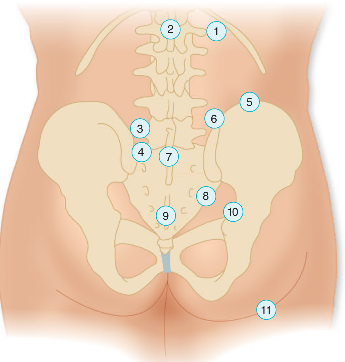
| Point | Location | Tenderness suggests |
|---|---|---|
| 4 | Dorsum of sacrum | Sacral bone pathology |
| 7 | L5-S1 spinous processes | Lumbosacral disc disease or spina bifida occulta |
| 8 | Between PSIS and PIIS - sacroiliac ligaments | Sacroiliac sprain; L5/S1 disc pathology |
| 9 | Sacrococcygeal junction | Coccygeal injury (sprain or fracture) |
| 10 | Sacroiliac notch region | L4/L5 disc rupture or sacroiliac sprain |
- Adams and Victor's Principles of Neurology, 12th Ed., Fig. 10-2, p. 218
Differential Diagnosis
1. Sacral Insufficiency Fracture
- Most common in elderly women with osteoporosis, or after radiation therapy
- Presents as low back/sacral pain that is present at rest but markedly worsened by direct pressure or percussion over the sacrum
- Often bilateral, H-shaped pattern on bone scan ("Honda sign")
- MRI or bone scan is far more sensitive than plain X-ray
- Grainger & Allison's Diagnostic Radiology describes sacral insufficiency fractures as a well-recognized complication of generalized osteoporosis, visible on MRI as bilateral signal changes flanking the sacral ala
2. Sacroiliac (SI) Joint Dysfunction / Sacroiliitis
- Pain typically at or below the posterior superior iliac spine (PSIS)
- Tenderness on direct pressure over the SI joint/sulcus is a cardinal sign
- SI joint pain accounts for 15-30% of chronic low back pain cases
- Provocation tests (FABER/Patrick, Gaenslen's, compression/distraction) help confirm
- Etiologies: osteoarthritis, seronegative spondyloarthropathy (ankylosing spondylitis, psoriatic arthritis), infection, pregnancy-related
- Adams and Victor: "In cases of sacroiliac joint disease, abduction of the upside leg against resistance reproduces pain in the sacroiliac region" (p. 218)
3. Lumbosacral Disc Disease (L5-S1)
- Tenderness over the interspinous ligaments or articular facets between L5 and S1 is consistent with lumbosacral disc disease
- May be accompanied by radiation to the leg (S1 root: posterior calf, heel, lateral foot)
- Adams and Victor, p. 218: "Tenderness over the interspinous ligaments or over the region of the articular facets between the fifth lumbar and first sacral vertebrae is consistent with lumbosacral disc disease"
4. Coccydynia
- Pain specifically at the sacrococcygeal junction on firm pressure
- Occurs notably on sitting; patient shifts from buttock to buttock
- Caused by fall onto coccyx, prolonged sitting, or childbirth trauma
- Goldman-Cecil Medicine: "Coccydynia is manifested by pain in the coccyx area when pressure is applied to the area. This most notably occurs on sitting."
5. Bone Tumor / Metastasis / Myeloma
- Localized deep bone tenderness on percussion is a classic sign of bone infiltration
- Adams and Victor (p. 218): "Localized tenderness is seldom pronounced in disease of the spine because the involved structures are so deep, but there are exceptions - tumor in the spinal bones and epidural abscess, and pathologic or traumatic compression fracture"
- Red flags: age >50, prior malignancy, unexplained weight loss, night pain
- Workup: ALP, PSA (prostate), serum protein electrophoresis (myeloma), Ca2+, MRI
6. Sacral Osteomyelitis / Epidural Abscess
- Rare but serious; deep percussion tenderness + fever, elevated ESR/CRP
- Risk factors: IV drug use, immunosuppression, recent spinal procedure
7. Sacroiliac Ligament Sprain
- After trauma or pregnancy; tenderness over the SI ligaments (between PSIS and PIIS)
- Pain with SI provocation maneuvers
8. Referred Visceral Pain
- Uterine, rectal, or pelvic organ pathology can refer to the sacral area
- However, referred pain typically does NOT produce focal bony point tenderness
- Firestein & Kelley's Textbook of Rheumatology: "visceral pain radiates to the sacral area from lower abdominal diseases"
Key Clinical Clues
| Feature | Think of |
|---|---|
| Elderly + osteoporosis | Sacral insufficiency fracture |
| Young + morning stiffness + bilateral | Ankylosing spondylitis / sacroiliitis |
| Trauma (fall on buttocks) | Sacral fracture or coccydynia |
| Pain below PSIS, + provocation tests | SI joint dysfunction |
| Night pain + constitutional symptoms | Malignancy / osteomyelitis |
| Radiates to posterior leg | L5-S1 disc disease (S1 root) |
| Pain only on sitting, at coccyx | Coccydynia |
Suggested Workup
- History: Age, onset, trauma, constitutional symptoms, posture-related vs. constant
- Examination: Localize the exact point of tenderness (see figure above); provocative SI joint tests; neurological exam of lower limbs
- Imaging:
- X-ray sacrum/pelvis: first step (fractures, spondyloarthropathy)
- MRI pelvis/sacrum: best for soft tissue, stress fracture, infection, tumor
- Bone scan: if insufficiency fracture suspected and MRI unavailable
- Labs (if red flags): CBC, ESR, CRP, ALP, calcium, PSA, SPEP, HLA-B27
Note: The fact that pain is present only on hard touch and absent at rest is reassuring against acute fracture (which usually hurts constantly) and more consistent with SI joint dysfunction, ligamentous pathology, or an early/chronic bone lesion. However, sacral insufficiency fractures in the elderly can sometimes be surprisingly mild at rest.
If you share more details - age, gender, trauma history, associated symptoms, radiation pattern - a more targeted differential can be offered.
What is your age and did this follow any trauma or fall?
It looks like you're asking the patient (yourself?) these questions back to me — but I'm Orris, the AI assistant. I should be asking you these questions to help narrow down the diagnosis!
Let me ask properly:
Asking for Preferences
What is your age and gender? · Did this pain follow any trauma, fall, or injury? · Any associated symptoms? (select all that apply)
Good - this gives a much clearer picture. Here is a focused assessment:
Assessment: Under 30, Minor/Indirect Trauma, No Other Symptoms
Most likely diagnosis: Sacrococcygeal or Sacral Ligament Sprain / Contusion
Given your profile:
- Young age - rules out insufficiency fracture, malignancy, degenerative disease
- Minor/indirect trauma - the most common cause of localized sacral tenderness in young people is a direct contusion or sprain of the sacrococcygeal ligaments/periosteum
- Pain only on hard touch, absent at rest - this "deep tenderness only" pattern is classic for a bone contusion or periosteal bruise, where the injury is sub-threshold at rest but the nerve endings in the periosteum respond sharply to mechanical pressure
- No radiation, no stiffness, no fever - makes disc disease, sacroiliitis, and infection very unlikely
What Likely Happened
A minor bump, awkward landing, prolonged sitting on a hard surface, or even a misstep can cause:
| Condition | What it is |
|---|---|
| Sacral/coccygeal contusion | Bruising of the bone's outer layer (periosteum) - very pain-sensitive on pressure |
| Sacrococcygeal ligament sprain | Micro-tear of ligaments around the lower sacrum/coccyx junction |
| Periosteal bruise | Direct trauma to the sacral dorsum - tender to firm palpation only |
What You Can Do
Conservative management (usually resolves in 2-6 weeks):
- Avoid direct pressure - use a coccyx/donut-shaped cushion when sitting
- NSAIDs (e.g., ibuprofen 400 mg with food) for 5-7 days to reduce periosteal inflammation
- Cold pack for first 48 hours, then warm compresses
- Avoid activities that reproduce the pain (prolonged sitting on hard surfaces, impact sports)
When to Seek Medical Review
Go see a doctor if:
- Pain starts occurring at rest or at night
- Pain worsens over 2 weeks instead of improving
- You develop numbness, tingling, or weakness in the legs
- Fever or chills appear (rules out infection)
- The mechanism of injury was more forceful than initially recalled (rules out fracture)
Bottom line: In a person under 30 with minor trauma and no systemic symptoms, sacral tenderness only on firm pressure is almost always a benign contusion or ligamentous sprain. No imaging is needed at this stage unless it fails to improve or new symptoms develop. Rest, cushioning, and anti-inflammatories should be sufficient.
This is a shared conversation. Sign in to Orris to start your own chat.